In modern medicine, MRI is often used both during primary and preventive examinations, and before and after surgical interventions.
Using nuclear magnetic resonance, radiologists obtain a three-dimensional image of internal organs and systems, which allows them to make an accurate diagnosis and determine how best to treat the patient.
The effectiveness of the procedure is beyond doubt, but not all patients are confident in its safety and are aware of exactly how magnetic resonance imaging affects the body.
Contraindications for MRI
The procedure is not indicated in the following cases.
- When using a pacemaker - regardless of the severity of the pathology and the need for diagnostics. There is only one advanced model of pacemaker, the wearing of which does not interfere with MRI scanning.
- The presence of metal inserts or the use of prostheses. Any metal objects can damage and even destroy the magnetic tomograph.
- Pregnancy. Although the procedure is safe, it is used with caution in pregnant women. This diagnosis is safer than CT and X-rays, but the effect of the procedure on the fetus has not been fully studied.
- Claustrophobia. Fear of a confined space is a contraindication, since the load on the psyche of such patients during the procedure is great. If necessary, anesthesia may be used.
With the exception of the listed conditions, MRI is allowed for everyone. Diagnosis is prescribed with caution to young children who will not be able to remain still during the entire process.
The influence of power on the human body
Modern tomographs operate using magnetic fields ranging from 0.2 to 12 Tesla (T). The quality of the resulting images depends on this indicator. In clinical practice, devices with a power exceeding 3.5 Tesla are not used. The limit of the maximum safe magnetic field strength for humans has been established experimentally and is equal to 4 Tesla. When this threshold is exceeded, the transmission of nerve impulses is inhibited. The use of fields with a power of 6 Tesla or more leads to an increase in blood pressure in 60% of subjects.
A relationship has been established between the amplitude of ECG waves and the time spent in the magnetic voltage zone. Thus, when exposed to a field of 2.0 T, after half an hour a twofold increase in the height of the indicators on the film is observed. However, the described effect produces only a constant high-intensity field. Moreover, during experiments it was proven that increasing the amplitude of ECG indicators by 4 times does not have a significant effect on the functioning of the cardiovascular system.
Radiofrequency waves, which are used in MRI, are theoretically capable of heating biological tissue. But experiments using a magnetic field and radio frequencies have shown that even long-term scanning cannot lead to an increase in temperature by more than 1 degree.
MRI of the head
How long does an MRI take?
The diagnostic method helps to examine the required organ by applying a magnetic field that creates an image of the body part being studied. Using MRI, an image of the target area is obtained, which shows possible pathologies. The scan in most cases lasts no more than half an hour.
The duration of the procedure varies depending on the complexity and scope of the study. Thus, diagnostics of the brain takes about 20 minutes, the spine – 25 minutes, a comprehensive examination of the whole body lasts up to an hour. Preparation lasts about 15 minutes.
Preparing for an MRI with contrast
Preparation largely depends on which organs and systems will be examined.
The bladder is better visible when full, so you should refrain from urinating an hour before the test, and drink 2 glasses of water 20 minutes before the test.
If the gastrointestinal tract is examined, it is recommended to follow a low-carbohydrate diet for several days and exclude foods that contribute to excess gas formation: brown bread, legumes, milk, raw vegetables and fruits. The last full meal should be 6-8 hours before the procedure, but a light snack is allowed.
MRI of the head, neck, and joints does not require special preparation.
Clothing is preferable to a loose fit, without metal parts.
It is better to leave hairpins, piercings, jewelry, and watches at home.
How often can I get an MRI?
In essence, an MRI machine is a magnet, which, when activated, excites hydrogen atoms in the human body. True to its name, the survey produces a strong magnetic effect without causing damage. Since the diagnosis is harmless, it is carried out with the frequency necessary to adjust treatment, observing short time intervals between hospital visits. In most situations, a consultation and referral for the procedure should be done. MRI is a measure of monitoring the condition of the body, therefore it is often used in the postoperative period. To prevent pathologies, MRI of different areas is done at prescribed intervals. For example, brain scans should be done twice a year.
Advantages and disadvantages of the MRI method
The undoubted advantages of the method include the following factors:
- the study is carried out quickly enough, the doctor receives the result immediately;
- The quality of the images is high, the projections are clear, it is possible to “rotate” the three-dimensional image of the organ, which allows the doctor to accurately determine its condition. The images obtained with an MRI machine are much more contrasting than with ultrasound or CT machines;
- The magnetic field used during the study is safe for humans, although its magnitude exceeds the Earth’s own magnetic field tens of times. Simply put, an MRI machine is a magnet. And since it is a magnet, this adds disadvantages to the method, which are discussed below;
- During the examination, the patient is not exposed to radiation, as with an x-ray;
- the research method is non-invasive, that is, there is no need to make incisions on the patient’s body and give him anesthesia, which is harmful to the heart;
- The study can be done in many public and private clinics; today many centers are equipped with such devices.
The result is a three-dimensional image showing the organ under study from different angles and from all sides.
Despite such serious advantages, the MRI method has a number of disadvantages and limitations associated with the principle of its operation:
- Since the MRI machine is a magnet, patients with pacemakers, artificial valves, metal prostheses and rods in the bones, in general, everything that can be attracted by a magnet, are not allowed to be examined. The magnetic field is strong enough to rip a metal fragment out of a person's body. Therefore, even tattoos with a ferromagnetic component will be a contraindication for research;
- The patient must lie absolutely still to obtain a high-quality image. Even due to the movement of the chest during breathing, inaccuracies appear in the images, which can be removed with subsequent computer processing. This causes certain difficulties when performing research for a child or pregnant women who, due to their age or position, find it difficult to lie still. Therefore, an anesthesiologist is often invited to the child to perform the procedure under anesthesia;
- the effectiveness of lung examination is quite low due to the patient’s breathing;
- does not detect formations such as stones, some types of bone pathologies;
- high cost of the device, requirement for the premises - it must be shielded from interference;
- The size of the tomograph itself sometimes becomes a limitation: people who are too fat and people weighing more than 130 kg may not fit into the device.
MRI should be performed with caution in patients with claustrophobia and in pregnant women, although pregnancy is not a contraindication. Often pregnant women are prescribed an MRI instead of an X-ray.
How many times a year can an MRI be done without consequences?
There is no clear answer to this question. Since the electromagnetic field is not capable of harming the human body, MRI can be prescribed an unlimited number of times, depending on diagnostic needs.
How often are MRIs with contrast allowed?
The doctor can use contrast, which allows him to better see the areas of interest in the image. MRI with contrast helps to identify soft tissue pathologies in the early stages. The substance, administered intravenously or taken orally, stains blood vessels. Although the components of the substance are safe, some people have individual intolerance, so contrast enhancement is prohibited for them.
How often can an MRI of the brain be done?
MRI of the brain should be done in the following cases:
- somatic disorders accompanied by headache or dizziness;
- if you suspect a brain tumor;
- in case of damage to the blood flow in the head or stroke;
- MRI of the occipital part of the head is performed in case of disruption of the spinal cord;
- for pain and after surgery.
MRI of the head is done as often as required by treatment or recovery after surgery. Although the number of such checks is unlimited, the doctor rarely prescribes more than two procedures per year. The need for scanning should be based on the nature and characteristics of the pathology.
Is it possible to perform tomography of the same organ several times?
You can repeat an MRI of one organ if necessary for treatment. The procedure does not harm the body. Repeated examination is a tool for monitoring the course and development of the disease, the passage of therapy or the recovery process after surgery. The image allows you to track changes occurring in the body. In some cases, it is necessary to conduct several examinations within one day. Since tomography is harmless, it is allowed to be performed in parallel with other examinations. If necessary, you can do an MRI twice in a row: before and immediately after surgery.
How does research affect the body?
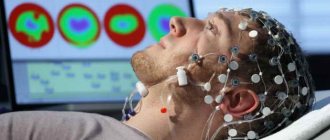
MRI is performed using special equipment (tomograph). The device emits a constant and/or alternating magnetic field. It is especially important how water molecules in tissues react to the latter. Hydrogen atoms inside these compounds are arranged in a specific way and change the polarity of the substance. When radio waves pass through, the orientation of the molecules undergoes a transformation with the release of energy. The tomograph's receiving coils capture state conversion, which a computer program converts into images. Body tissues are magnetized to varying degrees, depending on the amount of fluid they contain. This feature ensures high accuracy and detail of scanning results.
MRI does not carry radiation, does not stimulate the formation of free radicals in the body, and does not change the function or structure of cells. There were no cases of harmful effects of the procedure recorded.
How often can a child’s body scan be done?
Magnetic resonance imaging is widely used in pediatrics. Since the procedure is safe, even a small patient does not feel discomfort. There are practically no side effects, so MRI is often recommended for the child.
The downside is the need to remain motionless to obtain an objective picture. To achieve this, a short general anesthesia is used. To avoid negative consequences of the anesthetic, MRIs should not be performed frequently in children. Anesthesia is not a requirement for the procedure, as many children are able to remain still for several minutes while the scanning process takes place. In such cases, repeated examinations can be carried out over a short period.
Before screening, you should conduct a preventive preparatory conversation with the child: explain the purpose and how the process will take place. The psychological factor is the basis in any interaction between children and doctors. Let us note that science does not fully know the effect of a magnetic field on the developing organism of children under three years of age.
History of MRI development
MRI (magnetic resonance imaging) is a method for studying the condition of human internal organs and blood vessels. The year of its active introduction into medicine is considered to be 1973, when the American chemist Paul Christian Lauterbur published an article in the journal Nature in which he explained how it is possible to obtain an image using the nuclear magnetic resonance effect.
Lauterbur, in collaboration with British physicist Peter Mansfield, refined this idea. The result of their joint scientific research was an MRI machine. For their contributions to medicine, they were awarded the Nobel Prize in Medicine in 2003.
How necessary is the procedure to study intracranial structures?
How many times a brain MRI can be done depends on the diagnosis and the purpose of the diagnosis. Typically the procedure is prescribed for:
- identifying pathologies (inflammatory, tumor, vascular and other nature);
- clarification of the extent of the lesion (the amount of destruction of the brain matter, the presence of metastases, the size of neoplasms, necrotic foci, the degree of vascular stenosis, etc.);
- monitoring the effectiveness of therapy.
Brain pathologies and the specifics of their treatment in most cases eliminate the need for multiple tissue examinations in a short period of time. Diagnosis of diseases and collection of information about them involves 1 MRI procedure. In rare cases, it may have to be repeated to obtain detailed information. The minimum interval for monitoring the effectiveness of therapy is 3 months.
MRI is a highly informative and safe procedure, the potential harm of which is negligible compared to the benefits. Repeated studies can be carried out as many times as necessary to make a correct diagnosis or exclude a medical error.
How often can an MRI be performed?
Knowing about the absolute harmlessness of this examination, some especially suspicious patients, worried about the course of their disease, consider it necessary to regularly prescribe an MRI. But at the same time, they are afraid and interested in how many times a year an MRI can be done so that it in no case entails the development of any pathologies. To do this, it is worth considering in detail the types of procedures and categories of patients.
Conducting in contrast
To obtain higher quality images for certain diseases, MRI is performed using a contrast agent made from gadolinium salts. The contrast agent makes it possible to recognize pathological changes in the initial stages and various neoplasms of the smallest size. In most cases, contrast is used to perform MRI of the vessels of the head.
Preparations made on the basis of gadolinium salts are non-toxic and absolutely harmless to the human body. However, some patients may have individual intolerance to certain components of the contrast agent, which will require a complete refusal of the procedure or reducing its amount to a minimum. In this case, you can do an MRI without contrast or try to replace the examination with another one that can provide an equally reliable result necessary to establish a diagnosis.