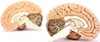
The cerebellum of the human brain is responsible for the normal functioning of muscle fibers, their tone, coordination of movements, balance and adaptation in space. It is divided into a middle part (represented by a worm) and two hemispheres, which are located on the sides. On the ventral side, a small formation is adjacent to the hemisphere - a shred.
The cerebellum contains the processes of neurons, and the cortex is located on its surface. The cerebellar cortex is a superficial layer that has 3 layers: molecular, ganglionic and granular. Under the cortex is white matter formed by myelinated nerve fibers: some fibers (afferent) go to the cerebellar cortex from the spinal cord and medulla oblongata, others (climbing) - from the cortex to the subcortical nuclei.
We will try to understand the structural features of the organ, as well as its functional responsibilities, in more detail.
Where is the cerebellum located?
Many people are interested in what the cerebellum is. So it consists of almost 50% of the neurons of the entire central nervous system, despite this it is small in size and weight (up to 150 g). Located in the cranial cavity between the temporal lobes. Due to this localization, the small brain communicates with other parts of the central nervous system, which are responsible for the functioning of the entire organism.
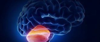
The cerebellum is located under the cerebral hemispheres behind the brain stem and pons. Due to this arrangement, it manages to perform its functions.
Anatomy of the cerebellum
The structure of the cerebellum resembles the structure of the cerebral hemispheres. Let us consider each of the structural units and functions of the cerebellum in more detail.
Worm
The worm is a narrow strip. A small element runs along the side, called the amygdala, and is responsible for motor activity and balance. The amygdala is divided into upper and lower parts, grooves are located on the sides, they are assigned the function of separating the worm from the hemispheres.
The outer surface of the worm is covered with gray matter, which controls body posture, muscle fiber activity, and balance. A disorder in its functioning causes problems with walking and standing.
Shares
The lobes of the cerebellum are separated by large grooves, one of which is in contact with the surface of the hemispheres. Their totality is considered part of the small brain.
The vermis and lobes of the hemispheres are located at the same level. They are distinguished: tongue, central particle, apex, slope, letter, tubercle, pyramid, sleeve, nodule.
realizing them through the entire structure of the central nervous system.
The cerebellum takes part in various activities of the body:
- motor,
- somatic,
- vegetative,
- sensory,
- integrative, etc.
The cerebellum consists of two hemispheres and has a cortex of gray matter. The cortex contains cells with numerous dendrites that receive impulses from many sources associated with muscle activity: proprioceptors of tendons, joints and muscles, as well as from the motor centers of the cortex. Therefore, the cerebellum integrates information and coordinates the work of all muscles involved in movement or maintaining a posture. The cerebellum is absolutely necessary for coordinating fast movements such as running, typing on a keyboard, and talking.
The cerebellum is anatomically and functionally divided into archi-, paleo- and neocerebellum.
- Archicerebellum
(ancient cerebellum) - the flocculomedullary lobe belongs to it, has the most pronounced connections with the vestibular system, which explains the importance of the cerebellum in the regulation of balance. - Paleocerebellum (old cerebellum) - consists of sections of the cerebellar vermis, pyramid, tongue, parafloccular section and receives information mainly from the pro-prioreceptive systems of muscles, tendons, periosteum, and joint membranes.
- Neocerebellum (new cerebellum) - includes the cortex of the cerebellar hemispheres and parts of the vermis; it receives information from the cortex, mainly along the fronto-pontine-cerebellar pathway, from the visual and auditory rehearsal systems. This indicates its participation in the analysis of visual and auditory signals and in the organization of appropriate reactions.
The cerebellar cortex creates the conditions for rapid information processing
Functions of the organ and what it is responsible for
The small brain plays the role of evaluator when making decisions. The functions of the cerebellum of the human brain are the reception and storage of information about the results of this action and possible consequences.
The cerebellum performs the following functions:
- maintaining the tone of muscle fibers and control over body posture;
- adjustment of slow movements and their coordination in relation to body position;
- control of the process of performing movements that require quick reactions.
To study as best as possible the characteristics of this organ and why it is needed, scientists conducted a number of studies on animals. The cerebellums were removed, and as a result, the following development was observed:
- astasia - the animal felt unsure in space, it spread its limbs wide and swayed;
- atony - disruption of the ability of normal functioning of muscle fibers, loss of the ability to flex and straighten limbs;
- asthenia - loss of the ability to exercise control over one’s movements;
- ataxia - sudden movements.
Consequences of disruption
One way or another, the cerebellum, like any structure of the nervous system, is capable of succumbing to various diseases and conditions, including infectious diseases, traumatic brain injuries or tumors. People who have survived various diseases subsequently ask themselves the question of how to train the cerebellum .
The development of cerebellar functions can be achieved by performing a number of simple exercises, including:
- Perform 15 tilts in a position where the feet are adjacent to each other with eyes closed.
- Raising and lowering the leg with bending of the knee joint with eyes closed. Must be repeated up to 20 times.
Static position when one foot is placed in front of the other. To do this, you need to close your eyes and stand for 20-30 seconds. The key to how to develop the cerebellum lies in the performance of these actions, which are imprinted in the brain and, after a short course of repetition, become fixed as reflexes. These exercises must be performed systematically for a month.
Symptoms of cerebellar damage
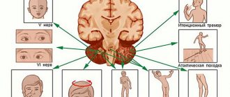
When the functioning of the small brain is disrupted, asthenia, ataxia and atony are more often observed. A variant of the norm is considered to be a situation where a person’s movements are coordinated, and in the process of their execution various groups of muscle fibers are actively involved, their contraction and relaxation occurs with a certain force at the right time. When the cerebellum is damaged, this process is disrupted.
Pathologies
Let us consider in more detail the characteristics of the pathologies that occur in disorders of the functioning of the cerebellum.
Ataxia
The characteristic symptom of the pathology is the patient’s unsteady gait. It seems to him that he is losing support under his feet, as a result he spreads his legs so as not to fall. When performing the Romberg test (consists of standing on your legs closed together), a positive result will be observed.
Dystonia
The tone of muscle fibers, mainly flexors and extensors, is disrupted. Performing any exercise leads to rapid muscle fatigue.
Dysarthria
This condition is characterized by impaired pronunciation, speech becomes slow, incomprehensible and slurred. It can also become chanted and fragmentary.
Adiadochokinesis
Damage to the cerebellum causes loss of the ability to analyze and process information received about the speed and strength of movements. There is a loss of the ability to perform them smoothly. The patient is asked to stretch his arms in front of him and return them. One of the upper limbs will lag behind (movements become asymmetrical).
Dysmetria
Characterized by loss of ability to perform operations related to precision. This deviation is explained by a violation of the coordination of muscle fibers - antagonists.
Diagnosis of organ problems
Identifying the problem, in the case of vermiform aplasia, using ultrasound examination is possible even during intrauterine development. Children are born with a large number of neurological disorders. They require mandatory treatment and a recovery course.
Identification of disorders of small brain structures occurs in a neurological office, through special exercises. If one of the hemispheres is damaged, a finger-nose test is performed, it allows you to determine the area that is damaged, and it is in that direction that the finger will deviate. If the ancient cerebellum or archicerebellum is damaged, there will be a loss of the ability to navigate in space.
To identify cerebellar ataxia caused by tumors, a comprehensive examination by specialized specialists is carried out. Examination of the cerebellum can be carried out using:
- performing a spinal tap;
- computed tomography;
- magnetic resonance imaging;
- Dopplerography;
- electronystagmography;
- DNA diagnostics.
Adenomas and cysts at the initial stage of development are detected using MRI of the brain.
Diseases and pathological conditions
Atrophic changes in the cerebellum
Signs of atrophy:
- headache;
- dizziness;
- vomiting and nausea;
- apathy;
- lethargy and drowsiness;
- hearing impairment; walking impairment;
- deterioration of tendon reflexes;
- ophthalmoplegia – a condition characterized by paralysis of the oculomotor nerves;
- speech impairment: it becomes inarticulate;
- trembling in the limbs;
- chaotic vibration of the eyeballs.
Cerebellar dysplasia in a child
Dysplasia is characterized by improper formation of the substance of the small brain. Cerebellar tissue develops with defects that originate in fetal development. Symptoms:
- difficulty performing movements;
- tremor;
- muscle weakness;
- speech disorders;
- hearing defects;
- blurred vision.
The first signs appear in the first year of life. Symptoms are most pronounced when the child is 10 years old.
Cerebellar deformity
The cerebellum can be deformed for two reasons: tumor and dislocation syndrome. The pathology is accompanied by impaired blood circulation in the brain due to compression of the cerebellar tonsils. This leads to impaired consciousness and damage to vital regulatory centers.
Cerebellar edema
Due to the enlargement of the small brain, the outflow and inflow of cerebrospinal fluid is disrupted, which causes cerebral edema and stagnation of cerebrospinal fluid.
Signs:
- headache, dizziness;
- nausea and vomiting;
- disturbance of consciousness;
- fever, sweating;
- difficulty holding a pose;
- unsteadiness of walking, patients often fall.
When arteries are damaged, hearing is impaired.
Cerebellar cavernoma
Cavernoma is a benign tumor that does not spread metastases to the cerebellum. Severe headaches and focal neurological symptoms occur: impaired coordination and accuracy of movements.
Late cerebellar degeneration
It is a hereditary neurodegenerative disease accompanied by the gradual death of the cerebellar substance, which leads to progressive ataxia. In addition to the small brain, the pathways and brain stem are affected. Late degeneration appears after 25 years. The disease is transmitted in an autosomal recessive manner.
The first signs: unsteadiness of walking and sudden falls. Speech gradually deteriorates, muscles weaken and the spine becomes deformed like scoliosis. 10-15 years after the first symptoms, patients completely lose the ability to walk independently and need help.
The influence of the cerebellum on autonomic functions
The small brain is endowed with the ability to influence the functioning of all body systems. Damage to the cerebellum leads to a decrease in blood pressure and the development of bradypnea. The tone of the respiratory muscles on the affected side will be decreased, while on the opposite side there will be an increase.
The tone of the intestinal muscle fibers changes, it will be reduced, and this causes a disruption in the removal of the contents of the intestines and stomach. There is a suppression of the absorption of nutrients. Metabolic processes accelerate, the level of glucose in the blood increases, which persists for a long period of time compared to the norm. Appetite worsens, the patient loses weight, and the process of tissue regeneration is suppressed.
In drawing a conclusion, I would like to focus on the fact that the cerebellum, despite its small size, plays a very important role in the functioning of the entire organism. Its defeat can cause severe disorders; in the absence of adequate therapy, this is fraught with irreversible consequences.
