Structure
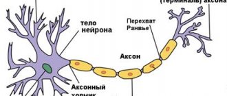
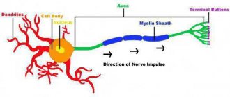
The general structure of a neuron is as follows: there is a body (soma), which contains the nucleus and other organelles, and processes - the axon and dendrites:
- axon
- this is the process along which the nerve impulse travels from a given cell to others. In other words, the axon is a signal output channel. - Dendrites
, accordingly, are channels for signal input, and there can be either a lot of them or very few. The number of dendrites depends on the type of neuron, and we will talk about this later.
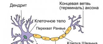
Rice. 1. Neuron diagram
Axons and dendrites
Axons are processes that can reach a length of more than a meter. To prevent the signal from “scattering” along the way from one cell to another, most axons in the body are covered with a myelin sheath, consisting of neuroglia cells (a general designation for the supporting cells of nervous tissue). The sheath provides insulation of one axon from the others and does not allow the electrical impulse to dissipate. Thanks to the myelin sheath, impulses travel along the axon more quickly. Dendrites are shorter and not covered with myelin.
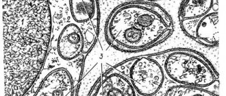
Endoplasmic reticulum (ER) and Golgi complex
The most important organelles, in addition to the nucleus, are the rough ER, which has ribosomes and carries out protein synthesis, and the Golgi apparatus, which synthesizes various organic substances and “packs” them into membrane vesicles. Why these systems are so important for the functional activity of a neuron will be clear later.
Functional characteristics of afferent, efferent and interneurons (the concept of “neuron”)
Neurons are specialized cells, capable of receiving, processing, encoding, storing, transmitting and reproducing information, organizing divisions into units, establishing contacts with other neurons and other organs. It is capable of generating electric potential and with their help, before information, through special endings - synapses. The neurotransmitters synthesized in its axoplasm contribute to the fulfillment of the function. Size 6-120 microns. There are up to 1011 neurons in the brain. The different types of brain organization are characterized by certain types of neural organization. Neurons form groups, nuclei, etc. Cellular accumulations of a gray image. Myelinated and non-myelinated fibers pass between the nuclei and cells: axons and dendrites. Structure: functions distinguish a trace part: the perceptive part - dendrites, soma, on which there is an axon hillock; transmitting part-axon. Soma: informative, trophic in relation to the processes. Sod-it: ribosomes, tigroid in-vo, CG, lysosomes, pigments-melanin and lipofuscin, mitochondria, neurotubes, nucleus with nucleolus. Dendrites - receive impulses from other nerve cells. The axon hillock is the generating point of the action potential. The axon conducts excitation to other cells. Types of neurons: by the number of processes: pseudounipolar (2 processes, merging near the body into 1), bipolar (1 axon, 1 dendrite), multipolar (several dendrites, 1 axon). By direction: afferent, efferent and intercalary. Depending on the nature of the influence: exciting and inhibitory. From activity: background-active (active without influence) and silent (active in response to irritation). From mediator: adrenergic, lolinergic, serotonergic, etc.
Afferent neurons - neurons that perceive information. as a rule, they are highly branched. This is typical for all levels of the central nervous system. In the dorsal horns of the spinal cord of the brain, small neurons sense neurons. Interneurons (interneurons) process the information received from neurons and transmit them to other interneurons or efferent neurons, forming a neural network). Strengthens the signal, lengthens the time the information is stored in the center. Insertion neurons can be excitatory (in the neocortex, facilitating the transfer of information from one group of neurons to another); and inhibitory (which are excited by direct signals or signals coming from the same center via feedback). Efferent neurons are neurons that transmit information from the nerve center to the executive or other centers of the nervous system. They connect different parts of the brain, providing interhemispheric and intrahemispheric connections, forming the function of the brain (fatigue, learning, etc.).
Neuroglia. Its types. Functional characteristics and physiology. role. Gliocyte pulsation and its significance.
Neuroglia is a collection of cells of nervous tissue, formed by special cells of various shapes. It was discovered by Virchow. Clusters of glia fill the spaces of m/u neurons, making up 40% of the brain volume. Glial cells are 3-4 times smaller than neurons in size. There are several types of glia, depending on the composition of cells: astrocytes, oligodendrocytes, microgliocytes. Astrocytes are multi-processed small cells with oval-shaped nuclei and a small amount of chromatin. 7-25 microns. Disp. in the gray one. They serve as a support for neurons, providing reparative processes, isolating nerve fibers, ensuring the transport of substances from capillaries to neurons and vice versa. Oligodendrocytes are cells that have a small number of processes. Smaller in size than astrocytes, they participate in the myelination of fibers, co-existing in the white matter, in the metabolism of neurons, and the trophism of neurons. Microglia are the smallest cells, multi-processed , capable of phagocytosis, capable of changing size. The change in the size of glial cells is rhythmic and is a very slow process. The “pulsation” frequency varies from 2 to 20 per hour. “Pulsation” occurs in the form of a rhythmic decrease in cell volume. Cell processes swell but do not shorten. The “pulsation” increases with electrical stimulation of glia; The latent period in this case is very long - about 4 minutes. Glial cells do not have impulse activity, like nervous cells, however, the glial cell membrane has a charge, which forms the membrane potential, which is distinguished by its great inertia. Glial cells are capable of transmitting pathogens, the spread of which from cell to cell will weaken. Due to the fact that glia are in close contact with neurons, the processes of excitability of the nervous system affect the electrical phenomena of glial cells. This influence may be due to the fact that the membrane potential of neuroglia depends on the concentration of K ions in the environment: in During the excitability of the neuron, a lot of K accumulates in the neuron, which leads to depolarization of the cell membranes.
Types of neurons
Now let’s look at what types there are according to the number of processes, and what are their features:
- Unipolar ones
have only one axon. These are sensitive cells in which excitation occurs, and they carry it further along the axon. - Pseudo-unipolar
. One small “tail” extends from the body, but it branches into two - one of them is an axon, and the other is a dendrite. Such neurons are found, in particular, in the nerve ganglia of the spinal cord. - Bipolar
. As the name suggests, they have two processes - an axon and a dendrite. They can be found in sensory organs such as the retina of the eye. - Multipolar
. They have one axon and many dendrites spreading around like tentacles. As a rule, this is how these cells are depicted in textbooks, and these are the majority of cells.
Important! Multipolar - predominate in the central nervous system (CNS). They receive signals from many neighboring neurons - each multipolar neuron can be connected to 1000 others!
Rice. 2. Neuron structure diagram
Functions of neurons
What functions does nerve tissue have? The nervous system, along with the endocrine system, coordinates the activities of the entire organism. Each neuron is part of a chain in the coordination of one or another physiological (or mental) process. Generally speaking, the main function of a neuron is to receive and transmit information.
This is true for any cell of the system under consideration, because this is exactly what it does - it receives information from some cells and transmits information to others in the form of nerve impulses. However, more specific functions are also distinguished for different neurons. Types of neurons by function:
- Afferent (sensitive): receive information directly from receptors, interacting between the outside world and our nervous system.
- Efferent (motor) : responsible for carrying out specific actions - muscle contraction, secretion of secretions by the gland.
- Associative (intercalary) : these are all “medium” neurons in the chain; there may be none at all or there may be several in one reflex arc. They are concentrated in the central nervous system and are responsible for processing information and, roughly speaking, making decisions by the nervous system about the actions of the body.
Rice. 3. Types of neurons
Efferent (centrifugal) nerve fibers
A reflex is a stereotypical reaction of the body in response to a stimulus, implemented with the help of the nervous system. Stimuli that cause reflexes can be of both physical (mechanical, electrical, temperature, sound, light, etc. stimuli) and chemical nature. The structural basis of the reflex is the reflex arc, which is a set of morphologically interconnected formations that ensure the perception, transmission and processing of signals necessary for the implementation of the reflex.
Unconditioned reflex.
1>2>3>4>5>6>7
1 Stimulus - irritation of receptors
2 Receptors are specialized formations designed for the perception by cells or the nervous system of stimuli or irritants of various nature. There are two types of receptors - sensory , i.e., providing the nervous system with the perception of various stimuli from the external or internal environment, and cellular chemical receptors - special membrane structures that ensure the perception of information carried by molecules of chemical substances - mediators, hormones, antigens, etc.
3 Afferent (centripetal) nerve fibers - nerve fibers (nerve cell processes) that conduct sensitive impulses from all tissues and organs of the body to the central nervous system. Afferent nerve fibers (lat. afferens, bringing) - centripetal nerve fibers - nerve fibers (nerve cell processes) through which excitation is transmitted from tissues to the central nervous system. When stimuli (environmental influences on the sense organs) act, potentials arise in the receptors that cause excitation of afferent sensory nerve fibers, which are further transmitted to the central nervous system. Stretch receptors are located in the ventricles of the heart. Afferent fibers from them go as part of the vagus nerves. Afferent fibers from lung stretch receptors also travel as part of the vagus nerves. Nervous structures responsible for the regulation of water-salt balance are localized in the diencephalon, especially in the hypothalamus and neighboring areas. In the frontal part of the hypothalamus there are numerous osmoreceptors, which are activated by an increase in the intracellular concentration of salts when cells lose water and serve as a sensitive thirst apparatus. In addition, it is suggested that stretch receptors in the walls of large veins near the heart are also involved in the regulation of water balance and the sensation of thirst when water is lost from the extracellular space. The hypothalamus is an important center for transmitting information from vagus nerve afferents associated with stretch receptors to the central nervous system.
4 A nerve center is a collection of neurons necessary to carry out a certain reflex or regulate a certain function.
The main cellular elements of the nerve center are numerous neurons, the accumulation of which forms the nerve nuclei. The center may include neurons scattered outside the nuclei. The nerve center can be represented by brain structures located at several levels of the central nervous system (for example, centers for regulating respiration, blood circulation, digestion).
Any nerve center consists of a core and periphery.
The nuclear part of the nerve center is a functional association of neurons, which receives basic information from afferent pathways. Damage to this area of the nerve center leads to damage or significant impairment of this function.
The peripheral part of the nerve center receives a small portion of afferent information, and its damage causes a limitation or reduction in the volume of the function performed
(Fig. 1).
Rice. 1. Diagram of the general structure of the nerve center
The functioning of the central nervous system is carried out thanks to the activity of a significant number of nerve centers, which are ensembles of nerve cells united through synaptic contacts and characterized by a huge variety and complexity of internal and external connections.
The following hierarchical departments are distinguished in the nerve centers: working, regulatory and executive (Fig. 2).
Rice. 2. Scheme of hierarchical subordination of different departments of nerve centers
The working department of the nerve center is responsible for carrying out this function. For example, the working section of the respiratory center is represented by the centers of inhalation, exhalation and pneumotaxis, located in the medulla oblongata and the pons; disruption of this department causes respiratory arrest.
The regulatory department of the nerve center is a center located in the cerebral cortex and regulates the activity of the working part of the nerve center. In turn, the activity of the regulatory section of the nerve center depends on the state of the working section, which receives afferent information, and on external environmental stimuli. Thus, the regulatory department of the respiratory center is located in the frontal lobe of the cerebral cortex and allows you to voluntarily regulate pulmonary ventilation (depth and frequency of breathing). However, this voluntary regulation is not unlimited and depends on the functional activity of the working part, afferent impulses, reflecting the state of the internal environment (in this case, blood pH, concentrations of carbon dioxide and oxygen in the blood).
The executive division of the nerve center is a motor center located in the spinal cord and transmits information from the working division of the nerve center to the working organs. The executive branch of the respiratory nerve center is located in the anterior horns of the thoracic spinal cord and transmits the orders of the working center to the respiratory muscles.
On the other hand, the same neurons in the brain and spinal cord can be involved in the regulation of different functions. For example, the cells of the swallowing center are involved in the regulation of not only the act of swallowing, but also the act of vomiting. This center provides all the successive stages of the act of swallowing: movement of the muscles of the tongue, contraction of the muscles of the soft palate and its elevation, subsequent contraction of the muscles of the pharynx and esophagus during the passage of the bolus. These same nerve cells ensure contraction of the muscles of the soft palate and its elevation during vomiting. Consequently, the same nerve cells enter both the swallowing center and the vomiting center.
Properties of nerve centers
The properties of nerve centers depend on their structure and mechanisms of excitation transmission at synapses. The following properties of nerve centers are distinguished:
- One-sidedness of excitation
- Synaptic delay
- Excitation summation
- Rhythm transformation
- Fatigue
- Convergence
- Divergence
- Irradiation of excitation
- Excitation concentration
- Tone
- Plastic
- Relief
- Occlusion
- Reverberation
- Prolongation
Unilateral conduction of excitation in the nerve center. Excitation in the central nervous system is carried out in one direction from the axon to the dendrite or cell body of the next neuron. The basis of this property is the features of the morphological connection between neurons.
The unilateral conduction of excitation depends on the structure of the synapse and the humoral nature of the transmission of impulses in it: the transmitter that transmits excitation is released only at the presynaptic terminal, and the receptors that perceive the mediator are located on the postsynaptic membrane;
Deceleration of excitation conduction (central delay). In the reflex arc system, excitation is carried out most slowly at the synapses of the central nervous system. In this regard, the central time of the reflex depends on the number of interneurons.
The more complex the reflex reaction, the longer the central time of the reflex. Its value is associated with the relatively slow conduction of excitation through sequentially connected synapses. The slowdown in the conduction of excitation is created due to the relative duration of the processes occurring in the synapses: the release of the transmitter through the presynaptic membrane, its diffusion through the synaptic cleft, excitation of the postsynaptic membrane, the emergence of an excitatory postsynaptic potential and its transition to the action potential;
Transformation of the rhythm of excitation. Nerve centers are capable of changing the rhythm of impulses arriving at them. They can respond to single stimuli with a series of impulses or to stimuli of low frequency with the occurrence of more frequent action potentials. As a result, the central nervous system sends a number of impulses to the working organ that is relatively independent of the frequency of stimulation.
This is due to the fact that a neuron is an isolated unit of the nervous system; at any moment a lot of irritations come to it. Under their influence, a change in the membrane potential of the cell occurs. If a small but long-lasting depolarization is created (long excitatory postsynaptic potential), then one stimulus causes a series of impulses (Fig. 3);
Rice. 3. Scheme of transformation of the excitation rhythm
Aftereffect is the ability to maintain arousal after the end of the stimulus, i.e. there are no afferent impulses, but efferent impulses continue to act for some time.
The aftereffect is explained by the presence of trace depolarization. If the trailing depolarization is prolonged, then against its background action potentials (rhythmic activity of the neuron) may arise within a few milliseconds, as a result of which the response is maintained. But this gives a relatively short aftereffect.
A longer aftereffect is associated with the presence of circular connections between neurons. In them, the excitation seems to support itself, returning along collaterals to the initially excited neuron (Fig. 4);
Rice. 4. Scheme of ring connections in the nerve center (according to Lorento de No): 1 - afferent pathway; 2-intermediate neurons; 3 - efferent neuron; 4 - efferent pathway; 5 - recurrent branch of axon
Making it easier to navigate or pave the way. It has been established that after excitation that arose in response to rhythmic stimulation, the next stimulus causes a greater effect, or to maintain the previous level of response, a smaller force of subsequent stimulation is required. This phenomenon is called "relief".
It can be explained by the fact that with the first stimuli of a rhythmic stimulus, the transmitter vesicles move closer to the presynaptic membrane and with subsequent stimulation the transmitter is more quickly released into the synaptic cleft. This, in turn, leads to the fact that, due to the summation of the excitatory postsynaptic potential, the critical level of depolarization is reached more quickly and a propagating action potential arises (Fig. 5);
Rice. 5. Facilitation scheme
Summation, first described by I.M. Sechenov (1863) and consists in the fact that weak stimuli that do not cause a visible reaction, with frequent repetition, can be summed up, create a suprathreshold force and cause an excitation effect. There are two types of summation - sequential and spatial.
- Consecutive summation at synapses occurs when several subthreshold impulses arrive at the centers along the same afferent path. As a result of the summation of local excitation caused by each subthreshold stimulus, a response occurs.
- Spatial summation consists of the appearance of a reflex reaction in response to two or more subthreshold stimuli arriving at the nerve center along different afferent pathways (Fig. 6);
Rice. 6. Property of the nerve center - spatial (B) and sequential (A) summation
Spatial summation, like sequential summation, can be explained by the fact that with subthreshold stimulation coming through one afferent pathway, an insufficient amount of transmitter is released to cause depolarization of the membrane to a critical level. If impulses arrive simultaneously through several afferent pathways to the same neuron, a sufficient amount of transmitter is released at the synapses, necessary for threshold depolarization and the occurrence of an action potential;
Irradiation. When a nerve center is excited, nerve impulses spread to neighboring centers and bring them into an active state. This phenomenon is called irradiation. The degree of irradiation depends on the number of interneurons, the degree of their myelination, and the strength of the stimulus. Over time, as a result of afferent stimulation of only one nerve center, the irradiation zone decreases, and a transition to the process of concentration occurs, i.e. limiting excitation to only one nerve center. This is a consequence of a decrease in the synthesis of mediators in interneurons, as a result of which biocurrents are not transmitted from this nerve center to neighboring ones (Fig. 7 and 8).
Rice. 7. The process of irradiation of excitation in nerve centers: 1, 2, 3 - nerve centers
Rice. 8. The process of concentration of excitation in the nerve center
The expression of this process is a precise coordinated motor response in response to stimulation of the receptive field. The formation of any skills (labor, sports, etc.) is due to the training of motor centers, the basis of which is the transition from the process of irradiation to concentration;
Induction. The basis of the relationship between nerve centers is the process of induction - guidance (induction) of the opposite process. A strong excitation process in a nerve center causes (induces) inhibition in neighboring nerve centers (spatial negative induction), and a strong inhibitory process induces excitation in neighboring nerve centers (spatial positive induction). When these processes change within the same center, they speak of sequential negative or positive induction. Induction limits the spread (irradiation) of nervous processes and ensures concentration. The ability to induce largely depends on the functioning of inhibitory interneurons—Renshaw cells.
The degree of development of induction determines the mobility of nervous processes and the ability to perform high-speed movements that require a rapid change of excitation and inhibition.
Induction is the basis of the dominant - the formation of a nerve center of increased excitability. This phenomenon was first described by A.A. Ukhtomsky. The dominant nerve center subjugates the weaker nerve centers, attracts their energy and thereby strengthens itself even more. As a result of this, stimulation of various receptor fields begins to cause a reflex response characteristic of the activity of this dominant center. A dominant focus in the central nervous system can arise under the influence of various factors, in particular strong afferent stimulation, hormonal influences, motivations, etc. (Fig. 9);
Rice. 9. Formation of a dominant due to spatial negative induction.
Divergence and convergence. The ability of a neuron to establish multiple synaptic connections with different nerve cells within the same or different nerve centers is called divergence. For example, the central axon terminals of a primary afferent neuron form synapses on many interneurons. Thanks to this, the same nerve cell can participate in various nervous reactions and control a large number of other neurons, which leads to irradiation of excitation.
The convergence of different pathways of nerve impulses to the same neuron is called convergence. The simplest example of convergence is the closure of impulses from several afferent (sensitive) neurons on one motor neuron. In the CNS, most neurons receive information from different sources through convergence. This ensures spatial summation of impulses and enhancement of the final effect (Fig. 10).
Rice. 10. Divergence and convergence
The phenomenon of convergence was described by C. Sherrington and was called Sherrington's funnel, or the common final path effect. This principle shows how, when various nervous structures are activated, the final reaction is formed, which is of paramount importance for the analysis of reflex activity;
Occlusion and relief. Depending on the relative position of the nuclear and peripheral zones of different nerve centers, the phenomenon of occlusion (blockage) or relief (summation) may appear during the interaction of reflexes (Fig. 11).
Rice. 11. Occlusion and relief
If mutual overlap of the nuclei of two nerve centers occurs, then upon stimulation of the afferent field of the first nerve center, two motor responses conditionally arise. When only the second center is activated, two motor responses also occur. However, with simultaneous stimulation of both centers, the total motor response is only three units, not four. This is due to the fact that the same motor neuron belongs to both nerve centers simultaneously.
If there is an overlap of the peripheral parts of different nerve centers, then when one center is irritated, one response occurs, and the same is observed when the second center is irritated. When two nerve centers are simultaneously excited, three responses occur. Because motor neurons that are in the overlap zone and do not respond to isolated stimulation of the nerve centers receive, with simultaneous stimulation of both centers, a total dose of the transmitter, which leads to a threshold level of depolarization;
Fatigue of the nerve center. The nerve center has low lability. It constantly receives from many highly labile nerve fibers a large number of stimuli that exceed its lability. Therefore, the nerve center works at maximum capacity and gets tired easily.
Based on the synaptic mechanisms of excitation transmission, fatigue in the nerve centers can be explained by the fact that as the neuron works, the transmitter reserves are depleted and the transmission of impulses in the synapses becomes impossible. In addition, during the activity of a neuron, a gradual decrease in the sensitivity of its receptors to the transmitter occurs, which is called desensitization;
Sensitivity of nerve centers to oxygen and some pharmacological substances. Nerve cells carry out intense metabolism, which requires energy and a constant flow of the required amount of oxygen.
The nerve cells of the cerebral cortex are especially sensitive to lack of oxygen; after five to six minutes of oxygen starvation they die. In humans, even short-term restriction of cerebral circulation leads to loss of consciousness. Insufficient oxygen supply is more easily tolerated by the nerve cells of the brain stem; their function is restored 15-20 minutes after the complete cessation of blood supply. And the function of spinal cord cells is restored even after 30 minutes of lack of blood circulation.
Compared to the nerve center, the nerve fiber is insensitive to lack of oxygen. Placed in a nitrogen atmosphere, it stops excitation only after 1.5 hours.
Nerve centers have a specific reaction to various pharmacological substances, which indicates their specificity and the originality of the processes occurring in them. For example, nicotine and muscarine block the conduction of impulses in excitatory synapses; their action leads to a drop in excitability, a decrease in motor activity and its complete cessation. Strychnine and tetanus toxin turn off inhibitory synapses, which leads to increased excitability of the central nervous system and increased motor activity, up to general convulsions. Some substances block the conduction of excitation in nerve endings: curare - in the end plate; atropine - in the endings of the parasympathetic nervous system. There are substances that act on certain centers: apomorphine - on the emetic; lobelia - for respiratory; cardiazol - on the motor cortex; mescaline - on the visual centers of the cortex, etc.;
Plasticity of nerve centers. Plasticity is understood as the functional variability and adaptability of nerve centers. This is especially pronounced when different parts of the brain are removed. The impaired function can be restored if some parts of the cerebellum or cerebral cortex have been partially removed. The possibility of a complete restructuring of the centers is evidenced by experiments on stitching together functionally different nerves. If you cut the motor nerve that innervates the muscles of the limbs, and its peripheral end is sutured with the central end of the cut vagus nerve, which regulates the internal organs, then after some time the peripheral fibers of the motor nerve degenerate (due to their separation from the cell body), and the fibers of the vagus nerve grow into the muscle . The latter form synapses in the muscle characteristic of the somatic nerve, which leads to a gradual restoration of motor function. In the first time after restoration of the innervation of the limb, skin irritation causes a reaction characteristic of the vagus nerve - vomiting, since excitation from the skin travels through the vagus nerve to the corresponding centers of the medulla oblongata. After some time, skin irritation begins to cause a normal motor reaction, since a complete restructuring of the activity of the center occurs.
Efferent fiber
Efferent (centrifugal) nerve fibers
Efferent (centrifugal) nerve fibers are nerve fibers through which excitation is transmitted from the central nervous system (from the cell) to the tissues. Efferent (centrifugal) nerve fibers are processes of efferent (motor) neurons.
EFFECTOR
EFFECTOR (in physiology) is the final element of a reflex arc (muscle, gland), a change in the state of which serves as an indicator of the implementation of the reflex (for example, muscle contraction, secretion of secretion by the gland). Non-reflex stimulation of the effector is also possible (by chemicals through the blood, lymph).
in physiology, an effector organ is often called an executive organ or target organ that carries out certain “orders” of the central nervous system or endocrine glands. For example, in the case of a reflex withdrawal of the hand from a hot stove, the effector organ is the hand. When ACTH is released into the blood, the effector organ is the adrenal cortex. And in the case of a stress-induced increase in the concentration of adrenaline in the blood plasma and an increase in the flow of sympathetic stimulation impulses from the central nervous system, the effector organs are all organs that have sympathetic innervation or possess adrenergic receptors (heart, bronchi, muscles, etc.). The effector end (or effector terminal, effector synapse) is the distal end of the axon through which the neuron directly contacts the organ or tissue that it stimulates or inhibits.
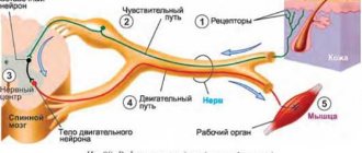
1 . The activity of the body is a natural reflex reaction to a stimulus. Reflex is the body’s reaction to irritation of receptors, which is carried out with the participation of the central nervous system. The structural basis of the reflex is the reflex arc (a sequentially connected chain of nerve cells that ensures the implementation of a reaction, a response to irritation).
The reflex arc consists of six components: receptors, afferent (sensitive) path, reflex center, efferent (motor, secretory) path, effector (working organ), feedback.
Reflex arcs can be of two types: simple - monosynaptic reflex arcs; complex – polysynaptic reflex arcs.
A feedback loop is a component that establishes a connection between the realized result of a reflex response and the nerve center that issues executive commands. With the help of this component, the open reflex arc is transformed into a closed one.
The reflex arc, in its structure and purpose of its elements, represents the regulation circuit described above. It includes the following links:
1) sensory receptors (sensors) that perceive stimuli from the external or internal environment,
2) afferent, or sensory, nerve conductors (channels of input signals),
3) nerve centers (control apparatus), consisting of afferent, intermediate, or intercalary, and efferent neurons, i.e. nerve cells receiving, processing and issuing information,
4) efferent, or motor, nerve conductors (output channels),
5) effectors, or executive bodies (control objects).
However, for optimal regulation, information about the effector's reactions to control signals is necessary, and therefore a feedback channel is an obligatory link in the reflex act. Thus, it is better to call the structural basis of the reflex not a reflex arc , but a reflex ring.
Nerve impulse transmission
How is impulse transmitted between cells? This question is more interesting than it seems at first glance. Now we will understand why neurons need highly developed ER and Golgi apparatus. The point of contact between an axon and a dendrite, the body of another neuron, or an effector is called a synapse. In addition to communication between cells, the synapse also recodes the signal, changing its various characteristics (frequency, amplitude). Let's take a closer look at the scheme of how synaptic transmission of a nerve impulse works:
- An electrical nerve impulse excites the membrane of a neuron
. The most excitable part on the body of such cells is the axon hillock, and the general excitation of the cell begins from there. It is transmitted further along the axon membrane and reaches its end - the presynaptic membrane. - Inside the axon there are vesicles with neurotransmitters
- biologically active organic substances. Remember about the ER and the Golgi apparatus? It is these systems that are responsible for the synthesis and transport of mediator substances to the axon terminal. - The synaptic cleft is the space between the axon of this cell and the dendrite of the next one.
Under the influence of electrical excitation, the presynaptic membrane releases transmitters into the space of the synaptic cleft. There they bind to the corresponding receptors on the postsynaptic membrane, thereby triggering the excitation of the next link in the nerve chain.
Important! At the level of neurophysiology, these processes look quite complex, and a deep understanding requires knowledge of not only biology, but also chemistry and physics. However, the main thing you need to understand is that there are two membranes, the connection between which is carried out by mediators (literal translation - intermediaries). The effect—excitation or inhibition of the next cell—depends on which mediator is released.
To summarize: neurons are the cells of the nervous system. Their coordinated work ensures the coordination of all functions of the body, from movement and work of internal organs to higher mental processes. Neurons transmit information through electrical impulses that travel along the axon of one cell and are transmitted to another cell using neurotransmitters at the synapse. This seemingly simple mechanism underlies the functioning of the nervous system. The video below will also help you better understand the functioning of the entire nervous system.
Previous
Anatomy Vital capacity of the lungs, tidal volume of the lungs of an adult, norm in liters, table, depth of breathing, types of lung volumes, functional residual capacity of the lungs
Next
AnatomyCircles of blood circulation briefly and clearly, diagram of arteries, blood vessels of the large and small circle, sequence of blood movement through the veins