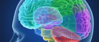
The terminal section is the most massive part of the brain - in an ordinary person it occupies about 78% of the total mass of the organ. It is divided into two parts by a central groove, in the depth of which there is a large commissure - the corpus callosum.
Despite its complex structure, the telencephalon is an interconnected system responsible for emotions, planning, memory, decision making, limb movements and perception of information from the environment. It carries out these and other functions with the help of a developed network of cells in the cerebral cortex. To understand the functions of the cerebral hemispheres, you must first understand their structure.
The structure of the cerebral hemispheres
The surface of the terminal part of the central nervous system is covered by the cortex, which occupies about 44% of the volume of the cerebral hemispheres. The area of this structure in an ordinary person is approximately 2200 cm², and most of it lies in the deep grooves or, as they are also called, the cerebral convolutions. Due to the presence of grooves and convolutions, the area of the cortex increases significantly.
The size and shape of the convolutions depend on the individual characteristics of a person - as is known, the brains of different people and even the hemispheres of one individual are visually different from each other. This phenomenon among experts is called “functional asymmetry of the cerebral hemispheres.”
According to observations, this feature affects the human psyche: for example, some people find it easier to study the exact sciences, while others use a more creative approach when solving pressing problems.
In the cortex, preparation for conscious movements occurs, speech is formed, thinking occurs, and the most significant information is memorized. It is also responsible for conditioned reflexes - acquired responses of the body to change.
The cortex is formed from a collection of cell bodies of neurons that form its layers. With their help, the cerebral hemispheres carry out their functions. The number of layers of bark in different areas is not equal: depending on the location of the zone and its type, there can be from 2 to 6.
Experts distinguish 4 types of surfaces in the cortex: ancient (paleocortex), old (archicortex), new (neocortex) and intermediate cortex, which consists of intermediate ancient and intermediate old cortex.
According to recent estimates, the number of cortical neurons varies between 10-14 billion units. They are connected to each other using synapses - special connections that allow you to instantly transmit impulses from one neuron to another. Signal transmission through the synapse occurs chemically with the help of active chemical elements or electrically through the passage of ions.
Under the cortex is white matter. It is formed by a cluster of axon bundles of cortical neurons, which are covered with myelin. The chemical structure of the membrane of nerve cell processes allows impulses to be transmitted between neurons 5-10 times faster than through unmyelinated connections.
Below the white matter, in the trunk there are centers of unconscious reflexes and controlling structures of internal organs and organ systems.
Hemisphere areas
The entire surface of the cerebral cortex is conventionally divided into several zones. Each of them performs specific functions. The boundaries of the zones are indicated by the most prominent convolutions.
Zones are not separate areas of the brain in which only specific mental and physiological processes occur, since they constantly interact with each other, which is confirmed by numerous studies in the field of psyche.
Topographically, the following areas of the cerebral cortex are distinguished:
- Occipital. Responsible for the perception and storage of data received from the organs of vision.
- Temporal. Its functions are based on the perception, analysis and reproduction of speech and sounds, the concept of auditory information, and data coming from the organs of taste and smell. Participates in memorizing information, namely accumulates it.
- Parietal lobe of the PD brain. The functional centers of the environmental analyzer are located in this area. It is responsible for the location of body parts in space.
- Frontal. It is the largest area through which the brain performs the following functions:
- movement and regulation of directed actions;
- letter;
- speech, namely the pronunciation of individual sounds, timbre, intonation;
- programming complex behavioral reactions, decision making, planning, analysis of the results obtained, as well as spontaneous behavior;
- The frontal zone contains the olfactory nerve center.
Thus, all zones: parietal, occipital, frontal and temporal, are involved in the perception of information from the environment, and also determine human behavior during the most significant changes.
Some areas can perform several functions at once. This becomes especially noticeable when the corresponding areas of the brain are damaged as a result of TBI - over time, their function is partially restored, as neighboring ones take over the work of the lost centers.
Brain
GENERAL OVERVIEW OF THE BRAIN
Brain,
is placed in the cranial cavity and has a shape that generally corresponds to the internal contours of the cranial cavity.
Its superolateral, or dorsal, surface, in accordance with the cranial vault, is convex, and the lower, or base of the brain, is more or less flattened and uneven. The brain can be distinguished into three large parts: the cerebrum
,
the cerebellum
and
the brain stem.
The largest part of the entire brain is occupied by
the cerebral hemispheres
, followed in size by
the cerebellum
, the remaining, relatively small, part is
the brain stem
.
Superior lateral surface of the cerebral hemispheres.
Both hemispheres are separated from each other by
the longitudinal medullary fissure ,
running in the sagittal direction.
In the depths of the longitudinal fissure, the hemispheres are interconnected by a commissure - the corpus callosum
, and other underlying formations.
In front of the corpus callosum, the longitudinal fissure is through, and behind it passes into the transverse fissure of the brain
, separating the posterior parts of the hemispheres from the underlying cerebellum.
The inferior surface of the cerebral hemispheres
(Fig. 272). From the lower surface of the brain, visible:
- underside of the cerebral hemispheres
- inferior side of the cerebellum,
- inferior surface of the brain stem
- nerves leaving the brain.
The anterior part of the lower surface of the brain is represented by the frontal lobes of the hemispheres
.
On the lower surface of the frontal lobes, olfactory bulbs are noticeable, to which thin nerve filaments approach from the nasal cavity through the openings of the ethmoid bone, which together form the first pair of cranial nerves - the olfactory nerves
. Usually, when the brain is removed from the skull, these threads are torn off from the olfactory bulbs.
The olfactory bulbs continue posteriorly into the olfactory tracts
, each ending in two roots, between which there is an elevation called
the olfactory triangle.
Directly behind the latter, on both sides, there is
perforated
in front of it , so named because of the presence of small holes here through which vessels pass into the medulla.
In the middle between both anterior perforated spaces lies the optic
chiasm
, shaped like the letter "X".
A gray tubercle
is located behind the optic chiasm ; its apex is elongated into a narrow tube, the so-called
funnel,
to which
the pituitary gland
.
Behind the gray mound there are two spherical, white elevations - mastoid bodies
.
Behind them lies a rather deep interpeduncular fossa, bounded laterally by two thick ridges converging posteriorly and called the cerebral peduncles
.
The bottom of the pit is pierced with openings for vessels, and therefore is called the back end of the fermented substance
.
the third
emerges on both sides -
the oculomotor nerve .
On the side of the cerebral peduncle one can see the thinnest of the cranial nerves,
the trochlear nerve
,
the fourth
pair, which, however, does not arise from the base of the brain, but from its dorsal side, from the so-called superior medullary velum.
Behind the cerebral peduncles there is a thick transverse shaft - the bridge,
which, tapering from the sides, plunges into the cerebellum.
The lateral parts of the bridge closest to the cerebellum are called the middle cerebellar peduncles ;
on the border between them and the bridge itself, the V pair emerges on both sides -
the trigeminal nerve
.
Behind the bridge lies the medulla oblongata
, between it and the posterior edge of the bridge on the sides of the midline the beginning of the VI pair -
the abducens nerve
, even further to the side at the posterior edge of the middle cerebellar peduncles two more nerves emerge side by side on both sides: VII - pair -
facial nerve
and VIII pair -
vestibulocochlear.
Between the pyramid and the olive medulla oblongata, the roots of the XII pair - the hypoglossal nerve
.
The roots of the IX, X and XI pairs - glossopharyngeal, vagus, accessory
- emerge from the groove behind the olive.
EMBRYOGENESIS OF THE BRAIN
Neural tube
very early it is divided into two sections corresponding to the brain and spinal cord.
Its anterior, expanded section, representing the rudiment of the brain, as noted, is divided by constrictions into three primary brain vesicles
lying one behind the other:
anterior
,
middle
, and
posterior
.
This three-vesicle stage progresses to the five-vesicle .
giving rise to the five main parts of the brain (Fig. 273).
At the same time, the brain tube bends in the sagittal direction. First of all, in the region of the middle bladder, a dorsally convex cephalic bend ,
and then, at the border with the spinal cord rudiment, a dorsally convex
cervical bend .
Between them, a third bend is formed in the region of the posterior bladder, convex to the ventral side -
the pontine bend .
Through this last bend the posterior medullary vesicle
is divided into two sections.
Of these, the posterior one turns into the medulla oblongata during final development, and from the anterior part, the pons
and
cerebellum
.
Rice.
273. Brain development (diagram).
a - five brain vesicles: / - telencephalon; 2 —
diencephalon;
3
- midbrain;
4
- the hindbrain itself as part of the rhombencephalon; 5 - medulla oblongata;
between the 3rd and 4th bladder - the isthmus; b
— brain development (according to R. D. Sinelnikov).
hindbrain
separated from the midbrain vesicle lying in front of it by a narrow constriction -
the isthmus of the hindbrain .
The common cavity
of the rhomboid brain
, which has the shape of a rhombus in a horizontal section, forms the IV ventricle, communicating with
the central canal
of the spinal cord.
Its ventral and lateral walls become greatly thickened due to the development of the cranial nerve nuclei in them, while the dorsal wall remains thin. In the region of the medulla oblongata, most of it consists of only one epithelial layer, fused with the pia mater.
The walls
of the middle cerebral
bladder thicken as the brain matter develops more evenly in them.
Ventrally, the cerebral peduncles arise from them, and on the dorsal side - the roof of the midbrain (see Fig. 273). The cavity of the middle bladder
turns into a narrow channel -
aqueduct
connecting the 3rd and 4th ventricles.
The anterior medullary vesicle undergoes more significant differentiation and modifications in shape.
, which is divided into the posterior part,
the diencephalon
, and the forebrain,
the telencephalon
.
The lateral walls of the diencephalon, thickening, form the thalamus
. In addition, the side walls
protruding to the sides, they form two optic vesicles, from which the retina and optic nerves subsequently develop.
The dorsal wall of the diencephalon remains thin, in the form of an epithelial plate, fused with the soft shell
.
Posteriorly, a protrusion arises from this wall, due to which the pineal body arises.
The hollow legs of the optic vesicles are retracted from the ventral side into the wall of the anterior medullary vesicle, as a result of which a depression, a
optic pocket
.
Behind the optic recess, another funnel-shaped depression appears, the walls of which give rise to a gray mound, a funnel
and
the posterior
(nervous)
lobe of the pituitary gland
. The cavity of the diencephalon forms the third ventricle.
Finite brain
is divided into a middle, smaller part and two large lateral parts -
the cerebral hemispheres,
which in humans grow very strongly and at the end of development are significantly larger in size than the rest of the brain.
The cavity of the middle part, which is the anterior continuation of the cavity of the diencephalon (III ventricle), communicates on the sides through the interventricular foramina
with the cavities of the hemispheres, which in a developed brain are called
lateral ventricles
. The anterior wall, at the beginning of the first month of embryonic life, forms a thickening from which the corpus callosum subsequently develops.
At the base of each hemisphere, inside, a protrusion is formed, from which the striatum
.
Part of the medial wall of the hemispheres remains in the form of a single epithelial layer, which is rolled into the vesicle by a fold of the pia mater
.
On the lower side of each hemisphere, already in the 5th week of embryonic life, a protrusion is formed - the rudiment of the olfactory brain
.
With the development of gray matter (cortex
), and then the white in the walls of the hemisphere, the latter increases and forms the so-called
cloak
, lying above the olfactory brain and covering not only the thalamus, but also the dorsal surface
of the midbrain
and
cerebellum
.
As the hemisphere grows, it increases first in the frontal lobe, then in the parietal and occipital lobes, and finally in the temporal lobe. This gives the impression that... as if the cape were rotating around the thalamus, first from front to back, then down, and finally curving forward towards the frontal lobe. As a result, on the lateral surface of the hemisphere, between the frontal lobe and the temporal lobe approaching it, a fossa, fossa lateralis cerebri, is formed, which, when the named lobes of the cerebrum approach each other, turns into a fissure - the lateral fissure of the brain
. At the bottom of it, a small special lobe of the brain is formed - an island.
the lateral ventricles of the brain develop and perform the indicated “rotation” together with it.
, as well as part of
the striatum
(
caudate nucleus
), which explains the similarity of their shape with the shape of the hemisphere: the ventricles have anterior, central and posterior parts and a lower part that curves downward and forward (see Fig. 295), and the caudate nucleus has the presence of a head, a body and a tail that curves downwards and forwards.
Rice. 276. Lower surface of the cerebrum.
/ - gyri orbitales; 2
- gyrus rectus;
3,4 -
gyri occipito-temporales medialis et lateralis;
5 - gyrus parahippocampalis; 6
- gyrus occipitotemporalis medialis;
7 - isthmus gyri cinguli; 8 -
cuneus;
9 -
gyrus temporalis medius;
10 —
tri-gonum olfactorium;
11 -
tr.
olfactorius; 12 -
bulbus ol-factorius;
13 -
sul.
olfactorius; 14 -
sulci orbitales;
/5—uncus gyri parahippocampalis; 16
- sul.
temporalis inferior; 17 -
sul.
hippocampi; 18 -
sul.
occipitotemporalis; /9 - sul. calcarinus; 20
- sul.
cotlateralis; 21 -
sul. parietooc-cipitalis.
Furrows and convolutions
(Fig. 274, 275, 276) arise due to uneven growth of the brain itself, which is associated with the development of its individual parts.
Thus, in place of the olfactory brain, the olfactory sulcus, hippocampal sulcus and cingulate sulcus appear. At
the border of the cortical ends of the cutaneous and motor
analyzers
(see the concept of the analyzer and the description of the grooves below) -
the central groove ;
on the border of the motor analyzer and the premotor zone, which receives impulses from the viscera, there is
the precentral sulcus ;
in place of the auditory analyzer -
the superior temporal sulcus ;
in the area of the visual analyzer -
the calcarine and parieto-occipital sulci.
All these grooves, which appear earlier than others and are characterized by absolute constancy, belong to the primary
grooves
.
The remaining grooves, which have names and also arise in connection with the development of analyzers, but appear somewhat later and are less constant, belong to the secondary grooves
.
By the time of birth, all grooves are present - primary and secondary. Finally, numerous small grooves that do not have names appear not only in uterine life, but also after birth. They are extremely variable in time of appearance, place and number; these are tertiary grooves
. The degree of their development determines the diversity and complexity of the brain relief.
The growth of the human brain in the embryonic period and in the first years of life, while the body is rapidly growing, adapting to a new environment, acquiring the ability to walk upright and the formation of a second, verbal, signaling system, occurs very intensively and ends by the age of 20. In newborns, the brain (on average) weighs 340 g in boys and 330 g in girls, and in an adult - 1375 g in men and 1245 g in women.
SEPARATE PARTS OF THE BRAIN
Based on embryonic development, as already indicated, the brain is divided into sections located, starting from the caudal end, in the following order:
1 rhomboid or hindbrain
, which in turn consists of: a)
the medulla oblongata
and b)
the hindbrain itself
;
2) midbrain
;
3) forebrain
, in which they distinguish: a)
diencephalon
and b)
telencephalon.
All of these sections, except the cerebellum and telencephalon, make up the brain stem
.
Diamond brain
Medulla
Medulla,
(Fig. 277, 278), represents a direct continuation of the spinal cord into the brain stem and is part of the rhombencephalon.
It combines the structural features of the spinal cord and the initial part of the brain, which justifies its name. It has the appearance of a bulb, (hence the term “bulbar disorders”); the upper expanded end borders the bridge
, and the lower border is the exit point of the roots of the first pair of cervical nerves or the level of
the large foramen of the occipital bone
.
1. On the anterior (ventral) surface of the medulla oblongata, three important anatomical landmarks are visible, located to the sides of the midline in this order:
- anterior median sulcus;
- pyramids;
- olives
the anterior median groove runs along the midline , constituting a continuation of the groove of the same name in the spinal cord. On its sides, on both sides, there are two longitudinal strands - pyramids, which seem to continue into the anterior cords of the spinal cord. The bundles of nerve fibers that make up the pyramid partly intersect with similar fibers opposite side, forming the decussation of the pyramids , after which they descend in the lateral funiculus on the other side of the spinal cord, forming the lateral pyramidal tract , partly remain uncrossed and descend in the anterior funiculus of the spinal cord on their side forming the anterior pyramidal tract ...
Pyramids are absent in lower vertebrates and appear along as
the new cortex develops; therefore, they are most developed in humans, since the pyramidal fibers connect the cerebral cortex, which has reached its highest development in humans, with the nuclei of the cranial nerves and the anterior horns of the spinal cord
Lateral to the pyramid lies an oval eminence - o l and v
a . )
2 .
On the posterior (dorsal) surface of the medulla oblongata (see Fig. 278) stretches
the posterior median groove
- a direct continuation of the spinal cord groove of the same name.
On its sides lie the posterior cords . .
In the upward direction, the posterior cords diverge to the sides and go to the cerebellum, becoming part of the
inferior cerebellar peduncles
the rhomboid fossa
below .
Each posterior cord is divided by the intermediate groove into a medial, thin fascicle,
and a lateral,
wedge-shaped fascicle.
At the lower corner of the rhomboid fossa, the thin and wedge-shaped fascicles acquire thickenings -
a thin tubercle
and
a wedge-shaped tubercle .
These thickenings are caused by the nuclei of the gray matter, similar to the fascicles,
the gracile nucleus
and
the cuneate nucleus
.
In these nuclei, the ascending fibers of the spinal cord (thin and wedge-shaped bundles) passing in the posterior cords end. The lateral surface of the medulla oblongata, located between the posterior lateral sulcus
and
the anterior lateral sulcus
, corresponds to the lateral funiculus.
from the posterior lateral sulcus
behind the olive.
Rice. 278. Brain stem; back view.
1
- pulvinar (posterior part of thalamus): 2 - pedunculus cerebellaris superior;
3
- pedunculus cerebellaru medius;
4 -
pedunculus cerebeilaris inferior;
5 - fasc. graciis; 6 -
fasc.
cuneatus; 7 ~ tuberculum gracilum, 8—
tuberculum cuneatum;
9 -
apertura meaiana ven-tricnli quarti;
10 -
plexus chorioideus and tela chorioidea ventriculi quarlti (cut and turned away, through the incision into the aid cavity of the fourth ventricle);
11 -
n. trochlearis;
12 -
collicuius inferior of the roof of the midbrain;
13 -
collicuius superior of the roof of the midbrain;
14 -
corpus geniculatum mediale;
15 -
corpus pineale.
Internal structure of the medulla oblongata
.
All parts of the brain consist of two types of substance: white and gray.
Gray matter of the medulla oblongata.
The medulla oblongata arose in connection with the development of the organs of gravity and hearing, as well as in connection with the gill apparatus related to respiration and blood circulation. Therefore, it contains nuclei of gray matter related to
- balance,
- coordination of movements,
- regulation of metabolism,
- breathing
- blood circulation
1. Olive Kernel
, has the appearance of a convoluted plate of gray matter, open medially and causes protrusion of the olive from the outside.
It is associated with the dentate nucleus of the cerebellum and is an intermediate nucleus of balance,
most pronounced in humans, whose vertical position requires a perfect gravitational apparatus.
2. Reticular
formation
the interweaving of nerve fibers and the nerve cells lying between them. Responsible for regulating the tone of the nervous system.
3. Core four
pairs of lower cranial nerves (XII - IX), related to the innervation of the derivatives of the branchial apparatus and viscera.
4. Vital breathing centers
and
blood circulation
associated with
the nuclei of the vagus nerve
. Therefore, if the medulla oblongata is damaged, death can occur.
5
. nuclei of the thin and wedge-shaped fasciculi. They contain the second neurons of the proprioceptive sensitivity pathways.
The white
matter
of the medulla oblongata contains
long
and
short fibers
.
Long fibers
medulla oblongata are divided into ascending and descending.
Descending pathways
are divided into
pyramidal
and
extrapyramidal
. The pyramidal tracts in the medulla oblongata make a partial decussation.
Ascending pathways
– these are the paths of different types of sensitivity.
The fibers form a medial loop
, which decussates in the medulla oblongata. Thus, in the medulla oblongata there are two intersections of long pathways: the ventral motor one, the pyramidal chiasm, and the dorsal sensory one, the lemniscus chiasm.
Short pathways
include bundles of nerve fibers that connect individual nuclei of the gray matter, as well as the nuclei of the medulla oblongata with neighboring parts of the brain.
hindbrain
hindbrain
consists of two parts: ventral -
the bridge
and dorsal -
the cerebellum
.
Bridge.
Bridge
It is a thick white shaft from the base of the brain, bordering at the back with the upper end
of the medulla oblongata
, and at the front with
the cerebral peduncles
.
The lateral border of the bridge is an artificially drawn line through the roots of the trigeminal and facial nerves. Lateral to this line are the middle cerebellar peduncles
, plunging into the cerebellum on both sides.
The dorsal surface of the brain is not visible from the outside, since it is hidden under the cerebellum, forming the upper part of the rhomboid fossa (the floor of the fourth
ventricle).
The ventral surface of the pons is fibrous in nature, with the fibers generally running transversely and directed into the middle cerebellar peduncle
.
Along the midline of the ventral surface runs a gently sloping groove, the basilar groove ,
in which
the basilar artery
.
Internal structure of the bridge.
On cross-sections of the bridge, you can see that it consists of two parts: 1)
the anterior (ventral) part of the bridge
and 2)
the posterior (dorsal) part of the bridge
.
The boundary between them is a thick layer of transverse fibers - the trapezoidal body
, the fibers of which belong to the auditory pathway.
Ventral part of the bridge
contains longitudinal and transverse fibers, between which the nuclei of gray matter are scattered -
the pons' own nuclei
.
Longitudinal fibers belong to the pyramidal tracts
, which are connected with the own nuclei of the bridge, from where the transverse fibers that go to
the cerebellar cortex originate - the pontocerebellar path .
This entire system of pathways connects
cerebral
cortex with
the cerebellar cortex
. The more developed the cerebral cortex, the more developed the pons and cerebellum. Naturally, the bridge turns out to be most pronounced in humans, which is a specific feature of the structure of his brain.
In the dorsal part of the bridge
there is
a reticular formation ,
which is a continuation of the same formation of the medulla oblongata, and on top of the reticular formation is
fossa
lined with ependyma with the underlying
nuclei of the cranial nerves ( VIII
- V pairs). The pathways of the medulla oblongata continue here.
Cerebellum
Cerebellum,
is a derivative of the hindbrain, which developed in connection with gravity receptors. Therefore, it is directly related to the coordination of movements and is an organ of the body’s adaptation to overcoming the basic properties of body weight - gravity and inertia.
The development of the cerebellum in the process of phylogenesis went through 3 main stages, corresponding to changes in the methods of movement of the animal.
The cerebellum first appears in the class of cyclostomes, in lampreys, in the form of a transverse plate. In lower vertebrates (fish), paired ear-shaped parts (archicerebellum) and an unpaired body (paleocerebellum), corresponding to the worm, are distinguished; in reptiles and birds the body is highly developed, and the ear-shaped parts turn into rudimentary ones. Cerebellar hemispheres arise only in mammals (neocerebellum). In humans, due to upright walking with the help of one pair of limbs (legs) and the improvement of grasping movements of the hand during labor processes, the cerebellar hemispheres achieve the greatest development, so that the cerebellum in humans is more developed than in all animals, which constitutes a specific human feature of its structure.
The cerebellum is located under the occipital lobes of the cerebral hemispheres and lies in the posterior cranial fossa. It distinguishes voluminous lateral parts, or hemispheres of the cerebellum ,
and the middle narrow part located between them -
the worm .
At the anterior margin of the cerebellum is the anterior notch, which encloses the adjacent portion of the brain stem. At the posterior margin there is a narrower posterior notch that separates the hemispheres from each other.
The surface of the cerebellum is covered with a layer of gray matter that makes up the cerebellar cortex and forms narrow convolutions - the leaves of the cerebellum
, separated from each other by
the cerebellar sulci
.
.
Among them, the deepest
horizontal cerebellar fissure
runs along the posterior edge of the cerebellum, separating
the superior surface of the hemispheres
from
the inferior surface of the cerebellum
.
.
With the help of the horizontal and other large grooves, the entire surface of the cerebellum is divided into a number of
cerebellar lobules
.
Among them, it is necessary to highlight the most isolated small lobe - the flocculus
, which lies on the lower surface of each hemisphere at the middle cerebellar peduncle, as well as the part of the vermis associated with the flocculus -
the nodule
.
The flocculus is connected to the nodule by means of a thin strip - the stalk of the flocculus ,
which medially passes into a thin semilunar plate -
the inferior medullary velum
.
Internal structure of the cerebellum.
In the thickness of the cerebellum there are paired nuclei of gray matter, embedded in each half of the cerebellum among its white matter (Fig. 281).
On the sides of the midline, in the area where the tent
, lies the most medial nucleus -
the nucleus of the tent .
Lateral to it is
the spherical nucleus
, and even more lateral is
the corky nucleus
.
Finally, in the center of the hemisphere is the dentate nucleus
, which has the appearance of a gray sinuous plate, similar to the nucleus of an olive.
Rice. 281.
Cerebellar nuclei (diagram).
/ - tent core; 2
- spherical nucleus;
3 -
corky core;
4
- dentate core.
Similarities of the dentate nucleus
The cerebellum with the
olivary
nucleus , is not accidental, since both nuclei are connected by pathways (
olivocerebellar fibers) ,
and each gyrus of one nucleus is similar to the gyrus of the other. Thus, both nuclei together participate in the implementation of the equilibrium function (see Fig. 280, 281).
The named cerebellar nuclei have different phylogenetic ages:
- tent core
refers to the most ancient part of the cerebellum -
the flocculus
associated with the vestibular apparatus; - globular and corky nuclei
- to the old part that arose in connection with the movements of the body, and
- dentate nucleus
- to the youngest, developed in connection with movement with the help of limbs.
Therefore, when each of these parts is damaged, various aspects of motor function are disrupted, corresponding to different stages of phylogenesis, namely:
- when the flocculonodular system and tent core are damaged, the balance of the body is disrupted.
- When the worm and the corresponding cork-shaped and spherical nuclei are damaged, the functioning of the muscles of the neck and torso is disrupted,
- with damage to the hemispheres and dentate nucleus - the work of the muscles of the limbs.
The white matter of the cerebellum on a section has the appearance of small leaves of a plant, corresponding to each gyrus, covered on the periphery with the cortex of the gray matter. As a result, the overall picture of white and gray matter on a section of the cerebellum resembles a tree ( tree of life
; the name is given based on its appearance, since damage to the cerebellum is not an immediate threat to life). The white matter of the cerebellum is composed of various types of nerve fibers. Some of them connect the gyri and lobules, others go from the cortex to the internal nuclei of the cerebellum, and, finally, others connect the cerebellum with neighboring parts of the brain. These last fibers come as part of three pairs of cerebellar peduncles:
- Inferior peduncles, cerebellum
(to the medulla oblongata).
, the posterior spinocerebellar tract
goes to the cerebellum - from the nuclei of the posterior funiculi of the medulla oblongata and
olivocerebellar fibers -
from the olive.
The first two tracts end in the cortex of the vermis and hemispheres. In addition, there are fibers from the nuclei of the vestibular nerve, ending in the nucleus of the tent. Thanks to all these fibers, the cerebellum receives impulses from the vestibular apparatus and the proprioceptive
field , as a result of which it becomes the core of proprioceptive sensitivity, making automatic corrections to the motor activity of the rest of the brain.
As part of the lower legs, there are also descending pathways in the opposite direction, namely: from the tent core
to the lateral vestibular nucleus (see below), and from it to
the anterior horns of the spinal cord
, the vestibulospinal tract. Through this pathway, the cerebellum influences the spinal cord. - Middle cerebellar peduncles
(to the bridge).
They contain nerve fibers from the pontine nuclei to the cerebellar cortex. The pathways that arise in the pons nuclei to the cerebellar cortex, the pontocerebellar tract ,
are located in continuation of the corticopontine tracts,
corticopontine fibers
, ending in the pons nuclei after the decussation. These pathways connect the cerebral cortex with the cerebellar cortex, which explains the fact that the more developed the cerebral cortex, the more developed the pons and cerebellar hemispheres are, which is observed in humans.
3. Superior cerebellar peduncles
(to the roof of the midbrain).
They consist of nerve fibers running in both directions: 1) to the cerebellum - the anterior spinocerebellar tract
and 2) from
the dentate nucleus of the cerebellum
to
the tegmentum of the midbrain
-
the cerebellar-tegmental tract
, which, after decussation, ends in
the red nucleus
and the
thalamus
.
Along the first paths, impulses from the spinal cord go to the cerebellum, and along the second, it sends impulses to the extrapyramidal
system, through which it itself influences the spinal cord.
Functions
The main function of the cerebral cortex is to reproduce and accumulate information obtained during the learning process. It also hosts all higher mental processes, such as thinking, speech and memory.
According to neuropsychological research, the right and left hemispheres of the brain do slightly different things. Thus, the right side is responsible for sensory, figurative perception, memorizing images, music and their reproduction from memory.
This information is also used by the left hemisphere, which is engaged in comprehension, logical explanation and “criticism” of the work of the right side.
The functions of the right side of the brain remain at the level of perception of incoming information through analyzers of sensory-image properties and receptors, bypassing their physical qualities. For example, it is responsible for recognizing conventional and alphabetic symbols without understanding them.
A higher organizational level, where the analysis and evaluation of the content of signs occurs, is associated with the work of the left hemisphere. Its function is to determine the cause-and-effect relationship, the interaction of events that have occurred with each other, processing and understanding of incoming information that comes from the outside world with the help of words, sounds, and speech.
Left hemisphere
Each of the cerebral hemispheres is equally important for performing the basic functions of the human nervous system and his mental abilities. For example, such a concept as an “analytical mindset” does not depend on the size of a particular part of the brain - anyone can study the exact sciences and achieve success in them.
The left hemisphere is involved in the following functions:
- logical solution;
- mastering foreign languages;
- control of word pronunciation;
- ability to read, write, remember phrases.
In the left half there are nerve centers with the help of which a person can perceive the literal meaning of what is said.
The motor functions of the left hemisphere of the brain come down to the fact that it controls the limbs of the right side of the body. That is, when raising the right arm or leg, the left half of the brain gives the order.
Right hemisphere
It has long been believed that the right hemisphere is more developed in women, but this is not true in the cortex. The argument was that the weaker sex is more emotional compared to the opposite, and, as is known, it is precisely this manifestation of the psyche that is the main function of this side of the brain. It is also responsible for intuition, assessment, reproduction and transmission of non-verbal information.
According to psychology, those people who often “work” with the right hemisphere are distinguished by a subtle perception of music, painting and other forms of art, despite the fact that the left side is responsible for teaching these abilities.
Other functions carried out by the right hemisphere of the brain include imagination, visualization and understanding of allegorical definitions. It is also responsible for attractiveness, paranormal perception of information, fantasies, religion and dreams.
By analogy with the left part, the right one controls the movements of the limbs on the left side of the body.
Function creates the center
According to Ivan Petrovich Pavlov: “Function creates the center!” In early childhood, the boundaries of the cortical centers are diffuse and less differentiated, and only as life experience is gained, a gradual concentration of functional zones occurs, and therefore in children of the first years of life, focal cortical symptoms are weakly expressed and general cerebral symptoms often predominate.
4. Significant differences in the localization of simpler and more complex functions. The simpler the function, the more accurately it is localized. Conversely, the most complex functions are determined by the integrative activity of the entire brain, therefore the concept of “cortical center” (section of the cerebral cortex, fields of the cerebral cortex, areas of the cerebral cortex, parts of the cerebral cortex) is in most cases relative and conditional. Simple cortical functions include sensory function, motor function, visual function, auditory function, vestibular function, olfactory function, and gustatory function. Complex cortical functions include speech, writing, reading, counting, praxis, gnosis, thinking, and memory.
Nervous activity of the cerebral hemispheres
Another function performed by the structures of the cortex is the implementation of higher nervous activity in humans. It is carried out using neurons. For example, a clear manifestation of this feature is a person’s ability to learn and receive information from different sources.
The terminal section of the central nervous system plays a large role in the adaptation of the body to the environment - it is here that, through the formation of numerous synoptic nerve connections, conditioned responses of the body in response to external changes are formed.
These reflexes, acquired during a person’s life, are built on the basis of unconditioned reflexes under the influence of certain factors. Conditioned reflexes in a person can be formed both from their own and other people’s experience, for example, in the process of learning and assimilating third-party material. A striking example of this is schooling, where students receive the necessary knowledge from textbooks.
In a global sense, the functions of higher nervous activity are usually distinguished from the work of the entire nervous system, since it is responsible for the coordinated work of various parts of the body with each other. The functioning of the higher nervous activity of the central nervous system is associated with neurophysiological processes that occur in the cortex and the subcortical structures closest to it.
Speech analyzer, Wernicke's center, Broca's center, speech function - sensory center
The speech function is provided by the sensory center ( Wernicke's center ), which is located in the posterior part of the superior temporal gyrus. When Wernicke's center is damaged, sensory aphasia occurs. The speech function is also provided by the motor center ( Broca's center ), which is located in the posterior parts of the inferior frontal gyrus. When Broca's center is damaged, motor aphasia occurs. With pathology at the junction of the temporal and occipital lobes, amnestic aphasia and semantic aphasia are formed. Speech areas of the cerebral cortex.
