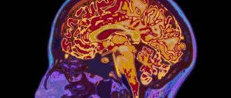
The human brain (BM) consists of many neurons that are interconnected by special connections. Taking into account the characteristics of the GM, it is considered the most complex structure. Its main components are lipids. They account for 60%. The GM is fed by the circulatory system, which delivers nutrients and oxygen. The brain, the structure and functions of its parts with the distinctive characteristics of the hemispheres are studied using tables as part of the biology curriculum in the 8th grade.
Posterior
This organ is primarily made up of cells called neurons.
These nerve cells produce electrical impulses that enable the nervous system to function. The work of neurons is ensured by cells called neuroglia - they make up almost half of the total number of cells in the central nervous system.
Neurons, in turn, consist of a body and processes of two types: axons (transmitting impulses) and dendrites (receiving impulses). The bodies of nerve cells form a tissue mass, which is commonly called gray matter, and their axons are woven into nerve fibers and represent white matter.
To protect it, nature has created a whole arsenal of various means. Externally, the parts of the brain are protected by the cranium, under which there are three more membranes of the brain:
- Solid. It is a thin film, one side adjacent to the bone tissue of the skull, and the other directly to the cortex.
- Soft. It consists of loose tissue and tightly envelops the surface of the hemispheres, going into all the cracks and grooves. Its function is to supply blood to the organ.
- Arachnoid. It is located between the first and second membranes and exchanges cerebrospinal fluid (cerebrospinal fluid). Liquor is a natural shock absorber that protects the brain from damage when moving.
Next, let's take a closer look at how the human brain works. According to morpho-functional characteristics, the brain is also divided into three parts. The lowermost section is called rhomboid. Where the rhomboid part begins, the spinal cord ends - it passes into the medulla oblongata and posterior (pons and cerebellum).
Next comes the midbrain, which unites the lower parts with the main nerve center - the anterior section. The latter includes the telencephalon (cerebral hemispheres) and diencephalon. The key functions of the cerebral hemispheres are the organization of higher and lower nervous activity.
This small superficial layer of gray matter (up to 4.5 mm) is the youngest formation in the central nervous system. It is the cerebral cortex that is responsible for the work of higher nervous activity in humans.
Research has made it possible to determine which areas of the cortex were formed relatively recently during evolutionary development, and which were present in our prehistoric ancestors:
- neocortex - the new outer part of the cortex, which is its main part;
- archicortex - an older formation responsible for human instinctive behavior and emotions;
- The paleocortex is the most ancient area involved in the control of autonomic functions. In addition, it helps maintain the internal physiological balance of the body.
Frontal lobes
The largest lobes of the cerebral hemispheres, responsible for complex motor functions. In the frontal lobes of the brain, planning of voluntary movements occurs, and speech centers are also located here. It is in this part of the cortex that volitional control of behavior is exercised. If the frontal lobes are damaged, a person loses control over his actions, behaves antisocially and simply inappropriately.
Occipital lobes
Closely related to visual function, they are responsible for the processing and perception of optical information. That is, they transform the entire set of light signals that enter the retina into meaningful visual images.
Parietal lobes
They carry out spatial analysis and process most sensations (touch, pain, “muscle feeling”). In addition, it promotes the analysis and integration of various information into structured fragments - the ability to feel one’s own body and its sides, the ability to read, count and write.
Temporal lobes
In this department, audio information is analyzed and processed, which ensures the function of hearing and the perception of sounds. The temporal lobes are involved in recognizing the faces of different people, as well as facial expressions and emotions. Here information is structured for permanent storage, and thus long-term memory is realized.
In addition, the temporal lobes contain speech centers, damage to which leads to the inability to perceive spoken language.
Insula
It is considered responsible for the formation of consciousness in a person. In moments of compassion, empathy, listening to music and the sounds of laughter and crying, active work of the insular lobe is observed. This is also where the processing of feelings of aversion to dirt and unpleasant odors, including imaginary stimuli, takes place.
The parts of the brain and their functions completely control our life processes. The human brain consists of 25 billion neurons. This incredible number of cells forms the gray matter. The brain is covered by several membranes:
- soft;
- hard;
- arachnoid (cerebrospinal fluid circulates here).
Liquor is cerebrospinal fluid; in the brain it plays the role of a shock absorber, a protector from any impact force.
Both men and women have exactly the same brain development, although their weight is different. More recently, the debate has subsided that brain weight plays some role in mental development and intellectual abilities. The conclusion is clear - this is not so. The weight of the brain is approximately 2% of the total weight of a person.
The cavities inside the brain are called ventricles. Paired cranial nerves go to different sections.
Each part of the brain does its own job. The table below clearly demonstrates this. The brain, like a computer, clearly performs its tasks, receiving commands from the outside world.
The table reveals the functions of the brain sections schematically and succinctly.
Below we will look at the parts of the brain in more detail.
The picture shows how the brain works. The most significant part is occupied by the cerebral hemispheres, despite this, all parts of the brain and their functions play a huge role in the functioning of the body. There are five main departments:
- final (of the total mass is 80%);
- posterior (pons and cerebellum);
- intermediate;
- oblong;
- average.
At the same time, the brain is divided into three main parts: the brain stem, the cerebellum, and the two cerebral hemispheres.
The surface layer of the brain is the cortex, it is 3 mm thick and covers the hemispheres. The structure consists of vertical nerve cells with processes. The cortex also contains efferent and afferent nerve fibers, as well as neuroglia. The parts of the brain and their functions are discussed in the table, but what is the cortex? Its complex structure has horizontal layering. The structure has six layers:
- external pyramidal;
- external granular;
- internal granular;
- molecular;
- internal pyramidal;
- with spindle cells.
Each has a different width, density, and shape of neurons. Vertical bundles of nerve fibers give the cortex vertical striations. The area of the cortex is approximately 2,200 square centimeters, the number of neurons here reaches ten billion.
The cortex controls several specific functions of the body. Each share is responsible for its own parameters. Let's take a closer look at the functions associated with calving:
- temporal – controls the sense of smell and hearing;
- parietal – responsible for taste and touch;
- occipital – vision;
- frontal – complex thinking, movement and speech.
Each neuron contacts other neurons, there are up to ten thousand contacts (gray matter). Nerve fibers are white matter. A certain part unites the hemispheres of the brain. White matter includes three types of fibers:
- association ones connect different cortical areas in one hemisphere;
- commissural connect the hemispheres to each other;
- projection ones communicate with lower formations and have analyzer paths.
Considering the structure and functions of parts of the brain, it is necessary to emphasize the role of gray and white matter. The hemispheres inside have basal ganglia (gray matter), their main function is the transmission of information. White matter is located between the cerebral cortex and the basal ganglia. There are four parts here:
- between the grooves in the gyri;
- in the outer places of the hemispheres;
- included in the inner capsule;
- located in the corpus callosum.
The white matter located here is formed by nerve fibers and connects the gyral cortex with the underlying sections. The subcortical nuclei form the subcortex of the brain.
The telencephalon controls all the vital functions of the body, as well as the intellectual abilities of a person.
The brain is one of the constituent organs of the central nervous system. Doctors are still studying it. It consists of 25 billion neurons, which are presented in the form of gray matter.
Rice. 1. Sections of the brain.
In addition, this organ of the nervous system is covered with the following types of membrane:
- soft;
- hard;
- arachnoid (cerebrospinal fluid circulates in it - cerebrospinal fluid, which serves as a kind of shock absorber and protects against shock).
The functions of generator and impulse transmission are performed by neurons. There are ventricles (cavities) inside the brain, from which paired cranial nerves extend to different parts of the human body. There are a total of 12 such pairs in the body.
The main organ of the nervous system consists of three parts:
- two hemispheres;
- trunk;
- cerebellum.
It also has five departments:
- final, constituting 80% of the mass;
- intermediate;
- rear;
- average;
- oblong.
Each section consists of a specific set of cells (white and gray matter).
White matter is presented in the form of nerve fibers, which can be of three types:
- association – connect cortical areas in one hemisphere;
- commissural - connects the two hemispheres;
- projection – connect the cortex with the underlying formations.
Gray matter consists of neuron nuclei, their functions include the transmission of information.
Rice. 2. Lobes of the cerebral cortex.
| Department | Structure | Functions |
| Finite | Located from the occipital to the frontal bone. It consists of two hemispheres, which have many grooves and convolutions. On top they are covered with a bark consisting of lobes. | The right hemisphere is responsible for the left side of the body, and the left hemisphere is responsible for the right side. The temporal lobe of the cerebral cortex regulates hearing and smell, the occipital lobe regulates vision, the parietal lobe regulates taste and touch; frontal – speech, thinking, movement. |
| Intermediate | Consists of the hypothalamus and thalamus. | The thalamus is a mediator in the transmission of stimuli to the hemispheres and helps to adequately adapt to changes in the environment. The hypothalamus regulates the functioning of metabolic processes and endocrine glands. Manages the work of the cardiovascular and digestive systems. Regulates sleep and wakefulness, manages food and drink needs. |
| Rear | It consists of the cerebellum and the pons, which is presented in the form of a white thick cushion located above the oblongata. The cerebellum is located behind the pons and has two hemispheres, the inferior and superior surfaces and the vermis. | This section provides a conductive function during the transmission of impulses. The cerebellum controls the coordination of movements. |
| Average | Located from the anterior edge of the bridge to the optic tracts. | Responsible for hidden vision, as well as the work of the orienting reflex, which ensures the body turns in the direction of the heard sharp noise. |
| Oblong | Presented as a continuation of the spinal cord. | Controls coordination of movements, balance, regulates metabolic processes, breathing, blood circulation. Controls the process of coughing and sneezing. |
| Part | Structure | Function |
| Oblong (stem section) | Extension of the spinal cord located on the trunk. It has a white substance on the outside and a gray substance on the inside. Gray matter is contained in the form of nuclei. | Conductive, nutritional, protective, control of respiratory rate, control of heart rate, control of vital reflexes responsible for sneezing, swallowing, hunger. |
| Average | Connects the forebrain and hindbrain. Contains parts called quadrigeminal tuberosities. | Primary or subcortical centers of hearing and vision. Thanks to this, a person in the field of vision touches new objects or sound sources that appear. There are also centers responsible for muscle tone. |
| Intermediate | It consists of: thalamus, epithalamus, hypothalamus. The thalamus contains the centers of almost all sensory senses. The hypothalamus is the part of the intermedia that connects to and controls the pituitary gland. | Vision, tactile and taste sensations, sensations of body temperature and the environment, memory, sleep. |
| Cerebellum (hindbrain) | The subcortical part of the brain that has grooves. It consists of two hemispheres, which are held together by a worm. | Regulates coordination of movement, the ability to maintain the body in free space. |
| Cerebral hemispheres ( telencephalon) | It is formed from two parts (right and left), divided into grooves and convolutions, due to which the surface increases. They consist of a large amount of gray matter, which is located on the outside, and white on the inside. | Vision (occipital lobe), skin-articular sensitivity and muscle tone (parietal lobe). Memory, thinking, consciousness, speech (frontal lobe) and hearing (temporal lobe). |
Structure
The main organ of the nervous system consists of three parts:
TOP 4 articles that are read along with this
- two hemispheres;
- trunk;
- cerebellum.
It also has five departments:
- final, constituting 80% of the mass;
- intermediate;
- rear;
- average;
- oblong.
Each section consists of a specific set of cells (white and gray matter).
White matter is presented in the form of nerve fibers, which can be of three types:
- association – connect cortical areas in one hemisphere;
- commissural - connects the two hemispheres;
- projection – connect the cortex with the underlying formations.
Gray matter consists of neuron nuclei, their functions include the transmission of information.
Rice. 2. Lobes of the cerebral cortex.
The following table will help you understand in more detail the structure and functions of the brain:
Finite brain
This part has the largest volume (80%) compared to the rest. It consists of two cerebral hemispheres, the corpus callosum connecting them, and the olfactory center.
The large hemispheres of the brain, left and right, are responsible for the formation of all thought processes. Here there is the greatest concentration of neurons and the most complex connections between them are observed. In the depths of the longitudinal groove, which divides the hemispheres, there is a dense concentration of white matter - the corpus callosum. It consists of complex plexuses of nerve fibers that intertwine different parts of the nervous system.
Within the white matter are clusters of neurons called the basal ganglia. The close location to the “transport junction” of the brain allows these formations to regulate muscle tone and carry out instant reflex-motor reactions. In addition, the basal ganglia are responsible for the formation and operation of complex automatic actions, partially repeating the functions of the cerebellum.
It is impossible to briefly describe the structure of the brain. To understand the parts of the brain and their functions, it is necessary to closely study their structure.
The telencephalon extends from the frontal to the occipital bone. Here we consider two large hemispheres: left and right. This section differs from others in the largest number of grooves and convolutions. The development and structure of the brain are closely interconnected. Experts have identified three types of bark:
- ancient (with olfactory tubercle, anterior perforated substance, semilunar subcallosal and lateral subcallosal gyrus);
- old (with the dentate gyrus - fascia and hippocambus);
- new (represents the entire remaining part of the cortex).
The hemispheres are separated by a longitudinal groove; in its depths there is the fornix and corpus callosum, which connect the hemispheres. The corpus callosum itself is lined with nerve fibers and belongs to the neocortex. The structure of the hemispheres is quite complex and resembles a multi-level system. Here we distinguish between the frontal, temporal, parietal and occipital lobes, subcortex and cortex.
Symptoms of tissue damage in the frontal lobe of the brain
Among the most severe lesions of the tissues of the frontal lobes are ischemic, atrophic changes in the tissue structure, stroke with a focus of ischemia or hemorrhage in this area. Impaired blood supply to this part of the brain negatively affects cognitive abilities. The main reasons for the deterioration and cessation of blood flow in this area:
- Exacerbation of arterial hypertension.
- Atherosclerotic damage to vascular walls.
- Developmental anomalies and acquired defects of elements of the circulatory system (vascular malformations, aneurysms).
- Disorder in the hemostasis system (increased thrombosis, blood clotting disorder).
Frontal lobe syndrome occurs due to damage to the tissues of the prefrontal region, which is responsible for multi-stage thought processes, behavior and cognitive abilities. General cerebral symptoms of pathology:
- Acute pain in the frontal plane of the head.
- Nausea, often leading to bouts of vomiting.
- Dizziness, confusion.
- Sometimes increased body temperature.
Frontal lobe syndrome is classified as an organic personality disorder in the international medical classification of diseases. It often develops against the background of other pathologies - Pick's disease and Alzheimer's disease, brain tumors, trauma to the head, damage to the vascular system in this area. Specific signs of the disease:
- Activation of rudimentary reflexes (sucking, grasping, searching). Occurs when a large area of tissue is affected.
- Loss of self-control, inability to plan and manage actions.
- Loss of awareness of one's own personality.
- Motor and speech dysfunction.
- Impossibility of abstract thinking and planning.
- Perseveration. Unconscious repeated repetition of one word, phrase, or action.
- Impaired concentration and memory functions.
Similar disorders are often observed in patients with Alzheimer's disease, who experience pathological structural changes in tissues - the formation of protein structures around neurons that interfere with and disrupt the relationships between nerve cells. Alzheimer's disease often causes dementia.
Medulla
The midbrain is located in the anterior pons and extends to the papillary bodies, as well as to the optic tracts. Here, clusters of nuclei are identified, which are called quadrigeminal tubercles. The structure and functions of the brain sections (table) indicate that this section is responsible for latent vision, the orientation reflex, gives orientation to reflexes to visual and sound stimuli, and also maintains the tone of the muscles of the human body.
The medulla oblongata is a natural extension of the spinal cord. That is why there are many similarities in the structure. This becomes especially clear if we examine the white matter in detail. Its short and long nerve fibers represent it. Gray matter is represented here in the form of nuclei. The parts of the brain and their functions (the table above) indicates that the medulla oblongata controls our balance, coordination, regulates metabolism, controls breathing and blood circulation. It is also responsible for such important reflexes of our body as sneezing and coughing, vomiting.
https://www.youtube.com/watch?v=W2_IGcFy2qE
The brainstem is divided into the hindbrain and midbrain. The trunk is called the middle, medulla oblongata, pons and diencephalon. Its structure consists of descending and ascending pathways connecting the trunk with the spinal cord and brain. This part monitors heartbeat, breathing, and articulate speech.
Only parts of the organ’s cortex, the limbic system and the cerebellum are responsible for memory functioning. The main influence is on areas located in the temporal zone of the left and right hemispheres.
Also, the main department for storing long-term information is the hippocampus.
He is responsible for cross-functional activities. Conveys motor sensations (coordination), tactile, and reflex sensations.
With the help of this area, a person can move in space without any problems.
The left hemisphere, in which the speech zones are located - motor and sensory - is mainly responsible for speech function.
The midbrain is located in the stem part. It is a conductor of signals from the front to various departments. Its main function is to regulate muscle tone. It is also responsible for the transmission of tactile sensations, coordination and reflexes. The functions of parts of the human brain depend on their location. For this reason, the midbrain is responsible for the vestibular apparatus. Thanks to the midbrain, a person can simultaneously perform several functions.
In the absence of intellectual activity, brain function is disrupted. People over 70 years of age are susceptible to this. When the middle part malfunctions, coordination failures occur and visual and auditory perception shifts.
It is located on the border of the spinal cord and the bridge and is responsible for vital functions. The oblong part consists of elevations called pyramids. Its presence is characteristic only of erectus. Thanks to them, thinking appeared, the ability to understand commands, and small movements were formed.
The pyramids are no more than 3 cm long; they are flanked by olive trees and posterior pillars. They have a large number of pathways throughout the body. In the neck region, motor neurons on the right side of the brain go to the left side and vice versa. Therefore, the loss of coordination occurs on the opposite side of the problem area of the brain.
The cough, respiratory and swallowing centers are concentrated in the medulla oblongata, and it becomes clear which part of the brain is responsible for breathing. When the ambient temperature drops, skin thermoreceptors send information to the medulla oblongata, which reduces the breathing rate and increases blood pressure. The medulla oblongata forms appetite and thirst.
Suppression of the function of the medulla oblongata may be incompatible with life. There is a violation of swallowing, breathing, and heart activity.
First of all, it regulates auditory and visual reflex activity (constriction of the pupil in bright light, turning the head towards a source of loud sound, etc.). After processing in the thalamus, the information goes to the midbrain.
Here its further processing takes place and the process of perception begins, the formation of a meaningful sound and optical image. In this department, eye movements are synchronized and binocular vision is ensured.
The midbrain includes the peduncles and quadrigeminal region (two auditory and two visual colliculi). Inside there is a cavity of the midbrain that unites the ventricles.
This is an ancient formation of the nervous system. The functions of the medulla oblongata are to ensure breathing and heartbeat. If this area is damaged, the person dies - oxygen stops flowing into the blood, which is no longer pumped by the heart. In the neurons of this department, protective reflexes such as sneezing, blinking, coughing and vomiting begin.
The structure of the medulla oblongata resembles an elongated onion. It contains the nuclei of gray matter: the reticular formation, the nuclei of several cranial nerves, as well as neural ganglia. The pyramid of the medulla oblongata, consisting of pyramidal nerve cells, performs a conducting function, uniting the cerebral cortex and the spinal region.
The most important centers of the medulla oblongata:
- breathing regulation
- regulation of blood circulation
- regulation of a number of functions of the digestive system
Diencephalon
The diencephalon serves as a kind of filter for neural signals - it receives all incoming information and decides where it should go. Consists of the lower and posterior parts (thalamus and epithalamus). In this department, the endocrine function is also realized, i.e. hormonal metabolism.
The lower part consists of the hypothalamus. This small, dense bundle of neurons has a tremendous impact on the entire body. In addition to regulating body temperature, the hypothalamus controls sleep and wake cycles. It also releases hormones that are responsible for the sensations of hunger and thirst. As a pleasure center, the hypothalamus regulates sexual behavior.
It is also directly connected to the pituitary gland and converts nervous activity into endocrine activity. The functions of the pituitary gland, in turn, are to regulate the functioning of all glands of the body. Electrical signals go from the hypothalamus to the pituitary gland of the brain, “ordering” the production of which hormones should be started and which ones should be stopped.
The diencephalon also includes:
- Thalamus - it is this part that performs the functions of a “filter”. Here, signals coming from visual, auditory, taste and tactile receptors undergo primary processing and are distributed to the appropriate departments.
- Epithalamus - produces the hormone melatonin, which regulates wakefulness cycles, participates in the process of puberty, and controls emotions.
It responds to external stimuli, is located at the end of the brain stem and is covered by the cerebral hemispheres. Thanks to it, a person can navigate in space, receive visual and auditory signals. Participates in the formation of all types of feelings.
All functions of the human brain are interconnected. Without an intermediate, the functioning of the entire organism will be disrupted. Damage to part of the midbrain leads to disorientation and dementia. If the connections between the lobes of the hemispheres are disrupted, speech, vision or hearing will be disrupted.
The diencephalon is also responsible for pain sensations. A malfunction increases or decreases sensitivity. This part forces a person to show emotions and is responsible for the instinct of self-preservation.
The diencephalon controls the production of hormones, regulates water metabolism, sleep, body temperature, and sexual desire.
The pituitary gland is part of the diencephalon and is responsible for height and weight. It regulates procreation, sperm and follicle production. Provokes skin pigmentation and increased blood pressure.
- thalamus;
- hypothalamus;
- pituitary;
- epithalamus.
The diencephalon is responsible for regulating metabolism and maintaining normal conditions for the functioning of the body.
The thalamus processes tactile and visual sensations. Detects vibration and responds to sound. Responsible for the change between sleep and wakefulness.
The hypothalamus controls heart rate, body thermoregulation, blood pressure, the endocrine system and emotional mood, produces hormones that help the body in stressful situations, and is responsible for feelings of hunger, thirst and sexual satisfaction.
The pituitary gland is responsible for sex hormones, maturation and development.
The epithalamus controls biological rhythms, secretes hormones for sleep and wakefulness, reacts to light when eyes are closed and secretes hormones for awakening, and is responsible for metabolism.
The parts of the brain and their functions (the table is presented above) include the diencephalon. If you look in more detail, it is worth saying that it consists of ventral and dorsal parts. The ventral region includes the hypothalamus, the dorsal region includes the thalamus, metathalamus, and epithalamus.
The thalamus is an intermediary that sends the received irritations to the hemispheres. It is often called the “visual thalamus.” It helps the body quickly adapt to changes in the external environment. The thalamus is connected to the cerebellum via the limbic system.
The hypothalamus controls autonomic functions. The influence goes through the nervous system, and, of course, the endocrine glands. Regulates the functioning of the endocrine glands, controls metabolism. The pituitary gland is located directly below it. Body temperature, cardiovascular and digestive systems are regulated. The hypothalamus also controls our eating and drinking behavior, regulates wakefulness and sleep.
Consequences of damage to the frontal lobe
When these brain structures are damaged, a person becomes carefree, he is characterized by changeable moods, meaningless, illogical actions and aspirations. Atrophy of the brain matter in this area is accompanied by a loss of motivation, which leads to passivity, indifference, apathy, and lack of interest in the world around us. The condition is often mistaken for laziness, not realizing that the cause of the change in behavior is due to the death of nerve cells in the forehead area.
The consequences of a frontal stroke develop in two types - abulic and disinhibited. In the first case, symptoms such as indifference, apathy, lack of initiative, loss of creative thinking and curiosity predominate. In the second, impulsive behavior is observed, actions lose their meaning and logic, and a person is not able to foresee the result of his actions.
The frontal lobes are structures that control an individual’s mental activity and behavior. When these parts of the brain are damaged, the individual loses the ability to weigh himself as a person, objectively perceive events and react to the situation, plan and take purposeful actions.
170
Rear
The hindbrain controls most of the autonomic and somatic reflexes. If it is disrupted, the chewing and swallowing reflex will cease to function. The cerebellum is responsible for muscle tone, coordination, and transmission of information throughout the cerebral hemispheres. If the functioning of the cerebellum is impaired, then movement disturbances appear, paralysis, nervous walking, and swaying occur. Thus, it becomes clear which part of the brain provides coordination of movement.
The posterior pons controls muscle contractions during movement. Allows the transmission of impulses between the cerebral cortex and the cerebellum, where the centers that control facial expressions, chewing centers, hearing and vision are located. Reflexes that are controlled by the bridge: coughing, sneezing, vomiting.
https://www.youtube.com/watch?v=zHEFeEQzT3k
The front and rear axles function with each other so that the whole body works smoothly.
The structure of the hindbrain includes the pons and the cerebellum. The function of the bridge is very similar to its name, since it consists mainly of nerve fibers. The cerebral pons is essentially a “highway” through which signals travel from the body to the brain and impulses travel from the nerve center to the body. Along the ascending pathways, the brain bridge passes into the midbrain.
The cerebellum has a much wider range of capabilities. The functions of the cerebellum are to coordinate body movements and maintain balance. Moreover, the cerebellum not only regulates complex movements, but also contributes to the adaptation of the motor system to various disorders.
For example, experiments using an invertoscope (special glasses that invert the image of the surrounding world) have shown that it is the functions of the cerebellum that are responsible for the fact that when wearing the device for a long time, a person not only begins to navigate in space, but also sees the world correctly.
Anatomically, the cerebellum follows the structure of the cerebral hemispheres. The outside is covered with a layer of gray matter, under which there is an accumulation of white matter.
The hindbrain includes the pons, which is located in front, and the cerebellum, which is located behind. Studying the structure and functions of parts of the brain, let's take a closer look at the structure of the pons: the dorsal surface is covered by the cerebellum, the ventral surface is represented by a fibrous structure. The fibers are directed transversely in this section.
The structure of the posterior bridge: the frontal section shows that there is a section of the anterior (large ventral) and posterior (small dorsal) parts. The boundary between them is the trapezoidal body, the transverse thick fibers of which are considered to be the auditory tract. The conduction function is entirely dependent on the hindbrain.