| Cranial nerves | |
| Diagram of the brain, brain stem and cranial nerves | |
| Latin name | nervus cranialis (plural: nervi craniales) |
| Catalogs |
|
Cranial nerves
(lat. nervi craniales) - twelve pairs of nerves extending from the brain stem. They are designated by Roman numerals in the order in which they are located, each of them has its own name.
In Russian-language sources, the term “cranial nerves” is often used. According to the anatomical terminology adopted in Sao Paulo in 1997, the term is designated as lat. nervi craniales - cranial nerves
[1]. In the 6th edition of Sinelnikov’s atlas of human anatomy, monographs devoted to human anatomy, the term is unified under the international anatomical classification. At the same time, the frequency of use of the combination “cranial nerves” is evidenced by the first phrase of the corresponding article in the Great Soviet Encyclopedia:
| cranial nerves, more correctly cranial[2] |
Sensitive species
This group includes the olfactory, visual and auditory nerves.
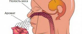
The olfactory nerves have processes located in the nasal mucosa. Starting from the nasal cavity, they cross the cribriform plate and approach the olfactory bulb, where the first neuron ends and the central pathway begins.
The visual pair consists of fibers extending from the retina, cones and rods. All nerves enter one trunk in the cranial cavity. First, they form a chiasm, and then the optic tract, which goes around the cerebral peduncle and sends fibers to the visual centers. One nerve contains about a million fibers (axons of retinal neurons) and, in addition, it has one sheath on the outside and another on the inside. The nerve enters the skull through the optic canal.
The eighth pair includes auditory cranial nerves - the remaining 12 pairs, except these three, are motor or mixed. In the auditory nerves, the fibers are directed from the middle ear to the nuclei. Each of them includes a vestibular and cochlear root. They arise from the middle ear and enter the cerebellopontine angle.
Cranial nerve pairs and their nuclei
The formation and development of nervous structures occurs already at the stage of intrauterine fetal formation - individual structures, for example, cranial nerves, begin their activity even before the moment of birth. Their final maturation takes more time, but, in general, most of the central nervous system of the newborn ensures its full interaction with the external environment.
In total, it is customary to distinguish 12 pairs of nerves. Their central part in the form of nuclei remains inside the brain. Whereas impulses reach the innervated organs through special fibers - a collection of nerve cell processes. Depending on what function these structures perform, experts traditionally distinguish several subgroups of pairs of cranial nerves:
- motor;
- sensitive;
- mixed.
The location of the exit of the nerve fiber is also of great importance - above the brain stem or below it. Doctors consider this criterion when diagnosing various pathologies of the cranial nerves.
Motor types
Another group of 12 pairs of cranial nerves includes the oculomotor, trochlear, accessory, hypoglossal and abducens nerves.
The third pair, that is, the oculomotor nerves, contain autonomic, motor and parasympathetic fibers. They are divided into upper and lower branches. Moreover, only the upper branches belong to the motor group. They enter the muscle that lifts the eyelid.
The next group includes trochlear nerves that move the eyes. If you compare all the cranial nerves - 12 pairs - then these are the thinnest. They originate from the nucleus on the tegmentum of the midbrain, then go around the cerebral peduncle and go to the orbit, innervating the superior oblique muscle of the apple of the eye.
The abducens nerves are related to the rectus oculi muscle. They have a motor nucleus in the fossa. Coming from the brain, they go to the superior orbital fissure, innervating the rectus ocular muscle there.
Accessory nerves originate from the medulla oblongata and cervical areas of the spinal cord. The individual roots are connected into one trunk, passing through the hole and dividing into outer and inner branches. The internal branch, which contains fibers involved in the innervation of the larynx and pharynx, is attached to the vagus nerve.
And the last of the 12 pairs of cranial nerves (a table of which, for convenience, is presented at the end of the article), related to the motor ones, are the hypoglossal nerves. The origin of this nerve is spinal. But, over time, its root moved into the skull. It is clear that this is the motor nerve of the tongue. The roots emerge from the medulla oblongata, then cross the carotid artery and enter the lingual muscles, dividing into branches.
Functions of cranial nerves
Olfactory nerve
Olfactory nerve
(
olfactory nerves
) (lat. nervi olfactorii) - the first of the cranial nerves, responsible for olfactory sensitivity.
Optic nerve
Optic nerve
(lat. Nervus opticus) - the second pair of cranial nerves, through which visual stimuli perceived by the sensitive cells of the retina are transmitted to the brain.
Oculomotor nerve
Oculomotor nerve
(lat. nervus oculomotorius) - III pair of cranial nerves, responsible for the movement of the eyeball, raising the eyelid, and the reaction of the pupils to light.
Trochlear nerve
Trochlear nerve
(lat. nervus trochlearis) - IV pair of cranial nerves, which innervates the superior oblique muscle (lat. m. obliquus superior), which turns the eyeball outward and downward.
Trigeminal nerve
V (trigeminal) nerve
(lat. nervus trigeminus) is mixed. Its three branches (ramus ophthalmicus - V1, ramus maxillaris - V2, ramus mandibularis - V3) through the Gasserian ganglion (ganglion trigeminale) carry information from the upper, middle and lower thirds of the face, respectively. Each branch carries information from the muscles, skin and pain receptors of each third of the face. In the Gaserian node, information is sorted by type, and information from the muscles of the entire face goes to the sensitive nucleus of the trigeminal nerve, located mostly in the midbrain (partially enters the pons); cutaneous information from the entire face goes to the “main nucleus” (nucleus pontinus nervi trigemini), located in the pons; and pain sensitivity is in the nucleus spinalis nervi trigemini, coming from the bridge through the medulla oblongata to the spinal cord.
The trigeminal nerve also belongs to the motor nucleus (lat. nucleus motorius nervi trigemini), which lies in the bridge and is responsible for the innervation of the masticatory muscles.
Abducens nerve
Abducens nerve
(lat. nervus abducens) - VI pair of cranial nerves, which innervates the lateral rectus muscle (lat. m. rectus lateralis) and is responsible for abduction of the eyeball.
Facial nerve
Facial nerve
(lat. nervus facialis), the seventh (VII) of the twelve cranial nerves, exits the brain between the pons and the medulla oblongata. The facial nerve innervates the facial muscles. Also included in the facial nerve is the intermediate nerve, which is responsible for the innervation of the lacrimal gland, the stapedius muscle and the taste sensitivity of the two anterior thirds of the tongue.
vestibulocochlear nerve
vestibulocochlear nerve
(lat. nervus vestibulocochlearis) is a nerve of special sensitivity responsible for the transmission of auditory impulses and impulses emanating from the vestibular part of the inner ear.
Glossopharyngeal nerve
Glossopharyngeal nerve
(lat. nervus glossopharyngeus) - IX pair of cranial nerves. Is mixed. Provides:
- motor innervation of the stylopharyngeus muscle (lat. m. stylopharyngeus), levator pharynx
- innervation of the parotid gland (lat. glandula parotidea), providing its secretory function
- general sensitivity of the pharynx, tonsils, soft palate, Eustachian tube, tympanic cavity
- taste sensitivity of the posterior third of the tongue.
Nervus vagus
Nervus vagus
(lat. nervus vagus) - X pair of cranial nerves. Is mixed. Provides:
- motor innervation of the muscles of the soft palate, pharynx, larynx, as well as striated muscles of the esophagus
- parasympathetic innervation of the smooth muscles of the lungs, esophagus, stomach and intestines (up to the splenic flexure of the colon), as well as the muscles of the heart. Also affects the secretion of the glands of the stomach and pancreas
- sensitive innervation of the mucous membrane of the lower part of the pharynx and larynx, the skin behind the ear and part of the external auditory canal, the eardrum and the dura mater of the posterior cranial fossa.
The dorsal nucleus of the vagus nerve, nucleus dorsalis nervi vagi, is located in the medulla oblongata lateral to the nucleus of the hypoglossal nerve.
Accessory nerve
Accessory nerve
(lat. nervus accessorius) - XI pair of cranial nerves. Contains motor nerve fibers that innervate the muscles responsible for turning the head, raising the shoulder and adducting the scapula to the spine.
Hypoglossal nerve
Hypoglossal nerve
(lat. nervus hypoglossus) - XII pair of cranial nerves. Responsible for the movement of the tongue.
Mixed species
This group includes the trigeminal, facial, glossopharyngeal and vagus nerves. The mixed nerves have ganglia similar to those of the spinal nerves, but they do not have anterior and posterior roots. In them, the fibers of the motor and sensory types are connected into a common trunk. They can also just be nearby.
The output of the 12 pairs of cranial nerves is different. Thus, the third, seventh, ninth and tenth pairs have parasympathetic fibers at the output sites, which are directed to the autonomic ganglia. Many of them are united by branches where different fibers pass.
The trigeminal nerve has two roots, where the larger one is sensory and the smaller one is motor. Innervation of the skin occurs in the parietal, ear and chin areas. Innervation also includes the conjunctiva and apple of the eye, the dura mater of the brain, the mucous membrane of the mouth and nose, teeth and gums, as well as the main part of the tongue.
The trigeminal nerves exit between the cerebellar peduncle, in the middle, and the pons. The fibers of the sensory root belong to the ganglion, which lies in the temporal pyramid near the apex, which was formed as a result of splitting the dura mater of the brain. They end in the nucleus of this nerve, which is located in the fossa, as well as in the nucleus of the spinal tract, which continues into the medulla oblongata and then goes to the spinal cord. The fibers of the motor nerve root originate from the trigeminal nucleus, which is located in the pons.
The maxillary, mandibular and ophthalmic nerves depart from the ganglion. The latter is sensitive, divided into nasociliary, frontal and lacrimal. The innervation of the 12 pairs of cranial nerves varies not only among the pairs themselves, but also among their derivative branches. Thus, the lacrimal nerve innervates the lateral canthus, transmitting secretory branches to the lacrimal gland. The frontal nerve, accordingly, branches on the forehead and supplies its mucous membrane. The nasociliary nerve innervates the eyeball, and the ethmoidal nerves depart from it, innervating the nasal mucosa.
The maxillary nerve, which passes into the pterygopalatine fossa and exits onto the anterior facial surface, is also sensitive. The superior alveolar nerves originate from it and pass to the teeth of the upper jaw and gums. The nerve on the cheekbones passes from the ganglion along the posterior nerves of the nose to its mucosa and nasopharynx. The nerve fibers here are sympathetic and parasympathetic.
The mandibular nerve belongs to the mixed type. It consists of a motor root. Its sensory branches include the buccal nerve, which supplies the corresponding mucosa, the auriculotemporal nerve, which innervates the skin on the temples and ears, and the lingual nerve, which supplies the tip and back of the tongue. The inferior alveolar nerve is mixed. Passing in the lower jaw, it ends on the chin, branching here into the skin and mucous membrane of the lower lip. Its branches are connected to the autonomic ganglia:
- auriculotemporal nerve - with the ear, innervating the parotid gland;
- lingual nerve - with a ganglion that supplies innervation to the sublingual and submandibular glands.
The facial nerves include motor and sensory cranial nerves. Mixed fibers create a taste sensation. Some fibers here innervate the lacrimal and salivary glands, while others innervate the anterior two-thirds of the tongue.
The facial nerve consists of motor fibers that begin in the upper part of the fossa. It includes the intermediate nerve with taste and parasympathetic fibers. Some are processes of the ganglion, ending in taste fibers of the vagus and glossopharyngeal nerves. And others begin in the salivary and lacrimal nuclei, located next to the motor nucleus.
The facial nerve begins in the cerebellopontine angle of the brain and then enters the facial canal through the auditory canal. The chorda tympani is located here and, passing through the cavity, connects to the lingual nerve. It includes gustatory and parasympathetic fibers that reach the submandibular ganglion.
The facial nerve leaves the temple bone and passes into the parotid gland, intertwining there. From here the branches spread out in a fan-shaped manner. At this time, all muscles related to facial expressions and some others are innervated. A branch in the neck from the facial nerve branches on it in the saphenous muscle.
The glossopharyngeal pair supplies the innervation of the lacrimal glands, the posterior part of the tongue, the inner ear and the pharynx. Motor fibers are directed to the stylopharyngeal muscle and pharyngeal constrictors, and sensory fibers for the parotid gland are directed to the ear ganglion. The nuclei of these nerves, unlike where the other nuclei of the 12 pairs of cranial nerves are located, are located in the fossa - the triangle of the vagus nerve.
Parasympathetic fibers begin in the salivary nucleus. The glossopharyngeal nerve, leaving the medulla oblongata, reaches the base of the tongue. The tympanic nerve begins from the ganglion, which has parasympathetic fibers that continue to the ear ganglion. Next begin the lingual, tonsil and pharyngeal nerves. The lingual nerves innervate the root of the tongue.
The vagus pair provides parasympathetic innervation in the abdominal cavity, as well as in the chest and neck. This nerve includes motor and sensory fibers. This is where the greatest innervation is. The vagus nerve has a double nucleus:
- dorsal;
- single path.
Coming out behind the olive on the neck, it moves with the neurovascular bundle and then branches.
Cranial nerve nuclei
| Character of the core | Nerve | Character of innervation | Innervation of the organ | Function |
| The magnocellular nucleus of the oculomotor nerve (paired) | III pair | motor | the levator superioris muscle, the superior, inferior and medial rectus muscles of the eye, the inferior oblique muscle of the eye | eye movement, raising the upper eyelid |
| Small cell nucleus of the oculomotor nerve (syn. Yakubovich nucleus), paired | III pair | parasympathetic | muscle constrictor pupil (lat. m.sphincter pupillae), ciliary muscle (lat. m.ciliaris) | constriction of the pupil and accommodation of the eye |
| Small cell unpaired nucleus of the oculomotor nerve (syn. nucleus of Perlia) | III pair | motor, convergence | medial rectus oculi muscle | simultaneous approach to the median plane (convergence) |
| Trochlear nerve nucleus | IV pair | motor | superior oblique muscle (lat. m. obliquus superior) | moving the eye to the side and down |
| The nucleus of the spinal tract of the trigeminal nerve (lat. nucleus tractus spinalis n.trigemini) | V pair | sensitive | face | superficial (pain and tactile) sensitivity |
| Core of deep sensitivity of the trigeminal nerve (lat. nucleus sensorius principalis n.trigemini) | V pair | sensitive | face | deep (proprioceptive) sensitivity |
| Motor nucleus of the trigeminal nerve (lat. nucleus motorius (masticatorius) n.trigemini) | V pair | motor | masticatory muscles (masseter, temporalis, lateral and medial pterygoid, mylohyoid muscles, anterior belly of the digastric muscle and muscle that tightens the soft palate) | chewing |
| The nucleus of the abducens nerve (lat. nucleus n.abducentis) | VI pair | motor | lateral rectus muscle (lat. m.rectus lateralis) | abduction of the eyeball to the side |
| The nucleus of the facial nerve (lat. nucleus n.facialis) | VII pair | motor | facial muscles | facial expressions |
| Core of the solitary tract (lat. nucleus tractus solitarius) | VII,IX,X pairs | sensitive | tongue (taste buds) | taste |
| Superior salivatory nucleus (lat. nucleus salivatorius superior) | VII pair | parasympathetic | lacrimal gland, submandibular and sublingual salivary glands | tearing, salivation |
| Anterior and posterior cochlear nuclei (lat. nuclei cochleares anterior et posterior) | VIII pair | sensitive | inner ear (auditory receptors) | hearing |
| Vestibular nuclei (superior, lateral, medial and inferior) (lat. nuclei vestibulares) | VIII pair | sensitive | inner ear (vestibular receptors) | vestibular apparatus |
| Double nucleus (lat. nucleus ambiguus) | IX, X and XI pairs | motor | muscles of the soft palate, pharynx and larynx | chewing, voice, articulation |
| Lower salivatory nucleus (lat. nucleus salivatorius inferior) | IX pair | parasympathetic | parotid gland | salivation |
| Sensitive nucleus of the glossopharyngeal and vagus nerves (lat. nucleus alae cinereae) | IX and X pairs | sensitive | oral cavity, middle and inner ear | overall sensitivity of these areas |
| Posterior nucleus of the vagus nerve (lat. nucleus dorsalis n.vagi) | X pair | parasympathetic | heart muscle, smooth muscles of the lungs, bronchi, stomach and intestines | heart rate, secretion of endocrine glands of the gastrointestinal tract, bronchial smooth muscle tone |
| Accessory nerve nucleus (lat. nucleus n.accessorii) | XI pair | motor | trapezoid and sternocleidomastoid (lat. m.sternocleidomastoideus) | turning the head, lifting the shoulder, scapula and acromial part of the clavicle upward (“shrug”), pulling the shoulder girdle back and bringing the scapula to the spine |
| The nucleus of the hypoglossal nerve (lat. nucleus n.hypoglossi) | XII pair | motor | muscles of the tongue and orbicularis oris | tongue movement, swallowing. |
Violations
All cranial nerves—12 pairs—may have dysfunction. The anatomy of the lesion sites appears at different levels of the nuclei or trunks. To make a diagnosis, an in-depth analysis of intracranial pathological processes is performed. If the lesion affects one side of the nuclei and fibers, then most likely we are talking about dysfunction of any of the affected 12 pairs of cranial nerves.
Neurology, however, also studies symptoms on the opposite side. Then damage to the pathways is diagnosed. It also happens that dysfunction of the nerves is associated with a tumor, arachnoid cyst, abscess, vascular malformations and other similar processes.
Simultaneous damage to the 12th pair of cranial nerves, that is, the hypoglossal, as well as the vagus and glossopharyngeal, is called bulbar palsy. This is a very dangerous disease, since there is a possibility of pathology of the most important centers of the brain stem.
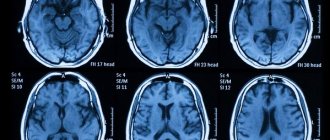
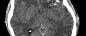
Knowledge of the topographic location of the cranial nerves allows one to correctly identify the narrow area of damage to each of them. To conduct research, special techniques are used. With the appropriate equipment, today it is possible to identify all the details of the condition of the fundus, optic nerve, diagnose the visual field and areas of loss. Computerized examination allows highly accurate localization of the affected area.
Mixed nerve fibers
Their complex structure—the presence of both sensory and motor nerve cells—allows these cranial nerves to perform many important functions for humans.
Thus, the exit point of the trigeminal nerve can be called the area between the middle cerebellar peduncle and the pons. The sensory fibers of the pair - the orbital with the maxillary, as well as the mandibular - connect with the motor ones, and then branch into many smaller branches. In total, their number reaches 15, and their activity covers almost the entire facial part of the cranial muscles.
The nucleus of the seventh cranial pair is located in the pons, or rather its tire. It is also called the facial nerve. It rushes through the layers of the pons towards the base of the brain, where the pair will be divided into several branches:
- large rocky;
- stirrup;
- drum string.
The glossopharyngeal nerve is no less complex in structure. Experts trace the pair from the inferior olive to the pharyngeal plexus and the thickness of the tongue. Many branches and interweavings with other cranial bundles of neurocytes allow it to perform a variety of functions. Whereas the vagus pair, intertwining with the fibers of the glossopharyngeal and accessory intracranial formation, as well as vessels, creates a complex neurovascular structure. Its functioning affects the organs of the abdomen and solar plexus.
Neurological examination and symptoms of disorders of individual cranial nerves
This technique is carried out in a certain order. The examination begins with the olfactory nerve. A cotton wool soaked in the irritant is brought to the nostrils one by one. The optic nerve is examined during an ophthalmological examination, based on which, in addition to direct damage, it is possible to identify even secondary changes. The pathology can be stagnant, dystrophic, inflammatory, or the nerve can be completely destroyed.
Lesions of the following three of the 12 pairs of cranial nerves (oculomotor, abducens and trochlear) cause diplopia and strabismus. Drooping of the upper eyelid, dilation of the pupil, and double vision may also occur.
Disturbances in the fifth pair, that is, in the trigeminal nerves, lead to a deterioration in sensitivity in the part of the face where they are present. This can be observed in the area of the temples, forehead, cheekbones, eyes, chin and lips. It happens that severe pain is felt, rashes and other reactions appear. Due to the fact that the facial nerves have many connections, this pair is characterized by a wide variety of pathological reactions.
If the auditory nerve is damaged, hearing deteriorates; the glossopharyngeal nerve impairs sensitivity in the inner ear; the sublingual nerve causes limited movement of the tongue. In the case of the vagus nerve, paralysis of the soft palate or vocal cord develops. In addition, the rhythm of the heart, breathing, and other visceral-vegetative functions may be disrupted.
Sensitive nerve structures
All brain nerves are important for the full functioning of the human body. If the predominant activity of a couple is the perception and transmission of information, then they are called sensory nerve fibers.
Thus, the olfactory nerve originates from the cells of the nasal mucosa. It then passes through the cribriform plate of the skull and moves deep into the brain to a structure called the olfactory bulb. Next, the nerve fiber forms a kind of tract, which passes into the olfactory triangle. The sensory structure ends in the tubercle of the cerebral cortex.
Another important sensory unit is the optic nerve. Its ganglion cells from the retina rush into the cranium, where the pair forms a kind of cross, from which the fibers run to the lateral geniculate structure of the brain. From here the optic nerve will continue to its final destination - the occipital part of the cerebral cortex.
Cells of the eighth pair of cranial nerves - auditory or vestibulocochlear. It is inherent not only to perceive and transmit the sounds of the surrounding world, but also to be responsible for the balance of the human body in space. Both parts of the nerve fiber - from the cochlear root and from the vestibular root, which begins from the vestibular ganglion, are directed to their final targets - the cerebellum, as well as the quadrigeminal cortex and the median geniculate body. The auditory nerve ends in the temporal gyrus.
Complex disorders and cranial nerves (12 pairs): anatomy, table
The functions of nerve fibers can be impaired either isolated or in combination, together with various pathologies of the lower skull. So, if all the nerves on one half of the cranial base are affected, then they talk about Garcin syndrome. When there is a tumor of the orbital bones and soft tissues, superior orbital fissure syndrome occurs. When both the hug and optic nerves are damaged, Kennedy syndrome occurs.
These and other diseases occur in both adulthood and childhood. For children, nerve damage that is associated with developmental defects is especially common.
Below is a structure that can help you better understand how the cranial nerves (12 pairs) work. Anatomy (the table is based on her knowledge) will help you navigate the intricacies of the functioning of their different groups.
Neuritis of the facial nerve diagnosis
Diagnosis of facial nerve neuritis is based on an analysis of the symptoms preceding and accompanying diseases of the nerve damage. Neurologists, neuropathologists, and reflexologists distinguish such forms of damage as neuritis of the facial nerve and neuropathy of the facial nerve. According to pathogenesis, neuritis is divided into primary and secondary.
Neuritis of the facial nerve must be differentiated from isolated damage to the motor nucleus of the nerve, a variety of common pathological processes in the area of the cerebellopontine angle. Damage to the nucleus of the facial nerve is accompanied by isolated paresis of the facial muscles of the same half of the face without autonomic and sensory disorders, which occurs mainly in the pontine form of poliomyelitis or poliomyelitis-like diseases, tick-borne encephalitis.
When the lower parts of the bridge are damaged, which occurs with tumors, encephalitis, vascular diseases, along with the nucleus of the facial nerve, the pyramidal tract is involved in the pathological process. In such cases, peripheral paresis of the facial muscles is combined with central hemiparesis of the opposite side (Millard-Hübler syndrome). If the nucleus of the abducens nerve is affected, then paresis of the external rectus muscle of the eye (Fauville syndrome) is added to the above symptoms.
Isolated neuritis of the facial nerve is differentiated from processes in the area of the cerebellopontine angle, which develop, for example, with arachnoiditis, tumors of the vestibulocochlear nerve, which is manifested by decreased hearing, deafness, paresis of the facial muscles, sometimes dysfunction of the trigeminal nerve, contralateral spastic hemiparesis.
Polyneuritis and polyradiculoneuritis often involve lesions of the facial nerve. Usually the lesion in these cases is bilateral, often asymmetrical, accompanied by limited or diffuse damage to other parts of the human peripheral nervous system.
Pathologies of sensitive cranial nerves
In most cases, lesions of sensitive neurocytes of cranial nerves are associated with their hypothermia or damage, sometimes with neuroinfections suffered by a person.
For example, hearing impairment is expressed in people’s complaints about a persistent deterioration in their perception of sounds from the outside world, up to persistent deafness. At the same time, patients may experience noise in the head of varying intensity. If disorders occur in the vestibular pairs, symptoms of dizziness, disorientation in space, nausea and even vomiting will appear.
Visual disturbances are no less common - due to stagnation, swelling or injury of the nerve fiber, a significant decrease in visual acuity occurs for patients. There may be areas of no image on the retina of the eyeballs and excessive light sensitivity.
Lesions of the olfactory pair are less common in medical practice, since they are the shortest among the cranial nerve fibers and are located deep in the brain. People may experience olfactory hallucinations or deterioration/loss of the ability to distinguish odors.
Pathologies of mixed cranial-cerebral formations lead to a decrease in a person’s quality of life - disruptions in cardiac and respiratory activity, food absorption, distortion of taste perception, unpleasant sensations in the facial part of the skull, up to persistent pain syndrome due to chronic inflammation, for example, of the trigeminal pair. Such changes in the innervation of the cranial nerves can only be dealt with by competently selected complex treatment, which will be selected by a doctor taking into account the etiology of the disorder.
Classification of cranial nerve formations
To facilitate understanding of the structural features, location and functional load of cranial-cerebral pairs, specialists created a brief classification of them:
| Numbering | Pair name | Function in the body |
| 1 | Olfactory | Human perception of a wide variety of odors |
| 2 | Visual | Transmission of perceived visual images to the occipital cortex of the brain |
| 3 | Oculomotor | Reaction of the pupils to light stimuli, movement of the eyeball in the socket |
| 4 | Block | Moving the eyes downwards or to the sides |
| 5 | Trigeminal | Sensitivity of the facial, oral and pharyngeal structures, as well as the transmission of commands to the muscle groups responsible for chewing food |
| 6 | Abductor | Moving the eyes outward |
| 7 | Facial | Activity of facial and stapedius muscles, performance of the salivary glands, as well as perception of information from the tip of the tongue |
| 8 | Auditory | Transmission of auditory signals from the structures of the inner ear to the auditory cortex |
| 9 | Glossopharyngeal | Detail of the levator muscle in the pharynx, the work of paired salivary glands, sensitivity of the throat, structures of the middle ear and auditory tube |
| 10 | Wandering | Innervation of structures from the pharynx to the intra-abdominal space and solar plexus |
| 11 | Additional | Ability to move the head in different directions - from shrugging the shoulders to bringing the angle of the scapula to the spine |
| 12 | Sublingual | Tongue movements, responsibility for the completeness of the act of swallowing and chewing |
The entire number of cranial nerves - twelve pairs of them - makes it possible to create a complex system of relationships between the brain and distant organs dependent on it.