Abducens optic nerve palsy is a syndrome caused by damage to the abducens nerve, leading to limited mobility or complete inability to move the eyeball outward.
In order to better understand the cause of this pathology, it is necessary to delve a little deeper into the anatomy.
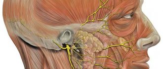
The abducens nerve regulates the mobility of the eye, retracting it to the outer edge of the eyelid.
Nerve fibers of this type control the direct lateral, or in other words, the external muscle. It is this that allows you to move the eyeball to the outside, move it to the sides, without turning your head.
The lateral rectus muscle of the eye is an antagonist of the intrinsic muscle, which moves the eye in the opposite direction, towards the center. These muscles, in the absence of damage, balance each other’s work.
Since the fibers of the abducens nerve are located superficially, they can be easily damaged as a result of injury, as a result of which they become pressed to the base of the skull and paresis develops.
Causes
The most common causes of gaze paresis in adults are:
- diabetes
- arterial hypertension
- atherosclerosis
- injury
- idiopathy.
Less common reasons:
- increased intracranial pressure
- giant cell arteritis
- presence of tumor inclusions
- multiple sclerosis
- stroke
- Chiari malformation
- hydrocephalus
- intracranial hypertension
- previous meningitis.
Among other things, paresis can also occur due to viral diseases such as diphtheria, syphilis, encephalitis, or as a result of complications after the flu.
In some cases, intoxication with ethyl alcohol, heavy metals or combustion products can provoke the appearance of pathology.
In pediatric practice, the most common causes are tumor diseases, injuries and idiopathies.
Reference! Children sometimes develop a benign and quickly recovering isolated abducens nerve palsy, which in some cases is a consequence of previous infections of the ear, nose or throat.
Symptoms and signs of damage
In the early stages of oculomotor nerve paresis, there are practically no symptoms, which complicates its diagnosis and further treatment. With a longer course of the disease, the following symptoms gradually begin to appear:
- Drooping of the upper eyelid (partial or complete);
- Lack of reaction (constriction/dilation) of the pupil;
- Diplopia (double vision due to loss of motor ability of the eye);
- Divergent strabismus (occurs due to lack of resistance of the upper and lower muscles of the eyeball);
- Loss of focusing and adaptation to changes in the distance between the eye and the object;
- Loss of motor ability;
- Protrusion of the eye.
With extensive damage, immobility can become only part of the entire symptomatology of the disease if other cranial nerves are also damaged. In addition, oculomotor nerve palsy itself may be a symptom of a more serious systemic disease. Most often, the lesion affects only one eye.
Symptoms
Normally, in a healthy person, the edge of the cornea is in contact with the outer edge of the eyelid junction.
In cases where this is not observed, nerve pathology is present. Symptoms of the pathology are:
- limited mobility of the eyeball
- secondary eye deviation
- dizziness
- spatial orientation disorder
- unsure gait
- diplopia (double images of one object)
- forced, involuntary position of the head.
With a mild form of paresis, the symptoms are mild and practically do not cause any discomfort. These signs are characteristic of both the right and left eyes.
Diagnostics
It is easiest to recognize the lesion, since this pathological process is characterized by drooping eyelids, dilated pupils and abnormal deviations of the eyes. Based on such signs, making a diagnosis will not be a problem, but often they are combined with each other in various combinations, so the doctor suspects the secondary nature of the disease. To differentiate paresis of the eye muscles from other possible ailments, the ophthalmologist will need to prescribe an examination, which consists of the following procedures:
- Fundus examination;
- Determination of visual acuity and degree of mobility of the eyeball;
- Light reflex test;
- Angiography (to identify vascular pathologies);
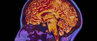
- Magnetic resonance imaging (checks brain tissue for abnormalities).
Sometimes a consultation with a neurologist may be required. If it was not possible to determine the cause of the pathology, then the patient should be registered with a doctor and periodically examined. To prevent the condition from getting worse, your doctor may recommend special sets of exercises and other treatment methods.
Treatment methods
Drug therapy
The medicinal method of treatment is the orbital-occipital method of administering the drug. Neuromidin is most often used for these purposes, since its use helps to both increase muscle contractility and reduce defects in connective muscles.
Reference! Additionally, after carrying out such a procedure, you need to lie down with your eyes closed for about 15 minutes.
Another option applicable at the initial stage of therapy is the use of botulinum toxin . Its introduction helps prevent contracture of the medial rectus muscle, and by reducing the size of the deviation, prismatic correction can be applied for a short period of time in cases where its use was previously impossible.
Most pathologies of the abducens nerve are associated with diseases of the central nervous system, on the basis of which appropriate treatment is prescribed.
If there is no improvement in the condition after drug therapy, and the lesion does not go away on its own, surgical intervention is used.
Rehabilitation
Rehabilitation measures that speed up recovery from abducens nerve paresis include a variety of physical procedures.
The impact on the affected nerve occurs through the use of low-frequency electromagnetic field pulses or through stimulation with electric current.
The procedure has a pronounced calming, anti-inflammatory and analgesic effect.
The main disadvantage of this method is the need to carry out long courses of procedures to achieve a visible effect; moreover, in some cases, it may be absent altogether.
A good result in the rehabilitation of abducens nerve pathology is the combined use of electrophoresis with a 15% solution of neuromidin. According to the standard scheme, the duration of one session is 15 minutes. The procedure is carried out daily for two weeks.
In addition to physical procedures, the doctor prescribes special gymnastics for the eyes, which should also be done daily.
With paresis of the abducens nerve, additional action is required on diplopia. For these purposes, Fresnel prisms , which are thin and flexible plates that are attached to the patient's glasses. Due to them, the symptoms of paresis are alleviated and binocular vision is supported.
Prisms exist with different angles and are selected individually.
In patients with more severe forms of deviation, the thickness of the prism used can have an extremely negative impact on vision, so in this case, occlusion is most often used - temporarily closing one eye.
Reference! Occlusion is almost never used in pediatric practice for such pathologies, as this can lead to the development of “lazy eye” syndrome.
The use of a Fresnel prism or occlusion requires a long period of observation, usually lasting from 9 months to 1 year. This is due to the fact that some types of paresis can be restored without surgical intervention.
Most often, these measures are carried out in combination with drug therapy, which can significantly speed up the recovery process.
Course of therapy
Treatment methods for paresis of the eye muscles in children are not particularly different from those in adults. However, it must be borne in mind that most congenital anomalies are corrected through surgery. If the operation is successfully performed, the extraocular muscles are partially or completely restored. If the problem is compression of the nerve pathway, then the main task is to eliminate the cause.
After eliminating the factor influencing the development of muscle paresis, treatment is adjusted towards restoring blood flow and damaged nerve fibers. For this purpose, exercises that strengthen the oculomotor muscles are often used. They serve as the basis for the treatment of minor injuries and are a good preventive measure. In severe cases of the disease, therapeutic exercises well complement the main course of therapy.
Drug therapy for paresis may include the following:
- Vitamin complexes;
- Preparations for strengthening the extraocular muscles and restoring their innervation;
- Eye drops;
- Medicines that improve blood circulation;
- Corrective glasses and bandages.
The pathology can be treated with medications only according to the regimen prescribed by the doctor, so as not to aggravate its course and not to impair vision, especially if the child is sick. It is recommended to combine drug therapy with other methods, namely:
- Steriopictures. By watching them, the extraocular muscles are trained and blood flow improves. The nerve tissues that innervate the muscles of the eye are extremely tense during the procedure, due to this the lost innervation is restored. The procedure must be carried out under the supervision of a specialist so as not to cause complications;
- Electropheresis. This physiotherapeutic procedure is carried out with a 1.5% solution of Neuromidin. The duration of one session of electropheresis usually does not exceed 20 minutes, and it acts directly on the synapses (junctions) of the muscle and nervous tissue of the eyeball. After a course of such therapy, the patient’s severity of paresis decreases and the innervation of the eye muscles improves.
It is impossible to eliminate some causes of paresis in children, for example, congenital anomalies, without surgical intervention. Their duration and degree of risk depend on the type of operation and the factor that influenced the development of the pathology. In the case of severe damage to the optic nerves, it will not be possible to completely eliminate the problem, but there will be a chance to save the child’s vision.
Due to paresis of the extraocular muscles, many complications develop, such as strabismus, ptosis, etc. In children, this pathological process is often a consequence of congenital anomalies. It may not appear immediately, but only over time. This is why it is important to see an ophthalmologist and other doctors, especially in the first years of a child’s life.
Abducens optic nerve palsy is a syndrome caused by damage to the abducens nerve, leading to limited mobility or complete inability to move the eyeball outward.
In order to better understand the cause of this pathology, it is necessary to delve a little deeper into the anatomy.
The abducens nerve regulates the mobility of the eye, retracting it to the outer edge of the eyelid.
Nerve fibers of this type control the direct lateral, or in other words, the external muscle. It is this that allows you to move the eyeball to the outside, move it to the sides, without turning your head.
The lateral rectus muscle of the eye is an antagonist of the intrinsic muscle, which moves the eye in the opposite direction, towards the center. These muscles, in the absence of damage, balance each other’s work.
Since the fibers of the abducens nerve are located superficially, they can be easily damaged as a result of injury, as a result of which they become pressed to the base of the skull and paresis develops.
Prognosis for recovery
In most cases, paresis of the abducens nerve, unlike pathologies of a number of other optic nerves, is a reversible condition.
If the cause of paresis lies in an infection, then after it is completely cured, the nerve function is restored on its own.
In some cases, when paresis is a consequence of serious skull injuries, inoperable tumor diseases , or occurs due to severe damage to the nerve itself, paralysis of the abducens ophthalmic nerve occurs and the pathology becomes incurable.
Description of the pathology, development mechanism
The oculomotor nerve is part of the third pair of cranial nerves and consists of viscemotor and somatomotor (motor) fibers. Its main function is to provide motor ability to the eyeball. The nerve controls the following systems:
- Ciliary muscles;
- Sphincter of the pupil (provides its ability to expand and contract depending on the lighting);
- Optical-kinetic nystagmus (the ability to follow moving objects);
- Muscles to regulate the movement of the upper eyelids;
- Vestibulospectral reflex (the ability of the pupil to move when the head turns);
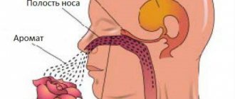
- Accommodation (change in the curvature of the eye lens depending on surrounding objects and phenomena).
Thus, damage to the optic nerve always entails a limitation in the functionality of the visual apparatus. The movement of the eye and pupil is limited or lost. In turn, the lesion has its own reasons for occurrence or acts as a sign of another disease. Men and women of all ages are at risk, but statistics show that children are more likely to suffer from oculomotor nerve palsy.
Treatment of paresis of the abducens ophthalmic nerve. Damage to the abducens (vi) nerve (n. abducens)
Clinical methods for studying the oculomotor system
are mainly used to diagnose the initial stages of paresis of the nerves innervating the eye muscles, especially in cases where patients complain of double vision, but there is a noticeable deviation of the eye.
In concomitant strabismus, the use of these methods is limited; they are used to determine the participation of the paretic component in its origin.
It should be remembered that with long-term strabismus, secondary changes in the muscles of the eye can simulate signs of paresis.
It is very difficult to differentiate these conditions, therefore, when identifying such signs, it is better to talk about muscle insufficiency, keeping in mind that this term can hide both its true paresis and secondary weakness.
In preschool children, the state of the oculomotor system is judged by the degree of eye mobility in different directions (determination of the visual field). In older children, coordimetry and “provoked” diplopia methods can be used for this purpose.
A simplified way to determine the field of view is as follows.
The patient sits opposite the doctor at a distance of 50-60 cm.
The doctor fixes the patient’s head with his left hand and invites him to alternately monitor with each eye (the second eye is covered at this time) the movement of an object in 8 directions (pencil, hand-held ophthalmoscope, etc.).
), which he holds in his right hand. Muscle deficiency is judged by the limitation of eye mobility in one direction or another. In this case, use the rules given in table. 14.
Table 14. Rules for determining the affected muscle when eye mobility is limited
Using this method, only severe limitations in eye mobility can be identified. A simplified method for determining the field of view allows you to obtain clearer results in case of insufficiency of the vertical muscles. If there is a visible vertical deviation of one eye, a simple abduction method can be used to identify the paretic muscle.
It is based on the fact that the lifting and lowering action of the oblique muscles is most pronounced in the adduction position of the eye, and the rectus muscles - in the abduction position. The patient is asked to look at an object, move it to the right and left, and observe whether the vertical deviation increases or decreases with extreme deviations of gaze.
The rules for determining the affected muscle are given in table. 15.
Table 15. Rules for determining the affected muscle by the method of abduction - adduction
Examples of determining the affected muscle using this method are presented in Fig. 47. A unilateral increase in eye deviation in the abduction or adduction position, as a rule, indicates muscle paresis, a symmetrical bilateral increase indicates vertical phoria.
Rice. 47. Paresis of the superior oblique muscle of the right eye. The upward deviation of the right eye is especially pronounced in the adduction position
Coordimetry method
is based on separating the visual fields of the right and left eyes using red and green filters colored in complementary colors. When these colors are superimposed on each other, the effect of completely blanking the image occurs. The method makes it possible to judge the localization in space of the image of both eyes during strabismus.
Let's look at Fig. 48. The right eye fixes the luminous point O. The visual line of the left, squinting eye is deviated from the point of fixation to the right by 20° and is directed to point O1.
Obviously, the image of point O will be located in the nasal half of the retina of the squinting eye, 20° from the central fovea.
As is known, with a normally functioning visual analyzer, an image located 20° inward from the central fovea of the retina is projected in space 20° outward from the point of fixation.
Rice. 48. Options for localizing an image with a squinting eye. Explanation in the text
By association with these habitual spatial relationships, the patient falsely places the image of point O with his left eye at the corresponding angle towards the temple, and it turns out to be located at point O2.
Now let’s put a red glass on the patient’s right eye, and a green glass on the left. Then we will direct a green beam of light to point O, give the patient a red flashlight and ask him to combine its beam with point O.
For this to happen, the patient must obviously direct a beam of red light to the O2 point. This principle is the basis of coordimetry.
To conduct the study, you must have a graphic screen (1), red (2) and green (3) flashlights and red-green glasses (4) (Fig. 49). The study is performed in a darkened room, on one of the walls of which there is a screen divided into small squares. The screen dimensions are 2X2 m, each square is 5.25X5.25 cm.
With such ratios, the study is carried out from a distance of 1 m. In this case, the side of each square is equal to three angular degrees. In the central part of the screen there are nine marks placed in the form of a square, the position of which corresponds to the isolated physiological action of the oculomotor muscles.
A patient wearing red-green glasses sits at a distance of 1 m from the screen.
Source: https://TherapyaDushi.ru/priznaki/parez-verhnej-pryamoj-myshcy-glaza-2.html