The vestibulocochlear nerve (VCN) is part of the system of the organ of the same name, which is responsible for the functioning of the hearing aid. This is a fiber of special sensitivity, which includes several roots. The vestibular one is responsible for impulses from the static apparatus, and the cochlear one conducts signals from the organ spiral to the labyrinth. In medicine, there are several methods for testing the vestibulocochlear nerve in order to identify its pathologies. After all, they affect many unpleasant symptoms: from dizziness to severe weakness and headaches.
Physiology
The red nuclei resulted from a large accumulation of neurons along the entire length of the midbrain. They are red in color because neurons have a large number of capillaries and iron-containing substances. The kernels consist of two parts:
- Parvocellular. In this part lies the beginning of the red nuclear olivary tract. This part began to develop in the brain due to the fact that a person began active movement on two limbs. Over the millennia it has developed more and more.
- Large cell. In this part lies the beginning of the rubrospinal tract. This part was always with the ancient man. In essence, it is a motor center.
Thanks to connections between the red nuclei and the cerebellum, the extrapyramidal system influences all skeletal muscles. In addition, they have projections to the nuclei of the spinal cord.
Location
The Greek term (mesencephalon) clearly indicates its location - inside the head, its etymology - from roots meaning "middle" and "brain".
The Greek term enkephalos literally means inside the head. The Russian language contains maximum information content - an indication of the main belonging to a complex complex of brain structures and its location.
As part of the brain stem, the SC is located between the intermedius (part of the hypothalamic-pituitary system) and the pons (sometimes called just the pons for simplicity).
A description of the anatomy of the midbrain, starting with the exact localization of the object in the complex interweaving of brain formations, indicates its location as follows:
- caudal direction – border with the metencephalon or hindbrain;
- rostral – boundary with the diencephalon (intermediate);
- the trunk of the large GM consists of the nuclei of the middle, bridge and diencephalon, as part of the structures of the trunk, the mesencephalon can be detected by focusing on the area of \u200b\u200bthe 1st and 2nd cervical vertebrae and the occipital fossa;
- an outdated understanding of the middle complex of important components, now separated into a separate structure - a specific layer between the pons and the diencephalon.
Previously, the mesencephalon was not given the same importance as in modern neurology; it was simply called a mound of medulla.
Studies carried out using special devices have shown that the reflex activity of the midbrain, as part of a complex system of interactions, makes it an indispensable part of the entire physiological functionality of the human body.
Without its independent structures, it is impossible to see and hear normally: the neurons of the quadrigeminal tubercles of the midbrain are responsible for eye movement, regulation of the lens and pupil lumen, start reflexes - as a guarantee of the survival of the body. Because they are the ones who react even to those signals that are not recognized by the auditory or visual center.
Functions of red kernels
Their main function is to ensure communication and transfer of information coming from the cerebellum and brain, more precisely its cortex, to all underlying structures. In a sense, this can be called the regulation of unconscious automatic movements. In addition to the main function, the red cores perform other, equally important tasks:
- Providing an open pathway between the extrapyramidal system and the spinal cord.
- Supports the active work of all skeletal muscles of the body.
- Coordination of movements with the cerebellum.
- Control of automatic movements, for example, changing body position during sleep.
The role of red nuclei
Their role is to ensure the transition of efferent signals from the nucleus itself to other neurons along a special path. After successful passage of the signal, the motor muscles of the limbs receive all the necessary information. Through a special tract, the red nuclei help facilitate the onset of active motor neuron activity, and the neurons also help regulate the motor abilities of the spinal cord.
But what happens if this path is damaged? After disruption of connections with the red nucleus of the midbrain, the syndromes presented below begin to develop, which in most cases are fraught with death.
Pathologies in violation
It all started when science received a description of severe muscle tension in animals. Tension was created by breaking the connections of the red core. This break is called decerebrate rigidity. Based on this observation, a conclusion was made, the essence of which is that when the connection between the red and vestibular nuclei is lost, severe tension occurs in the skeletal muscles, muscles of the limbs, as well as the muscles of the neck and back.
The above muscles are distinguished by their ability to counteract gravity, so it was concluded that this development of events is associated with the vestibular system. As it turned out later, the vestibular nucleus of Deiters is capable of triggering the work of extensor motor neurons. The activity of these neurons is significantly slowed down under the influence of the red nuclei and Deiters nucleus.
It turns out that active muscle work is the result of the joint work of the entire complex. In humans, decerebrate rigidity occurs as a result of traumatic brain injury. You can also encounter this phenomenon after a stroke. It should be understood that such a condition is a bad sign. You can find out about its presence by the following signs:
In case of injuries, severe infectious diseases, all kinds of internal damage to organs, including the brain, as well as tumor processes and aggression of the immune system - all this leads to disruption of the brain. Thus, if connections with the red nuclei are disrupted, decerebrate rigidity may occur, as well as disruption of the eyeball and eyelid muscles, the latter being an easier reaction of the body to the rupture of connections.
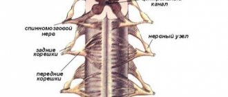
Anatomy and topography of the nerve ending
Anatomy of the vestibulocochlear nerve
Topography divides the fiber into two parts. The first is the cochlear part of the vestibular-cochlear nerve fiber, which is also called the vestibular. It integrates with afferent endings and transmits information from them. They are embedded in the semicircular channels of the elliptical and spherical sac. The second part is the cochlea, which transmits impulses from the spiral organ to the brain.
The anatomy of the parts of the vestibulocochlear nerve is as follows:
- The vestibule. The node of the same name is located here, deep in the auditory canal. It is also divided into 2 parts: the upper, directed to the receptive fields, and the lower, forming the spherical-saccular nerve. The ampullary nerve, which arises from the vestibulocochlear nerve, also originates here. The central roots of the cells of this node form the vestibular part, and in the cochlear zone they connect to the auditory canal. Inside, the nerve goes along with the intermediate endings and the branch of the facial nerve, and then diverges into the cranial cavity through the auditory canal. At the cerebellopontine angle the ending passes into the brain stem. The nuclei of the vestibular part of the vestibulocochlear nerve are also formed here. The vestibular part in the region of the vestibular field of the rhomboid fossa ends at the top in the ankylosing spondylitis nucleus, in the middle – in the Schwalbe nucleus, and at the back – in the Deiters nucleus.
- Cochlear zone. This part originates from the spiral ganglion formed by the neurons of the auditory pathway. Then it passes into the spiral canal of the cochlea rod. The peripheral roots end in the spiral organ. And the central ones run through the rod and the opening of the spiral path in the ear canal. The cochlear part of the apparatus is formed there. Then, together with the vestibular nerve ending, it passes into the cranial cavity through the auditory opening and goes to the brain stem. In the cerebellopontine part it enters the trunk and ends in the ventral and dorsal nuclei.
Topographically, the features of PUN do not differ from the characteristics of other sensitive fibers passing in this zone.
Claude syndrome
In 1912, when the famous transatlantic liner Titanic sank and the first metro line was opened in Hamburg, Henri Claude first described the syndrome, which was named after its discoverer. The essence of Claude's syndrome is that when the lower part of the red nuclei is damaged, the fibers from the cerebellum to the thalamus, as well as the oculomotor nerve, are damaged.
After the lesion, the patient’s eyelid muscles stop working, causing them to droop or one eyelid to droop on the side where the disorder occurred. Pupil dilation is also observed, and divergent strabismus appears. Body weakness and hand tremors are observed.
Claude's syndrome is caused by damage to the lower part of the red nucleus, through which the third nerve root passes. In addition, there are dento-rubral connections passing through the superior cerebellar peduncle. When these important connections are disrupted, a person begins to experience intentional tremors, hemiataxia, and muscle hypotonia.
Possible diseases
Symptoms of damage to the vestibulocochlear nerve
Damage to the vestibulocochlear nerve is accompanied by several symptoms. They are closely related to the part in which the violation is localized:
- If the vestibule is damaged, a person experiences dizziness, problems with stability, nausea, and vomiting. The pulse and breathing rhythm change, and blood pressure fluctuations are possible.
- If the cochlear part is damaged, the patient experiences severe tinnitus and hearing loss. Complete deafness is possible.
If both parts are damaged, all symptoms appear simultaneously and can be either severe or mild.
Causes of nerve damage
The most common diseases leading to pathologies of PUN include:
- Infections and viruses. Symptoms can be caused by influenza, colds, as well as measles, scarlet fever, syphilis, mumps and tuberculosis.
- Diseases of the heart and blood vessels. Strokes, heart attacks, atherosclerosis and myocarditis can also cause poor health.
- Intoxication. Symptoms appear when treated with antibiotics and other medications.
There are other diseases that are specific only to the ears, such as labyrinthitis. With this pathology, the inner ear is affected.
Rarely occurs acoustic neuroma involving PUN. It develops very slowly and provokes the gradual onset of all symptoms. Meniere's syndrome is also possible. It is associated with a disease of the same name, in which the production and outflow of fluid in the inner ear changes. From the side of the affected nerve, noise in the ear and a feeling of fullness appear. The person may suffer from attacks of dizziness and hearing loss.
To identify the exact cause, a full diagnosis is required. The patient is prescribed electroencephalography and examination by an ENT specialist or a neurologist. A CT and MRI of the ear area may be required.