Anterior branches (rr. ventrales, rr. anteriores) of the spinal nerves
just like the posterior ones, mixed in function, contain motor, sensory and autonomic (sympathetic) fibers.
Innervates the muscles and skin of the neck, chest, abdomen and limbs.
During the development of the limbs, a mixture of myotomes and dermatomes occurs, so the anterior branches of the cervical, lumbar, sacral and coccygeal nerves lose their initially metameric structure.
The segmental course of the anterior branches of the spinal nerves was preserved only on the trunk, where there was no displacement of the myotomes. This is where the intercostal nerves develop.
Intercostal nerves
The anterior branches of the thoracic spinal nerves form twelve pairs of intercostal nerves (nn. intercostales), which pass in the intercostal spaces (Fig. 5.9).
Figure 5.9.
Diagram of the spinal nerve in the thoracic region: 1 - n.
spinalis (spinal nerve); 2 - ramus dorsalis (posterior); 5 - ramus ventralis (anterior); 4 - back muscles; 5 - t. iniercostalis internus; 6 - t. intercostalis externus; 7 - oblique, transverse and rectus abdominis muscles; 8 - cutaneous branches Each intercostal nerve passes at the lower edge of the corresponding rib and is located below the artery and vein of the same name between the external and internal intercostal muscles.
The six superior intercostal nerves reach the edge of the sternum. Six lower intercostal nerves pierce the diaphragm between its costal teeth and pass between the internal oblique and transverse abdominal muscles. The XII intercostal nerve, subcostal (n. subcostalis), goes under the XII rib, lies on the surface of the m. quadratus lumborum (Fig. 5.9, 5.10).
Rice.
5.10. Intercostal nerves, on the posterior wall of the chest cavity (inside view): 1 - nervi intercostales;
2 - arteriae intercostates; 3 - venae intercostales; 4 - truncus sympathicus; 5 - mm. intercostales interni The muscular branches of the intercostal nerves (rr. musculares) innervate all the muscles developing from the ventral sections of the myotomes, i.e., the ventral muscles of the walls of the thoracic and abdominal cavities, as well as the back muscles of ventral origin (mm. serrati posteriores superiores et inferiores, mm. levatores costarum). The muscular branches of the VII-XII intercostal nerves innervate the diaphragm.
The skin of the chest and abdomen is innervated by the anterior and lateral cutaneous branches of the intercostal nerves (rami cutanei anteriores et rami cutanei laterales). The lateral branches extend to the lateral surface of the chest and abdomen along the anterior axillary line (Fig. 5.11).
Rice.
5.11. Segmental innervation of the skin of the body in front (rr. ventrales): C - nn.
cervicales; Th - nn. thoracici; L - n. lumbales Sensitive branches of the intercostal nerves innervate the parietal layers of the peritoneal pleura.
Anatomy
The median nerve is one of the largest nerves of the brachial plexus. It originates from the bundles of the brachial plexus, or more precisely, from the lateral and medial. In the shoulder area, it is conveniently located in the groove of the biceps muscle among all the other nerves. Then it descends along the front of the arm through the hole in the elbow to the forearm, where it is very conveniently located between the flexors of the fingers - deep and superficial. Then it passes into the lower section along the median groove and through the carpal tunnel enters the palm. In the region of the palmar aponeurosis, it divides into three terminal branches, which further create seven separate digital nerves.
The median nerve in the forearm innervates not only two of the pronators, but all the flexors. The exceptions are the flexor carpi ulnaris and half of the flexor profundus, which is responsible for the motor function of the fingers. As for the hand, here it is responsible for the muscles of the thumb and both lumbricals, the middle of the palm and the palmar side of the I-III and half of the IV fingers.
Plexus
In the cervical, lumbar, sacral and coccygeal regions, the anterior branches of the spinal nerves have lost their metameric structure as a result of displacement of the myotomes due to the development of the limbs. The anterior branches are connected to each other by loops (ansae), forming plexuses (plexus) (Fig. 5.12). The nerves arising from these plexuses can be mixed, sensory or motor. Therefore, the clinical picture of their damage consists of motor, sensory and autonomic disorders.
There are three plexuses:
cervical - cervicalis, shoulder - plexus brachialis and lumbosacral - plexus lumbosacralis, which is formed from pi. lumbalis, pi. sacralis. n. coccygeus. It should be taken into account that nп. coccygeus and the anterior branch of the fifth sacral nerve form the plexus coccygeus.
When studying each plexus, it is necessary to know: the sources of its formation (the anterior branches of which spinal nerves take part in its formation), the topography of the plexus, the functional characteristics of the nerves (the presence of mixed, cutaneous, muscle branches), division into short and long branches, zones of innervation.
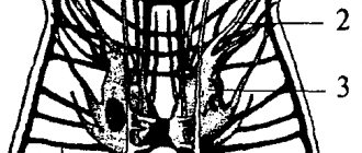
1 - plexus cervicalis (cervical plexus); 2 - plexus brachialis (brachial plexus); 3 - nn. intercostales (intercostal nerves); 4 - plexus lumbalis (lumbar plexus); 5 - plexus sacrococcygeus (sacrococcygeal plexus).
Cervical plexus
The cervical plexus (plexus cervicalis) is formed from the anterior branches of the four upper cervical nerves (C1-C4), which form three arcuate loops among themselves, located on the side of the transverse processes of the cervical vertebrae on the deep muscles of the neck between the prevertebral muscles medially (the beginning of the m. scalenus anterior et m. longus colli) and vertebrates (m. scalenus medius, m. levator scapulae, m. splenius cervicis) laterally. This plexus is joined by connecting branches from n. accessorius, hypoglossus et tr. sympathicus. The plexus is covered with m. sternocleidomastoideus.
The branches extending from the cervical plexus are divided into cutaneous (n. auricularis magnus - large auricular nerve, n. transversus coin - transverse nerve of the neck, n. occipitalis minor - lesser occipital nerve, nn. supraclaviculares - supraclavicular nerves), muscle (rr. musculares to the deep muscles of the neck and chest) and mixed (nn. phrenicus) (Fig. 5.12-5.15). Diagram of the cervical plexus and areas of innervation I, II, III, IV, V - cervical vertebrae; C1, C2, C3, C4, C5 - spinal nerves (cervical);
Rice.
5.12. Diagram of the anterior branches of the spinal nerves and the principle of formation of plexuses: 1 - plexus cervicalis (cervical plexus); 2 - plexus brachialis (brachial plexus); 5 - nn. intercosiales (intercostal nerves); 4 - plexus lumbatis (lumbar plexus); 5 - plexus sacrococcygeus (sacrococcygeal plexus)
Rice. 5.13. Diagram of the cervical plexus
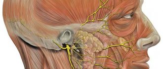
Rice. 5.14. Cutaneous branches of the cervical plexus innervation area
Branches arising from the cervical plexus:
1 - n. occipitalis minor (small occipital nerve) - skin of the lateral part of the occipital region; 2 - n. auricularis magnus (great auricular nerve) - skin in front and behind the auricle; 3 - n. transversus colli (transverse nerve of the neck) - skin of the neck above and below the hyoid bone; 4 - nn. supraclaviculares (supraclavicular nerves) - skin above and below the collarbone and suprascapular region; 5 - n. phrenicus (phrenic nerve) - sensory branches go to the pleura and pericardium, muscular branches - to the diaphragm; 6 - ansa cervicalis (cervical loop) - to the muscles below the hyoid bone; 7 - n. hypoglossus
Cutaneous branches of the cervical plexus:
1 - n. occipitalis major (greater occipital nerve) - posterior branch of the II cervical nerve. Innervates the skin of the back of the head; 2 - n. occipitalis minor (C2-C3) (small occipital nerve) - emerges from under the posterior edge of the m. sternocleidomastoideus and is directed behind the auricle to the skin of the lateral region of the back of the head. Innervates the skin of the lateral region of the neck; 3 - n. auriculans magnus (C3) (great auricular nerve) - goes around the posterior edge of m. sternocleidomastoideus near its middle and goes up to the auricle, ending at the bottom of the latter, as well as to the skin in front and behind the auricle; 4 - n. transversus colli (C3) (transverse nerve of the neck) - departs, like the previous one. Innervates the skin above and below the hyoid bone; 5 - nn. supraclaviculares (C3-C4) (supraclavicular nerves). Innervates the skin above and below the clavicle and the suprascapular region.
The muscular branches of the cervical plexus innervate the prevertebral muscles (m. rectus capitis anterior et lateralis, n. longus capitis et colli), the middle scalene muscle (m. scalenus medius) and the levator scapula muscle (m. leyator scapulae).
The lower root (radix inferior) (C1-C2) of the cervical plexus connects with the upper root (radix superior) of the hypoglossal nerve (n. hypoglossus), forming a cervical loop - ansa cervicalis, which innervates the muscles lying below the hyoid bone.
N. phrenicus (phrenic nerve) is connected by branches to the middle cervical and lower sympathetic node of the sympathetic trunk (tr. jaympatnicus), enters down the m. scalenus anterior and between a. et v. subclavia penetrates the chest cavity, where it is located in the upper and middle mediastinum. Between the pericardium and the mediastinal pleura, as part of the bundle of blood vessels of the pericardium in front of the root of the lung, it passes to the diaphragm (motor fibers), being a motor nerve, and gives off sensory branches to the pleura and pericardial sac.
On the right is n. phrenicus enters the diaphragm closer to the spinal column, on the left - at the border of its thoracic and costal parts. In addition, the sensory branches of the right phrenic nerve penetrate the abdominal cavity and go to the liver capsule, gall bladder and solar plexus called nn. phrenicoabdominales (phrenic-abdominal nerves).
The lateral surface of the neck (n . sternocleidomastoideus) has been removed
:
1 - clavicula; 2 - m. scalenus anterior; 3 - muscles below the hyoid bone; 4 - n. occipitalis minor (C2-C3); 5 - n. transversus colli (C2-C3); 6 - nn. supraclaviculares (C3-C4); 7 - n. hypoglossus; 8 - n. accessorius (anastomoses with C2). Innervates m. trapezius et m. sternocleidomastoideus; 9 - ansa cervicalis (anastomoses with C2-C3). Innervates the muscles below the hyoid bone; 10 - n. phrenicus (C3-C4-C5).
Rice. 5.15. The lateral surface of the neck (m. sternocleidomastoideus) is removed
Nerve function
Each of the nerves in the human body is responsible for specific functions. Thus, the median nerve provides flexion and extension of three fingers on the hand: thumb, index and middle. In addition, it is responsible for the opposition of the thumb and pronation of the forearm.
Muscle atrophy in case of injury is most often expressed in the tenor area. As a result, the palm flattens, and the adduction of the thumb makes the hand very similar to a monkey's paw. To independently identify damage to this nerve, it will be enough to detect anesthesia of the terminal phalanges of two of the fingers - the index and middle.
Very often, patients turn to the doctor with complaints that several fingers on their hand do not obey. They feel discomfort in the hand and develop median nerve neuropathy, or neuritis, as well as nerve damage. But what are these pathologies, what causes and symptoms do they have?
Median nerve: injury
Nerve damage is a fairly common pathology, which is caused by a complete or partial interruption of the nerve trunk. Closed injuries can occur due to compression of soft tissues by a foreign object, for example, if a person is under a rubble when hit with a blunt object. Tumors and bone fragments during a fracture can also injure the nerve. Open injuries can occur if a person cuts himself or receives a gunshot wound to the arm.
Nerve tissues regenerate very poorly, and with this kind of damage, Wallerian degeneration can very quickly develop in the distal part of the nerve - this is a process during which the nerve tissue is reabsorbed, and it is replaced by scar connective tissue. That is why no one can guarantee that the outcome of treatment will be favorable; ultimately, the patient becomes disabled.
Definition of Median Nerve Movement Disorder
To determine movement disorders due to compression or any other lesion of the median nerve, the doctor may recommend the following tests:
- if you clench your fist, then at this moment the index finger, as well as partially the thumb and middle finger, remain straightened, and the other two fingers on the hand are pressed so tightly that it can be difficult to unclench them even later;
- if the median nerve is affected, then the patient, when crossing his fingers, is not able to quickly rotate the thumb of the affected hand around the thumb of the healthy one, this test is called a “mill”;
- the patient will not be able to scratch the table with his index finger, he can only achieve friction with the distal phalanx of the finger, or he simply knocks with it, at this moment the hand lies on the table;
- if two palms are folded together, the index finger of the injured hand will not be able to scratch the healthy one;
- the patient is unable to abduct the thumb enough to form a right angle with the index finger.
If, after a visual examination, there are such disruptions in the movement of the fingers, then it is recommended to undergo a comprehensive examination.
Nerve Damage: Classes
The median nerve of the hand, depending on how damaged it was, can provoke several pathologies:
- Shake. In this case, no morphological and anatomical abnormalities were noticed. Sensitivity and movement functions return within 15 minutes after injury.
- Injury. This condition is due to the fact that the anatomical continuity of the nerve trunk is preserved, but the epineural membranes are torn, and blood enters the nerve. With such damage, motor function is restored only after a month.
- Compression. With this pathology, the severity of the disorders is observed, and it depends on the severity and duration of the compression; minor disturbances may be observed, but there are also serious cases that require only the intervention of a surgeon.
- Partial damage manifests itself in the form of loss of individual functions. In this case, the functions are not restored on their own; only surgery is needed.
- Complete break - in this condition, the nerve splits into two separate ends - peripheral and central. If serious measures are not taken, then in this case the median fragment is replaced by a small part of scar tissue. The functions will not be restored on their own, muscle atrophy will increase every day, and further trophic disorders will be observed. In this case, only surgery can help, but it also does not always give the desired results.
Neuropathy or neuritis of the median nerve can be diagnosed at an early stage, and if appropriate measures are taken, this pathology can be cured without any consequences.