Causes of the disease
The reasons for this condition are not fully understood and are debatable (discussed by scientists). There are a fairly large number of conditions that can imitate Tolosa-Hunt syndrome (orbital myositis, infectious inflammation of the outer wall of the cavernous sinus, systemic diseases, tumors of the brain and orbit, vascular transformations, etc.). Based on this, such a diagnosis should be a “diagnosis of exclusion” (that is, made by excluding other possible diseases).
Men and women are equally susceptible to this disease; a higher incidence is observed in old and senile age. Clinical manifestations arise rapidly, acutely, without previous symptoms or gradually increasing, after a viral infection, which can go unnoticed, after hypothermia, stress.
Often the provoking factor is never found.
Complications
Complications of Ramsay-Hunt syndrome:
Permanent hearing loss and facial muscle weakness In most cases, hearing loss and facial paralysis in Ramsay-Hunt syndrome are temporary. However, in some people they can become permanent. Eye Damage Weakness in the facial muscles caused by Ramsay-Hunt syndrome can make it difficult for the eyelid to close. Incomplete closure of the eyelid can damage the cornea, the front, convex, transparent part of the eye that provides protection. This damage can cause eye pain and blurred vision. Postherpetic neuralgia This painful condition is characterized by damage to nerve fibers by shingles infection. The signals transmitted by these nerve fibers are mixed and amplified, thereby causing pain that may persist for a long period of time after the signs and symptoms of Ramsay-Hunt syndrome have disappeared.
Clinical signs
In addition to pain, some patients may experience exophthalmos and swelling of the conjunctiva.
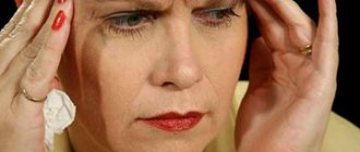
The disease, as a rule, begins with severe pain in the area behind the eyeball, in the temporal, frontal, and superciliary areas. A few days later (usually no later than 14 days, less often simultaneously), double vision, limited mobility of the eyeball and strabismus on the side of pain appear. In approximately 1/4 of patients, when all the nerves that pass through the superior orbital fissure are affected, a complete disturbance of movements of the eyeball in all leads develops.
More common are “incomplete” forms of Tolosa-Hunt syndrome, in which the branches of the oculomotor, trigeminal, and optic nerves in various combinations are involved in the pathological process. Some patients experience exophthalmos and swelling of the conjunctiva of the eyeball. The average duration of pain is about two months. Also, in isolated cases, an increase in body temperature to subfebrile levels and nonspecific changes in a clinical blood test may be observed.
What is Tolosa-Hunt syndrome and how does it manifest?
Tolosa-Hunt syndrome is diagnosed in both men and women equally, on both the left and right sides, but always only on one side, and extremely rarely on both at once. The age of patients ranges from 15 to 85 years. In most cases, it appears after 55 years, when the body is not as strong as in youth, and age-related changes occur in the vessels and joints.
The first signs of Tolosa-Hunt syndrome are in many ways similar in manifestations to classic migraine: a sharp pain appears in one half of the head, a distinct pulsation is felt in it, “shooting” in the eye. It is because of its disguise as other diseases that Tolosa-Hunt syndrome is sometimes called Chameleon.
Subsequently, on the side where acute pain manifests itself, damage occurs to the nerve endings located in the eye area, as well as arteries and veins, which leads to various disorders of the visual organs and changes in the functions of the eyelids.
Another name that is often found in this case is the symptom of the superior orbital fissure. However, it is not entirely true, since it does not fully reflect the essence of the disease.
Diagnostics
What is it necessary to differentiate this disease from? There are a huge number of reasons for the development of similar symptoms: tumors of the middle cranial fossa, pterygoid bone, pituitary gland, parasellar tumors, retrobulbar space-occupying processes, cavernous sinus tumors, cavernous sinus thrombosis, carotid artery aneurysms, periostitis, osteomyelitis, leukemic infiltration in the area of the superior orbital fissure.
Do not forget about diseases such as myasthenia gravis, diabetes mellitus, thyroid pathology, temporal arteritis, meningitis, multiple sclerosis, migraine with aura. All of the above conditions can lead to ophthalmoplegia and dysfunction of the cranial nerves. That is why a patient with syndromes of impaired movement of the eyeball and pain in one half of the face requires close attention. The research must be comprehensive and multifaceted. A consultation with an ophthalmologist is necessary to examine the fields and visual acuity and fundus. The neurologist must carefully collect anamnesis and conduct a full clinical examination.
Additional research methods include neuroimaging methods (CT and MRI of the brain and sella turcica), angiography, and echography of the orbits.
According to modern diagnostic criteria, Tolosa-Hunt syndrome can be diagnosed only in one case: when magnetic resonance imaging (MRI) of the brain reveals granulomatous inflammation of the outer wall of the cavernous sinus. Under ideal conditions, a biopsy should be performed for diagnosis. Recent studies have shown that half of patients with a confirmed diagnosis of Tolosa-Hunt syndrome have changes in the venous bed of the orbital region. If an MRI does not reveal a granuloma, it would be more correct to make a diagnosis of “superior orbital fissure syndrome” and manage the patient under dynamic observation. At the same time, there are clinical diagnostic criteria, which include the following manifestations and conditions:
- “burning” or “tearing” pain of a constant nature behind the orbit or in the orbital-temporo-frontal region;
- impaired mobility of the eyeball that occurred immediately or within 14 days after the onset of pain;
- damage to other nerves that pass through the superior orbital fissure (III, IV, VI cranial nerves, I branch of the trigeminal nerve, autonomic fibers);
- symptoms intensify over several days, even weeks;
- characterized by the presence of spontaneous remissions, often without residual effects;
- the disease may return after several months or years;
- a thorough examination does not find other causes that can cause this condition;
- regression of symptoms within 72 hours after the start of immunosuppressive therapy.
Symptoms
The two main signs of Ramsay-Hunt syndrome are:
Painful red rashes in the form of fluid-filled blisters in and around one ear. Weakness of the facial muscles or facial paralysis on the side of the affected ear. Typically, the rash and facial paralysis develop simultaneously. However, in some cases, the rash may appear before the facial paralysis and, conversely, the paralysis may develop before the rash. In some cases, no rash appears.
Ramsay-Hunt syndrome may be accompanied by the following manifestations:
- Earache
- Hearing loss
- Ringing in the ears (tinnitus)
- Difficulty closing one eye
- A feeling of imaginary rotation or movement of objects around (true dizziness)
- Change in taste perception or loss of taste sensation
Conditions under which you need to see a doctor
If you develop facial paralysis or develop a shingles-like rash on your face, you should consult a doctor. Starting treatment within seven days of the onset of signs and symptoms may prevent the development of long-term complications.
Treatment
The only pathogenetically significant method of treating Tolosa-Hunt syndrome is the use of steroids. The effect of their administration appears in the first three days, which is one of the criteria for the correct diagnosis. However, we should not forget that a good response to the use of steroids can also occur in other conditions that mimic Tolosa-Hunt syndrome, such as pachymeningitis, chordoma, lymphoma, aneurysm, carcinoma and others. That is why difficulties arise with the reliability of the diagnosis, which requires a comprehensive and thorough examination of the patient. The use of other medications is possible for the purpose of symptomatic treatment. Analgesics and anticonvulsants are used to reduce pain, general metabolic agents, and vitamin therapy.
Is it possible to prevent Tolosa-Hunt syndrome?
There is no way to prevent the development of Tolosa-Hunt syndrome as such, since it develops transiently and for an unknown reason. However, if primary symptoms appear, such as double vision, pain in the eye area, forehead, or orbit, it is necessary to consult a doctor for an examination.
When the syndrome is in remission, it is important to carry out secondary prevention: take hormonal medications, vitamins, strengthen the immune system, and avoid stressful situations. In addition, it is important to avoid hypothermia, infectious and inflammatory diseases.
Ganglionitis (ganglioneuritis) of the pterygopalatine ganglion
Ganglionitis (ganglioneuritis) of the pterygopalatine ganglion is one of the neurodental syndromes. This symptom complex is characterized by significant variability in clinical manifestations. The node has three main roots: somatic (sensitive) - from the second branch of the trigeminal nerve, parasympathetic - from the facial nerve and sympathetic - from the plexus of the internal carotid artery; the latter also has connections with the ciliary, auricular, upper cervical sympathetic nodes and cranial nerves, especially with the trigeminal and facial.
Etiology and pathogenesis.
The pterygopalatine node is affected by inflammatory processes in the main and maxillary sinuses, the ethmoidal labyrinth, since the node is closest to the upper or lower jaw. Toxic effects from tonsillitis, complicated caries and local trauma can cause the disease. Provoking factors are overwork, lack of sleep, loud noise, excitement, alcohol consumption, smoking.
Clinical picture
. Neuralgia of the pterygopalatine ganglion (Slader syndrome) is characterized by spontaneous sharp pain in the eye, around the orbit, in the root of the nose, upper jaw, and sometimes in the teeth and gums of the lower jaw. Pain can spread to the temple, auricle, back of the head, neck, shoulder blade, shoulder, forearm and even hand. Painful paroxysms are accompanied by pronounced vegetative symptoms, a kind of “vegetative storm” (redness of half the face, swelling of the facial tissues, lacrimation, copious discharge of secretions from one half of the nose). The attack lasts from several minutes to several hours, and sometimes 1-2 days. and more. Often painful paroxysms develop at night. One of the important diagnostic signs of damage to the pterygopalatine ganglion is the cessation of the attack after lubricating the posterior parts of the nasal cavity with a solution of cocaine and adrenaline.
The illness continues for months and even years. After an attack, a number of vegetative symptoms remain mild. The variety of clinical manifestations of damage to the pterygopalatine ganglion is explained by its individual anatomical features and numerous anastomoses with various nerve formations of the face and other areas.
Treatment.
In the acute period, the nasal cavity posterior to the middle turbinate is lubricated with a 3-5% cocaine solution. Novocaine is also used: cotton swabs moistened with a 2% novocaine solution are inserted into the nasal cavity. For severe pain, ganglion blockers (benzohexonium, pentamine) are prescribed. In severe cases, they resort to blocking the node using anesthetics. Treatment must be comprehensive. If the syndrome develops against the background of inflammatory processes in the paranasal sinuses, face, mouth and jaws, then anti-infective therapy (antibiotics, sulfonamides) is necessary. Treatment should be carried out against the background of desensitizing drugs (diphenhydramine, suprastin, pipolfen). A good therapeutic effect is achieved with hydrocortisone injections into the area of the node projection. Pachycarpine, antispasmodics, and antipsychotics are prescribed in combination with antidepressants. If the clinical picture is dominated by symptoms of irritation of the parasympathetic part, then anticholinergic drugs are used (platiphylline, antispasmodic, belladonna preparations, metacin, aprofen).
Endonasal electrophoresis of a 2% novocaine solution, UHF therapy, and diadynamic currents are used. After the acute phenomena have subsided, mud applications at low temperatures (36-37°C) are used on the affected side or in the form of a collar. Perform a light massage of the neck and face muscles. B vitamins and biogenic stimulants (aloe extract, PHYBS, vitreous) are indicated. Elderly and senile people are prescribed anti-sclerotic drugs, as well as drugs that improve cerebral and coronary circulation. In severe forms, they resort to blockade of the node using a 2% trimecaine solution or alcoholization. Node destruction is rarely used. The treatment provided does not always relieve patients from relapses of the disease, but the severity of clinical manifestations is significantly reduced.