Empty sella syndrome (abbreviated ETS) is a disease that is a weakness of the diaphragm of the brain and, as a result, penetration of the soft membranes of the brain into the space of the sella turcica, compression of the pituitary gland and a change in its height.
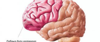
The sella turcica has the appearance of a depression, a “pit,” which determines the name of this syndrome. The sella turcica is the location and seat of the pituitary gland, which regulates most of the major endocrine glands. In the formation of an empty sella turcica, a role is played by prolapse of the soft membrane of the brain into the cavity of the sella turcica and compression of the pituitary gland, which seems to be flattened and pressed against the walls of this bone, which often leads to disruption of its function. Often the diagnosis of sella turcica is made after an MRI.
Medical is a clinic that provides a variety of services, including in the field of endocrinology. The examination and treatment regimen in each specific case is developed based on the characteristics of the disease and the general condition of the patient, and the possible presence of concomitant diseases.
general description
Empty sella syndrome is a syndrome in which the pituitary gland does not completely fill the sella turcica, and a protrusion of the meninges filled with cerebrospinal fluid penetrates into the empty space.
The dimensions of the sella turcica are usually increased. An empty sella turcica is found mainly in women (80%), more often after 40 years of age, and in multiparous women. About 75% of patients are obese. The term empty sella symptom was introduced by V. Bush in 1953 to refer to invagination of the subarachnoid space into the intrasellar region. This anatomical defect is observed in 10% of the population and in 9 cases out of 10 is not accompanied by symptoms of hypothalamic-pituitary dysfunction. In those cases when pathological symptoms appear, they speak of empty sella syndrome.
Causes:
- Primary syndrome: congenital defect of the sella diaphragm and increased intracranial pressure. It is more often detected in women over 40 years of age who are obese, as they have increased cerebrospinal fluid pressure.
- Secondary syndrome: reduction in the size of the pituitary gland after surgical treatment, irradiation, hemorrhage of the pituitary gland.
Symptoms of an empty sella turcica
Empty sella syndrome is manifested by disruptions in the endocrine and nervous systems, and disturbances in the functioning of the visual organs.
During stressful situations, neurological symptoms appear everywhere:
- Increasing headache is the most common symptom of the syndrome. The pain has no specific localization, does not depend on body position, and occurs at different times of the day;
- a jump in blood pressure along with shortness of breath and chills. May be accompanied by pain in the heart, diarrhea, fainting;
- panic fear, acute lack of air, emotional depression or, conversely, anger towards everyone around;
- Abdominal pain and leg cramps may occur;
- sometimes the temperature rises to low-grade levels.
An empty sella turcica of the brain can manifest itself as disturbances in the endocrine system, namely:
- weakened sexual function, enlarged mammary glands in men;
- excess weight - more than 70% with the syndrome suffer from obesity;
- the appearance of diabetes insipidus;
- reduction of the thyroid gland: swelling of the face, drowsiness, constipation, lethargy, swelling of the extremities, dry skin;
- enlarged thyroid gland: sweating, rapid heartbeat, trembling of hands and eyelids, emotional excitability;
- disruptions in the menstrual cycle or even infertility in the fairer sex;
- Itsenko-Cushing syndrome – deterioration of adrenal function. Accompanied by skin pigmentation, mental disorders, and excessive growth of body hair.
The sella turcica is located near the optic nerves. In the presence of the syndrome, they are compressed, thereby disrupting their blood circulation. Therefore, visual symptoms occur in almost absolutely all cases of the disease. Symptoms in this situation:
- deterioration of visual acuity;
- severe tearing;
- splitting of objects;
- the appearance of black dots;
- darkening of the eyes.
In most cases, the above symptoms have many other diseases. The sella turcica can be identified in the brain only after consultation with a specialist and after examination.
Get a free consultation Consultation on the service does not oblige you to anything
Incidence (per 100,000 people)
| Men | Women | |||||||||||||
| Age, years | 0-1 | 1-3 | 3-14 | 14-25 | 25-40 | 40-60 | 60 + | 0-1 | 1-3 | 3-14 | 14-25 | 25-40 | 40-60 | 60 + |
| Number of sick people | 3 | 5 | 7 | 13 | 15 | 18 | 21 | 6 | 10 | 14 | 16 | 26 | 36 | 42 |
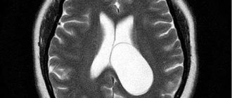
MRI and other diagnostic methods
Suspicion of the possible presence of empty sella syndrome arises in patients who have undergone repeated trauma to the skull, surgery or irradiation of the pituitary gland, and for women, aggravating factors are multiple pregnancies and long-term contraceptive therapy.
Most often, at the first stage, patients are referred for laboratory diagnostics of pituitary hormones (growth, thyroid-stimulating, adrenocorticotropic, prolactin, vasopressin), as well as target organs - adrenal cortisol, thyroid thyroxine, testosterone and estrogen. At this stage, it is not always possible to confirm the pathology, since many patients have normal hormonal levels.
The MRI method is recognized as the most informative. With its help you can detect:
- liquor fluid in the sella turcica;
- a reduced and flattened pituitary gland, similar to a crescent;
- displacement of the pituitary tissue to the bottom, posterior wall;
- signs of intracranial hypertension - dilated sinuses, ventricles of the brain;
- asymmetrical sagging of the tank above the saddle;
- displacement of the pituitary funnel to the side, forward or backward, its thinning and lengthening.
MRI of the pituitary gland
Treatment
Clinical recommendations are primarily aimed at monitoring the patient's condition. If there are no manifestations of the syndrome, there is no need for treatment. If a patient has complaints, it is recommended to determine the secretory function of the pituitary gland and hormonal correction if there is insufficiency.
Hyperprolactinemia in the primary form of the syndrome requires correction with bromocriptine.
Surgical treatment is considered only in one case - with cerebrospinal rhinorrhea.
Is it dangerous?
A change in the location of the optic nerves, which can lead to visual impairment the sella turcica .
Congenital anomalies of the diaphragm are quite common . But most people are not worried about anything; the endocrine gland performs its functions properly. Danger occurs only when pituitary dysfunction occurs.
If the syndrome is accidentally discovered and there are no symptoms, no treatment should be prescribed. A simple observation from a specialist is quite enough.
What diseases can it be associated with?
- Acromegaly is a neuroendocrine disease that develops with overproduction of growth hormone (somatotropin, growth hormone); The reason for this is usually pituitary adenoma (somatotropinoma), which is a benign tumor; simultaneous hyperproduction of somatotropin and prolactin by the tumor is common.
- Itsenko-Cushing's disease is a hypersecretion of adrenocorticotropic hormone, which develops mainly as a result of head contusions, concussions, traumatic brain injuries, encephalitis and other lesions of the central nervous system.
- Hyperprolactinemia - can develop as an independent or concomitant disease (against the background of hypothyroidism or adrenal insufficiency), is treated with hormone replacement therapy; may develop while taking certain medications, the abolition of which allows the condition to normalize.
- Hypopituitarism (Sheehan syndrome) - develops as a result of destruction of the adenohypophysis with a subsequent persistent decrease in the production of tropic hormones and disruption of the activity of peripheral endocrine glands; The main cause of hypopituitarism is circulatory disorders in the hypothalamic-pituitary region that develop after childbirth, complicated by massive blood loss, thromboembolism, and sepsis.
- Primary hypothyroidism - replacement therapy (mostly carried out for life) of hypothyroidism can provoke a secondary pituitary adenoma and, as a result, an empty sella turcica.
- Pituitary microadenomas (prolactinomas and somatotopinomas) - treatment of such with dopaminomimetics and somatostatin analogues, respectively, provoke changes in the location of the pituitary gland; the clinical picture corresponds to pituitary disorders associated with the hormonal activity of the pituitary tumor.
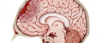
Anatomy and structure
The sella turcica (otherwise known as the pituitary fossa) is a rounded, hollow formation located inside the skull, next to the sphenoid bone. Normally, they are separated from the subarachnoid part by a hard plate - the diaphragm.
The optic nerve fibers and tracts cross above the sella, forming the chiasm (values in mm):
- length – 4-10;
- width – 3-11;
- thick – 5.
Sella turcica in the head, structure and location.
It is protected by the soft membrane of the brain, located on the border with the diaphragm, on top - with the base of the third cerebral ventricle, behind - with the pituitary gland, and on the sides it is surrounded by the carotid arteries.
In women, the depth of the pituitary fossa is about 9-12 mm, in men – 12-15 mm. Inside it is the pituitary gland (gland). It is connected to the hypothalamus by a stalk descending into the sella. Its diaphragm prevents the liquor fluid that surrounds the brain from leaking into the cavity.
The saddle also protects the pituitary gland from any mechanical damage.
Normally, the gland is of regular shape and fills the entire cavity. During pathological processes, the membrane of the brain descends and begins to put pressure on the pituitary gland, especially if the diaphragm is depleted or underdeveloped, cerebrospinal fluid gets inside. Then serious deviations arise in the functioning of the hypothalamus and endocrine system.
A healthy newborn has a cup-shaped pituitary fossa, cartilaginous structure, with a wide entrance. During the first three years, the saddle gradually increases in size. It reaches a size of 8-10 mm by five years. During adolescence, the pituitary fossa takes the shape of a saddle.
By the age of 13-15 its dimensions reach 9-12 mm. The sella turcica is fully formed by the age of 19. It can increase slightly more than the minimum size due to pituitary microadenoma or adenohypophysis hyperplasia.
If premature ossification of the sphenoid bone occurs, the volume of the pituitary fossa decreases. This is often preceded by rapid puberty.
Diagnostics
Neurologists and neurosurgeons deal with diseases of the brain structures. In this case, the pathology of the sella turcica can be an accidental finding during the search for other disorders of the central nervous system. If a patient consults a doctor with common complaints associated with the disease, a full diagnosis is carried out, including a survey, physical examination, instrumental and laboratory testing.
Before prescribing diagnostic procedures, the doctor carefully examines the medical history. Recent birth, congenital diseases, infections and other factors help make a preliminary diagnosis. During a physical examination, autonomic and ophthalmological symptoms of the disease can be detected. For an accurate diagnosis, hormone tests and instrumental studies are prescribed.
MRI
Magnetic resonance imaging allows doctors to obtain images of the smallest structures of the brain. In the picture you can see typical signs of altered anatomy of the sella turcica. Precise imaging capabilities also make it possible to assess the extent of damage to neurological structures and identify possible causes of the syndrome.
MRI of the sella region is a fast and safe procedure. This test is available in major hospitals and medical centers.
X-ray
X-ray of the sella turcica is a less accurate diagnostic method. Based on the results of such a study, doctors are not always able to determine the cause of the patient’s symptoms. In some cases, radiography may be recommended for preliminary diagnosis.