What is MRI of the pituitary gland?
Magnetic resonance imaging allows you to obtain a three-dimensional image of the gland and detect changes in it that are not detected using other research methods. Scanning based on magnetic resonance technology is safe, since the patient’s body is not exposed to x-ray radiation, is painless and informative, especially when diagnosing diseases of the central nervous system, when examination of small brain structures is required.
MRI of the pituitary gland shows changes in the tissue of the gland, even if the affected areas are very small or their visualization is difficult. The pituitary gland is clearly visible on MRI of the brain
, however, if necessary, a specialist may prescribe a more detailed study.
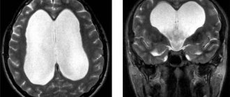
Empty sella syndrome on MRI
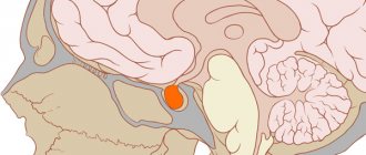
The abnormality, which doctors call sella turcica syndrome on MRI, is clearly shown on the anteroposterior image. The shape of the organ is more similar to a sickle, its thickness is no more than 3 mm. The image also clearly shows that at the bottom of the sella turcica there is a flatly trampled organ. In this case, the visual chiasm prolapses into the cavity of the bone formation.
Tumors of the area of the sella turcica and optic chiasm on images
The main types of tumors that are found in this area are microadenoma, macroadenoma, mesoadenoma, giant adenoma.
Microadenoma has the smallest dimensions, compared to others - a diameter of 10 mm.
Macroadenoma reaches 30 mm; there have been cases when it reached a size of 10 cm, but only if it extended beyond the sella turcica.
Mesoadenoma is a type of macroadenoma, reaches a diameter of 22 mm, but never extends beyond the sella.
With a diameter of more than 30 mm, the neoplasm is called a giant adenoma .
In addition to these tumors, there are also meningiomas, germinomas and others. All of them can be located not only in the sella turcica, but go beyond it and grow into all parts of the brain. Of course, any of them can be seen on MRI images.
Experts often notice the following deviations in images:
- changing the shape of the Turkish bottom;
- organ asymmetry;
- heterogeneous structure;
- displacement of the funnel from the midline.
It doesn't necessarily have to be a tumor. Along with this, it is worth conducting a number of studies and analyzes to get a complete picture of the examination.
Pituitary gland: what it is, description and functions
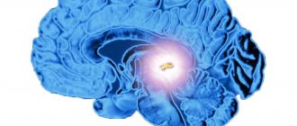
The pituitary gland is a small (up to 1 gram) gland located at the base of the brain. The hormones secreted by the pituitary gland are involved in regulating the functioning of the entire human endocrine system. Disturbances in the functioning of the pituitary gland affect the functioning of the entire body, cause changes in development, and can cause obesity, infertility, and other pathological conditions.
Pituitary tumors, among which adenoma is the most common, are well identified using MRI, which allows timely treatment to begin.
What to choose - brain tomography or pituitary MRI?
Brain tomography shows abnormalities in the organ in question, but with a small degree of probability. A targeted study allows you to obtain much more information to make the correct diagnosis.
The procedure is prescribed for suspected congenital abnormalities, cysts, inflammation, tumors, or disorders of the hypothalamic-pituitary system. Studies in this area show pathological lesions less than 1 cm and their structure. To increase the information content, an MRI with contrast is performed.
Hormones secreted by the pituitary gland
Pituitary hormones regulate the functioning of the thyroid gland and adrenal cortex. They are responsible for the growth of the body and reproductive function. Excessive production of somatotropin by the pituitary gland can cause gigantism and acromegaly (excessive growth of limbs, facial changes). The pituitary gland is responsible for the production of prolactin, which regulates lactation in women, and oxytocin, which is responsible for labor.
The normal functioning of the pituitary gland determines the timeliness of the onset of puberty in adolescents.
A few words about the pituitary gland
The pituitary gland is a small gland that controls the functioning of the endocrine organs. She is responsible for homeostasis. It is divided into two lobes - anterior and posterior; specialists diagnose both of them.
The neurohypophysis or posterior lobe is responsible for the accumulation of vasopressin (it, for example, is necessary for the proper functioning of blood vessels), the accumulation of oxytocin (contracts the muscles of the uterus). The anterior lobe produces TSH, which is responsible for the functioning of the thyroid gland, ACTH, which stimulates the adrenal glands, STH, which is responsible for growth, etc.
When is it necessary to do an MRI of the pituitary gland?
MRI of the pituitary gland is prescribed for frequent headaches and progressive disorders in the functioning of the brain, for visual impairment of unknown cause, weight changes (for example, for obesity not caused by excessive food consumption and a sedentary lifestyle) and other painful conditions.
Indications
- Traumatic brain injuries.
- Erectile dysfunction in men.
- Menstrual cycle disorders in women.
- Increased activity of certain groups of hormones, including high levels of prolactin.
- Suspicions of hormone-dependent diseases (acromegaly, Itsenko-Cushing syndrome).
- Infertility.
Contraindications
There are few contraindications to the study. Absolute contraindications:
- The presence of implants or electronic devices in the patient’s body, including a pacemaker, insulin pump, joint endoprosthesis, metal clips on blood vessels.
- Pregnancy (all trimesters – for MRI with contrast, 1st trimester – for MRI without contrast).
- Allergy to contrast agent.
The procedure is not performed if the patient weighs more than 120 kg, which is dictated by the technical requirements of the device.
Patients with heart, kidney, or liver failure should discuss with their doctor the feasibility of the study and possible risks if a contrast agent is to be used.
conclusions
MRI of the pituitary gland today provides the most complete picture of the condition of the pituitary gland and brain. Diagnostics helps to detect abnormalities in the early stages of development. Further consequences, and even the patient’s life, depend on this. MRI makes it possible to detect tumors as small as 4 mm, their exact location, type and effect on neighboring tissues.
Tomography has virtually no contraindications for use, but requires more careful preparation of the patient. Compliance with all recommendations predicts future diagnostic success.
Advantages of the method
The main advantage of MRI is its high information content, the ability to study tiny structures that are poorly distinguishable with other neuroimaging methods. The small number of contraindications, painlessness and non-invasiveness of the procedure also speak in its favor.
Magnetic resonance imaging can detect pathological changes in the gland at their very beginning, including inflammatory processes, benign and malignant neoplasms, and changes in the vascular bed. High image quality makes it possible to identify microscopic areas of lesions.
Contraindications
MRI is a safe procedure for the patient, even when contrast is used. However, unpleasant side effects can develop if direct contraindications are not followed:
- the presence of metal objects in the body - prostheses and implants. This category also includes ferrimagnetic fragments and Elizarov’s apparatus;
- presence of insulin pumps and pacemakers;
- severe condition caused by diseases of the pituitary gland and other pathologies or infections;
- excess weight - this is possible only if you weigh up to 120 kg (this category includes people with muscle mass, and not just fat deposits).
When using a contrast agent, contraindications such as heart failure, serious liver and kidney diseases are added. There are also relative contraindications for which MRI of the pituitary gland with contrast can be performed if the person’s condition is life-threatening:
- claustrophobia - the patient is afraid of closed spaces - the problem can be solved by using an open type apparatus or sedatives;
- first trimester of pregnancy.
The doctor should assess the risks and relevance of conducting a pituitary gland study.
How is MRI of the pituitary gland performed, how long does it take?
Special preparation is required only for MRI of the pituitary gland with contrast.
Before a routine examination, you will need to remove metal jewelry, hairpins, and watches. Wallets with credit cards, pens and glasses will also have to be left outside the room where the device is installed.
The patient is positioned on a movable table, which moves inside the tomograph for the examination. The entire study takes from 30 to 60 minutes, during which time you must lie still so that the results are not distorted (fixing belts may be used).
To make MRI of the pituitary gland more comfortable, it is recommended to use earplugs or special headphones, since the noise of the machine can cause discomfort.
How long does an MRI take?
The time for MRI is variable, depending on the area of interest and clinical tasks.
The magnetic field affects the body intermittently, according to the scanning protocol. It is difficult to say in advance how long an MRI will take. The research process includes:
- Preparation. Before MRI, a conversation is held with the patient to identify contraindications (early pregnancy, allergies to contrast, chronic renal failure for enhanced scanning). It is mandatory to complete medical documentation - filling out a questionnaire, signing a voluntary consent to a diagnostic procedure. Items containing metal (cell phone, keys, plastic cards, coins, jewelry, watches) should be deposited. The second stage of preparation is positioning the patient. The x-ray technician places the patient on the table, places the intensifying coils over the area of interest, and secures the limbs with straps to ensure immobility.
How long does an MRI take? Always different, scanning one area of the body lasts less than a volumetric examination, including assessment of several organs and systems
- Diagnostic procedure. The device takes a series of images, based on which the doctor evaluates the area of interest. During diagnostics in phases (arterial, venous, delayed), a contrast agent is introduced into the blood using an automatic injector, which makes it possible to identify hidden pathological processes, “highlight” the boundaries of the tumor, and the relationship with surrounding structures. The enhancer can be administered manually as a one-time injection.
- Description and display of results. Upon completion of the diagnostic procedure, the doctor who conducted the study fills out a standard form, indicating the characteristics of the organs and systems examined and the changes identified. If results are equivocal, peer review of MR images may be required, which will increase time. The report is collected within an hour after the scan.
MRI of the pituitary gland: what does it show with increased prolactin?
Magnetic resonance imaging of the pituitary gland is often prescribed for elevated levels of prolactin in the blood. The fact is that high prolactin can serve as a sign of the development of a pituitary adenoma, a benign tumor that can cause disturbances in the functioning of the entire body, including endocrine diseases.
MRI with elevated prolactin shows the presence or absence of a pituitary adenoma, other pathological changes in the tissues of the gland, and allows us to identify the reasons for the increase in hormone levels. Timely diagnosis of adenoma allows you to avoid such serious consequences as infertility and loss of vision.
Preparing for MRI of the pituitary gland with contrast
Magnetic resonance imaging of the pituitary gland with contrast agents is performed without any preliminary preparation. The study can be carried out at any time convenient for the patient. The main condition for safe MRI is the absence of foreign metal objects on the patient’s body. Magnetic fields generated during the examination can lead to heating of jewelry, piercings, and metal clothing fittings, with the risk of burns. It is strictly prohibited to carry mobile phones, electronic gadgets, credit and other magnetic cards, storage media, external batteries, etc. Otherwise, injury or burns may occur. The patient can leave anything unnecessary in the storage room.
Contrast drugs very rarely cause side effects - the most common of them is a mild allergy in the form of a small rash and itching on the skin. Severe allergic reactions to contrast are rare and also serve as a contraindication for its administration. In such cases, the study is performed without contrast.
What happens after the examination?
On a computer, a specialist uses a medical program to process the signals and create an image. Then the received data is evaluated and the pictures are printed. The results are given to the patient on disk and in printed form, or sent by e-mail.
In the meantime, a person can get dressed and pick up personal belongings. No rest is necessary after an MRI of the pituitary gland with contrast. Any food is allowed in food. If nausea or other reactions to the substance used occur, you should relax and not do work.
If the procedure was performed by a breastfeeding woman, the baby should not be given milk for 48 hours.
There are no other restrictions. If you experience severe discomfort, you should consult a doctor. However, in the absence of absolute contraindications, side effects are minimized.
During the contrast examination itself, some patients notice a metallic taste in the mouth almost immediately after the injection. In rare cases, a feeling of discomfort in the body occurs. Sometimes a skin reaction develops in the form of itching, redness and hives.
MRI of the pituitary gland using a contrast agent gives the most accurate results, but is not cheap. In most cases, the high price is justified by the effectiveness of the examination and the ability to quickly make a diagnosis after receiving the information.
Contrasting Features
The contrast method improves the clarity of the images used. MRI of the pituitary gland with contrast most often uses substances with gadolinium, which is part of the group of lanthanides - rare earth elements. The drug is administered in 2 ways:
- A single injection into a vein before the study, take 0.2 mg of the substance per 1 kg of weight.
- Drip injection using a dispenser - synchronization occurs during the scanning process. A similar injection method is used in dynamic MRI to improve diagnostic efficiency.
As soon as the substance enters through the veins, it begins to accumulate in areas of increased blood supply. This description includes tissues altered by a tumor or other pathological processes. During an MRI, the areas produce stronger magnetic waves.