Studying the functionality of individual brain structures allows us to identify existing abnormalities and prevent irreversible changes that can cause disability and, in some cases, death. The need for diagnostic techniques that would make it possible to identify certain pathologies has led to the emergence of several different procedures during which the condition of the main human organ is assessed.
Ultrasound is the most common and accessible method to assess the condition of the brain. This study is quite informative, non-invasive, and does not harm the patient’s health.
Features of diagnostics
Ultrasound of the vessels of the brain and cervical spine is a technique based on the influence of high-frequency sound. The study is safe.
The waves pass through the tissue structures of organs and are reflected from them. After which they are captured by a sensor, processed by specialized equipment and displayed on a computer monitor.
Positive features are:
- safety;
- the ability to undergo diagnostics an unlimited number of times if necessary;
- painlessness;
- minimal risk of introducing infectious agents;
- the ability to view and evaluate soft tissue;
- viewing organs in several projections;
- acceptable price.
The following methods are used for examination: echoscopy (ECHO), neurosonography, Dopplerography, duplex and triplex scanning.
to contents ^
When is it appointed?
Ultrasound examination of the neck and brain is prescribed for the following pathological conditions in adult patients:
- osteochondrosis of the upper body;
- disturbances in heart contractions;
- vegetative-vascular dystonia;
- symptoms of nervous disorders: fainting, ringing in the ears, problems with coordination, memory impairment, loss of sleep, dizziness, etc.;
- thrombosis;
- suffered strokes, heart attacks;
- increased intravenous pressure of chronic form;
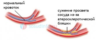
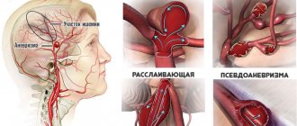
- aneurysm, stenosis, atherosclerosis;
- ischemia;
- constant weakness, excessive fatigue, glare in the eyes;
- high con cholesterol;
- traumatic brain injuries (including concussion);
- diabetes mellitus;
- a sharp deterioration in vision or its complete loss;
- poor sensitivity in the facial area;
- obesity;
- nicotine addiction;
- before planned heart surgery.
Ultrasound for children has the following indications: premature and difficult birth, hypoxia at birth and during intrauterine development. Poor weight gain by a child, neurological pathologies, diseases of internal organs, if the shape of the head and face is deviated from the norm.
Useful to know: MRI of the brain with contrast, how the procedure works and what you need to know about it
to contents ^
Indications for examination
Ultrasound of the brain is prescribed for adults and children in the presence of such alarming symptoms as:
- Constant headaches;
- Feeling of heaviness in parts of the head;
- Deterioration of visual and auditory functions;
- Memory impairment;
- Frequent fainting;
- Low blood pressure;
Elena Razumovna Lebedeva, a neurologist of the highest category, Doctor of Medical Sciences, Russian representative on the Council of Experts on Headaches at the European Academy of Neurology, tells how to determine why a headache hurts:
- Loss of balance that occurs suddenly and unexpectedly;
- Lacrimation, numbness of the upper extremities, spasms of the facial muscles that occur suddenly and go away on their own;
- Insomnia;
- Sensation of pulsation in the temples;
- Disorientation.
In addition, indications for ultrasound to assess the condition of the arteries of the brain and neck are:
- An upcoming operation involving intervention in the area of the heart muscle;
- A history of stroke or heart attack;
- Increased cholesterol levels;
- Long-term smoking;
- Diseases of the cervical spine;
- Previous injuries to the skull or neck area;
- Presence of diabetes mellitus;
- Persistent arrhythmia;
- Osteochondrosis;
- Suspicion of a tumor of the cerebral hemispheres;
- Vegetovascular dystonia.
What does the examination reveal?
Ultrasound helps:
- assess the carotid, vertebral, basilar arteries;
- detect inflammatory processes, foci of infection, internal hemorrhages;
- check the likelihood of vascular pathologies;
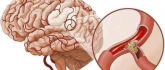
- find blood clots and the consequences of their occurrence;
- confirm or refute diseases that cause fainting, migraines, etc.;
- check the condition of the cervical arteries and veins;
- identify hydrocephalus;
- determine the causes of problems with blood circulation.
In addition, the diagnostic measure shows the condition of the brain vessels and the structures of their walls, and the patency of the arteries. Provides information about the speed of blood fluid in certain areas.
to contents ^
How is the examination carried out?
To perform an ultrasound of the head and neck, the patient is placed on a couch on his back. The neck area is freed from clothing and jewelry. To conduct the examination, the head is tilted back and turned away from the sensor. A gel is applied to the skin, this is necessary to remove air from under the sensor. The doctor moves the sensor over the area under study, stopping at the places where the vessels and arteries are projected. To increase the information content, functional tests may be required: the doctor can press the vessels with fingers or a sensor, ask the patient to breathe deeply. In some cases, vascular medications may also be required. The examination is painless and usually does not take more than 30 minutes. The big advantage of ultrasound is that it is harmless and has no contraindications. The examination is often prescribed even for newborn children and can be carried out as often as necessary.
Ultrasound involves conducting an examination in parallel with decoding. Such indicators as the maximum and minimum speeds of blood flow in the vessels, the ratio between the maximum and minimum speeds, and the nature of the blood flow should be recorded. The resistance index is also calculated for vessels. The obtained data are compared with the norm, which allows us to draw a conclusion about the patency of the vessels and the state of their lumen.
The diagnostic method is used not only to make a diagnosis, but also to monitor the dynamics of the disease and evaluate the effectiveness of the therapy.
You can undergo an ultrasound of the neck and head in Volgograd, Volzhsky and Mikhailovka at Dialine clinics. We use the most modern ultrasound machines, which, combined with the extensive experience and professionalism of our employees, allows us to carry out highly accurate diagnostics. You can sign up for an examination by phone or through the website. For maximum convenience of receiving medical services in our clinics, register in your personal account.
How to prepare for a diagnostic event
Before undergoing the examination, you need to prepare for it. During the day, you need to give up or reduce the consumption of alcohol-containing products, strongly brewed tea, coffee, and energy drinks. Drinks provoke hypertonicity of cerebral vessels, the results of the study will be inaccurate. Preparation also includes eliminating spicy and salty foods from the diet.
Before diagnosis, you need to avoid excessive physical and emotional stress. Protect yourself from stressful situations. It is also necessary to stop using painkillers and medications that relieve spasms. Five hours before the ultrasound, do not smoke: nicotine increases intravenous pressure, the examination results will be erroneous.
to contents ^
How is the diagnosis carried out?
You need to remove clothes above the waist, jewelry from the ears, neck, and piercings from the face. When conducting diagnostics, the patient should be in a calm state and feel comfortable.
The examination takes place in a supine or sitting position. The specialist palpates the head to identify bruises, injuries, and deformities. After this, the temples are lubricated with a special gel to improve signal conductivity, and ultrasonic sensors are installed. The data is displayed on a computer screen and analyzed.
Noise may occur during the examination. They do not indicate the presence of pathological conditions, but are only the sounds of blood fluid circulation in the vessels. After diagnosis, the specialist compares the received data with the variant norms, writes a conclusion, issues a protocol, and attaches photographs.
Useful to know: How long does the MRI procedure of the brain take?
An ultrasound examination usually does not last more than an hour. The procedure does not have any effect on the person’s further condition. Immediately after diagnosis, the patient can begin his normal activities.
to contents ^
How to do an ultrasound of the brain
Ultrasound examination of the head and neck vessels does not cause discomfort to a person and is not accompanied by painful sensations.
The event takes no more than an hour, after which the visitor to the diagnostic room can immediately return to their normal lifestyle. If any alarming symptoms appear during the session, you should report them to your healthcare provider.
An ultrasound scan of the brain involves the following nuances:
- the patient lies down on the couch or sits in a chair, takes a comfortable position;
- a diagnostic specialist examines and palpates the visitor’s skull for deformations, asymmetry, and hematomas;
- A special gel is applied to strategically important areas to increase conductivity, and an ultrasound sensor is installed on top of it. Depending on the purpose of the study, readings are taken from the temples, the back of the head, areas above the eye sockets, and areas of large vessels in the neck;
- while obtaining data, you must strictly follow the doctor’s instructions - he may ask you to breathe deeper or more often, hold your breath, pretend to cough, change your body position.
Having received the necessary information, the diagnostic room employee draws up a conclusion and attaches a transcript of the results to it.
Data collection may be accompanied by unusual sounds from the device, but these are not indicators of problems. There is no need to pay attention to them, it is better to try to relax.
Decoding the results
Normal indicators (interpretation of results):
- symmetrical distribution of gray matter;
- in the veins up to the sixth vertebra, the blood circulation speed is less than 0.3 m/s;
- clarity of the outlines of the ventricles, the average ability of the subcortical nuclei to reflect sound waves;
- absence of inclusions and foreign fluid between the hemispheres;
- aneurysms, blood clots, and neoplasms are not observed;
- the circumference of the vertebral arteries is at least two millimeters, the diameter corresponds to age;
- the vascular walls are homogeneous, without damage, clear, no more than 1.1 mm thick;
- there is no turbulent blood flow in places with no vein branching;
- malformation and compression are deviations from the norm.
An ultrasound scan of the brain provides not only a set of data, but also a preliminary diagnosis: meningitis, internal bleeding, cancer, etc. Ultrasound also confirms the death of the central nervous system organ.
Only qualified specialists can accurately decipher ultrasound data. You can't try to do this on your own. You can make a false diagnosis and start the wrong treatment. This will lead to adverse consequences.
to contents ^
By what principle is the received data decrypted?
Each vessel has its own parameters, on the basis of which calculations are made and the degree of their discrepancy with the norm is assessed.
Transcranial and extracranial Dopplerography of arteries compares digital data in each vessel segment:
- Artery wall thickness.
- Vessel diameter.
- Phases of blood flow, its features.
- The degree of symmetry of blood flow through identical arteries located on different sides.
- Diastolic blood velocity.
- Maximum systolic blood flow velocity.
- Pulsation and resistive indices, systole and diastole ratio.
- The degree of pathological narrowing of blood vessels (stenosis), the condition of the artery beyond its limits.
Dopplerography of veins evaluates the following parameters:
- Condition of the venous wall of the vessel.
- The nature of blood movement through the studied vessels.
- Vein diameter
Data obtained at rest and after functional tests are compared with normal values. Any deviations are marked with a special abbreviation of letters and numbers. Based on them, the neurologist prescribes further studies or forms a course of treatment.
Contraindications to manipulation
Ultrasound is used on any patient. There are no contraindications as such. However, there are limitations associated with the technical nuances of the diagnostic procedure. For example, ultrasound cannot replace angiography (a contrast method for studying blood vessels).
Ultrasound examination is difficult to study small capillaries. Some are not visible at all due to the thick bones of the skull. With atherosclerosis, the condition of the blood vessels often interferes with the passage of high-frequency waves.
Despite all the listed nuances, a highly qualified specialist is able to see everything that is necessary using ultrasound. Even on not the most modern and best equipment. Therefore, choosing a doctor when undergoing diagnostics must be approached seriously and responsibly.
Useful to know: What are diffuse changes in the bioelectrical activity of the brain: causes and treatment methods
to contents ^
Where to do
To undergo an ultrasound examination of the brain, you must go to the clinic at your place of residence. Experts will tell you which institution you can get the service for free. However, in this case you often have to wait several weeks or even months. If time is pressing, it is better to find a commercial clinic that has the appropriate equipment. Depending on the region, this procedure will cost from 800 to 2500 rubles.
Long-term studies have confirmed the safety and informativeness of brain ultrasound. Sessions are available even for newborn children, pregnant women and the elderly. Neurologists recommend, if possible, undergoing the procedure regularly for preventive purposes. This is especially true for people with high blood cholesterol, obesity, chronic headaches, hypertension, and decreased physical activity.


