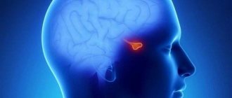
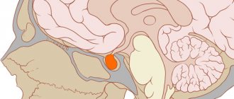
How does the body's hormonal system work and how is it controlled? The brain has a 3 appendage system known as the adenohypophyseal system. It includes 3 structures - the pituitary gland, the pineal gland, and the hypothalamus. We will explain and describe the location of the epiphysis appendage. This is a separate brain structure that was previously considered unnecessary and useless. But now we know that the pineal gland, or pineal gland, is needed to regulate biorhythms.
Structure
The pituitary gland consists of two large lobes of different origin and structure: the anterior one - the adenohypophysis (accounts for 70-80% of the mass of the organ) and the posterior one - the neurohypophysis. Together with the neurosecretory nuclei of the hypothalamus, the pituitary gland forms the hypothalamic-pituitary system, which controls the activity of the peripheral endocrine glands.
Anterior lobe (adenohypophysis)
Anterior pituitary gland
(lat. pars anterior), or
adenohypophysis
(lat. adenohypophysis), consists of glandular endocrine cells of various types, each of which, as a rule, secretes one of the hormones. Anatomically, the following parts are distinguished:
- pars distalis (most of the adenohypophysis)
- pars tuberalis (leaf-shaped outgrowth surrounding the pituitary stalk, the functions of which are unclear)
- pars intermedia, which is more correctly designated as the intermediate lobe of the pituitary gland.
Hormones of the anterior pituitary gland
:
- Tropic, since their target organs are endocrine glands. Pituitary hormones stimulate a specific gland, and an increase in the level of hormones secreted by it in the blood suppresses the secretion of the pituitary hormone according to the feedback principle. Thyroid-stimulating hormone (TSH) is the main regulator of the biosynthesis and secretion of thyroid hormones.
- Adrenocorticotropic hormone (ACTH) - stimulates the adrenal cortex.
- Gonadotropic hormones: follicle-stimulating hormone (FSH) - promotes the maturation of follicles in the ovaries, stimulation of endometrial proliferation, regulation of steroidogenesis..
- luteinizing hormone (LH) - causes ovulation and formation of the corpus luteum, regulation of steroidogenesis..
Pituitary adenomas develop from the adenohypophysis[2].
Posterior lobe (neurohypophysis)
Posterior pituitary gland
(lat. pars posterior), or
neurohypophysis
(lat. neurohypophysis), consists of:
- nerve lobe
.
It is formed by ependymal cells (pituicytes) and the axon endings of neurosecretory cells of the paraventricular
and
supraoptic
nuclei of the hypothalamus of the diencephalon, in which vasopressin (antidiuretic hormone) and oxytocin are synthesized, transported along the nerve fibers that make up the hypothalamic-pituitary tract to the neurohypophysis. In the posterior lobe of the pituitary gland, these hormones are deposited and from there enter the blood. - funnel
,
infundibulum
.
Connects the neural lobe with the median eminence. The pituitary infundibulum, connecting with the hypothalamic infundibulum, forms the pituitary stalk
.
The functioning of all parts of the pituitary gland is closely related to the hypothalamus. This position applies not only to the posterior lobe - the “receiver” and depot of hypothalamic hormones, but also to the anterior and middle sections of the pituitary gland, the work of which is controlled by hypothalamic hypophysiotropic hormones - releasing hormones [3].
Posterior pituitary hormones
:
- asparotocin
- vasopressin (antidiuretic hormone, ADH) (stored and secreted)
- vasotocin
- valitocin
- Glumitocin
- isotocin
- mesotocin
- oxytocin (stored and secreted)
Vasopressin performs two functions in the body:
- increased reabsorption of water in the collecting ducts of the kidneys (this is the antidiuretic function of vasopressin);
- influence on the smooth muscle of arterioles.
However, the name “vasopressin” does not quite correspond to the property of this hormone to constrict blood vessels. The fact is that in normal physiological concentrations it does not have a vasoconstrictor effect. Vasoconstriction can occur with the exogenous introduction of the hormone in large quantities or with blood loss, when the pituitary gland intensively secretes this hormone. With insufficiency of the neurohypophysis, diabetes insipidus syndrome develops, in which a significant amount of water (15 l/day) can be lost in the urine per day, as its reabsorption in the collecting ducts decreases.
During pregnancy, oxytocin does not affect the uterus, since under the influence of progesterone secreted by the corpus luteum, it becomes insensitive to this hormone. Oxytocin promotes the contraction of myoepithelial cells, which promote the secretion of milk from the mammary glands.
Intermediate (average) share
In many animals, the intermediate lobe of the pituitary gland is well developed, located between the anterior and posterior lobes. By origin it belongs to the adenohypophysis. In humans, it represents a thin layer of cells between the anterior and posterior lobes, extending quite deeply into the pituitary stalk. These cells synthesize their specific hormones - melanocyte-stimulating and a number of others.
STRETCH STRETCHES AS AN EXCHANGE PROBLEM
One of the most common diseases is the so-called endogenous hypercortisolism, or Itsenko-Cushing syndrome. This is a rare, multi-symptomatic endocrine disease of the hypothalamic-pituitary-adrenal system, manifested by excessive production of the hormone cortisol, which is produced by the adrenal cortex.
Often, patients with uncontrolled diabetes mellitus, arterial hypertension, obesity and other problems turn to cardiologists, therapists and endocrinologists; medications are selected and changed for them, but the therapy turns out to be ineffective. The patients' condition is deteriorating.
This should alert the doctor and raise the question of why the treatment is not helping. Is it due to some other pathological process?
There are other, more specific manifestations of neuroendocrine processes in such patients: fragility and fragility of bones (osteoporosis), short stature, sudden weight gain, skin changes and the appearance of stretch marks from pink to purple-bluish in color. Most often, such stretch marks (striae) appear on the thighs, abdomen, armpits, and in the area of the mammary glands.
A classic example of the formation of such stretch marks is pregnancy. However, in this case, similar processes occur outside of pregnancy due to the potentiation of the catabolic effect of cortisol on protein metabolism, disruption of the structure of the dermis, disorganization and reduction in the number of elastin fibers, and impaired collagenization, resulting in thinning of the skin. The presence of hematomas and discoloration of stretch marks is a consequence of increased capillary fragility and increased vascular permeability.
When examining such a “suspicious” patient, you may also see electrolyte changes in the blood, such as hypokalemia - low levels of potassium in the blood, which can contribute to the development of muscle weakness.
Vessels and nerves
The pituitary gland is supplied with blood from the superior and inferior pituitary arteries, which are branches of the internal carotid artery. The superior pituitary arteries enter the hypothalamic infundibulum and, penetrating the brain, branch into the primary hemocapillary network
;
these capillaries collect into portal veins, which are sent along the stalk to the anterior lobe of the pituitary gland, where they branch again into capillaries, forming the secondary capillary network
. The inferior pituitary arteries supply blood primarily to the posterior lobe. The superior and inferior pituitary arteries anastomose with each other. Venous outflow occurs into the cavernous and intercavernous sinuses of the dura mater.
The pituitary gland receives sympathetic innervation from the plexus of the internal carotid artery. In addition, many processes of neurosecretory cells of the hypothalamus penetrate into the posterior lobe.
Functions
In the anterior lobe of the pituitary gland, somatotropocytes produce somatotropin, which activates the mitotic activity of somatic cells and protein biosynthesis; lactotropocytes produce prolactin, which stimulates the development and function of the mammary glands and corpus luteum; gonadotropocytes - follicle-stimulating hormone (stimulation of ovarian follicle growth, regulation of steroidogenesis) and luteinizing hormone (stimulation of ovulation, formation of the corpus luteum, regulation of steroidogenesis); thyrocytes - thyroid-stimulating hormone (stimulation of the secretion of iodine-containing hormones by thyrocytes); corticotropocytes - adrenocorticotropic hormone (stimulation of the secretion of corticosteroids in the adrenal cortex). In the middle lobe of the pituitary gland, melanotropocytes produce melanocyte-stimulating hormone (regulation of melanin metabolism); lipotropocytes - lipotropin (regulation of fat metabolism). In the posterior lobe of the pituitary gland, pituicytes activate vasopressin and oxytocin in storage bodies. With hypofunction of the anterior pituitary gland in childhood, dwarfism is observed. With hyperfunction of the anterior pituitary gland in childhood, gigantism develops.
Pituitary insufficiency
A complex of symptoms caused by a deficiency of one or more pituitary hormones (deficiency of all - panhypopituitarism). Causes:
1) tumors - pituitary gland (adenomas, cysts), hypothalamus (craniopharyngioma, germinoma), optic chiasm area (meningioma, glioma), metastases (most often breast cancer);
2) trauma to the skull and iatrogenic damage (intraoperative, due to irradiation) of the hypothalamus, peduncle or pituitary gland;
3) vascular disorders - postpartum pituitary necrosis (Sheehan syndrome), pituitary stroke, internal carotid artery aneurysm;
4) inflammatory or infiltrative granulomatous changes (sarcoidosis, tuberculosis, syphilis, Langerhans cell histiocytosis, granulomatosis with vasculitis [Wegener]), lymphocytic (autoimmune) inflammation of the pituitary gland, encephalitis or meningitis;
5) congenital disorders or developmental abnormalities (hypoplasia, pituitary aplasia);
6) isolated deficiency of hormones - growth hormone or gonadotropins (in Kallmann syndrome, GnRH deficiency → hypogonadotropic hypogonadism with impaired sense of smell); isolated deficiencies of ACTH, TSH and PRL are very rare.
CLINICAL PICTURE AND NATURAL COURSE
The clinical picture depends on the age at which the deficiency developed, the etiology and duration of the disease, as well as the amount of hormonal deficiency of individual endocrine glands → table.
8.3-1. top Table 8.3-1. Symptoms of insufficiency of the anterior lobe of the pituitary gland depending on the deficiency of individual pituitary hormones
| Shortage | Symptoms |
| STG | insufficient growth (in children), decreased muscle mass, increased amount of subcutaneous fat (mainly visceral), decreased bone mineral density, hypoglycemia, hyperlipidemia. Symptoms of deficiency that develop in adulthood are mild. |
| ACTH | orthostatic hypotension, fainting due to stress factors, nausea and vomiting, anorexia, weight loss, lack of skin pigmentation, tendency to hypoglycemia (especially with concomitant GH deficiency) |
| TSH | secondary hypothyroidism - symptoms are less pronounced than with primary hypothyroidism |
| LH and FSH | amenorrhea, impotence, decreased libido, atrophy or absence of secondary sexual characteristics (sexual hair) |
| BPD | lack of lactation after childbirth |
With hemorrhagic stroke of the pituitary gland, the clinical picture depends on the severity of the bleeding: sudden headache, nausea, vomiting and disturbances of consciousness - due to increased intracranial pressure; visual impairment due to compression of the optic chiasm; paralysis of the extraocular muscles caused by hemorrhage into the cavernous sinus; symptoms of acute hypopituitarism; severe neurological disorders due to subarachnoid hemorrhage or hemorrhage into the ventricles of the brain; self-healing of the adenoma is possible due to its destruction - regression of symptoms of hormonal hyperfunction.
Acute ACTH deficiency, which can result from damage to the pituitary gland due to stroke, traumatic brain injury or neurosurgery, is an immediately life-threatening condition (due to adrenal crisis → section 11.1.3). Chronic ACTH deficiency and secondary hypocortisolism → section. 11.1.2 may occur with a sharp increase in the need for GCS under conditions of stress or infection.
Symptoms of post-traumatic hypopituitarism usually develop gradually, becoming fully apparent approximately one year after the injury. In ≈30% of patients with head trauma, damage to the supraoptic nuclei of the hypothalamus or posterior pituitary gland and vasopressin deficiency may also occur, leading to the development of diabetes insipidus → section. 8.1.
DIAGNOSTICS
It is necessary to look for clinical symptoms of secondary failure of peripheral endocrine glands (gonads, thyroid, adrenal glands) or signs of growth hormone deficiency (in patients before the end of the growth period). With secondary hypocortisolism, skin pigmentation is reduced (in contrast to skin hyperpigmentation with primary hypocortisolism). up
Additional research methods
1. Laboratory research:
1) a decrease in the level of hormones of the peripheral endocrine glands simultaneously with a low level of the corresponding tropic hormones of the pituitary gland or a decrease in the level of IGF-1 and GH;
2) insufficient secretory response of pituitary hormones in response to stimulating factors - insulin or somatoliberin - SRH (STH), corticoliberin - CRH (ACTH), gonadoliberin - GnRH (gonadotropins), thyroliberin - TRH (TSH and prolactin);
3) electrolyte disturbances in secondary hypocortisolism are usually not observed - in contrast to hyponatremia, hyperkalemia and hypovolemia that predominate in the picture of primary failure (the secretion of mineralocorticosteroids is not impaired, since it depends on the renin-angiotensin system, and not on ACTH).
2. MRI of the sella region: performed in each case of hypopituitarism to identify the cause of the damage.
3. Visual field examination: if a pathological process is suspected in the area of the optic chiasm.
Diagnostic criteria
Decreased release of pituitary hormones:
TSH and gonadotropins - clinical symptoms (hypothyroidism and hypogonadism), a decrease in the secretion of hormones from the peripheral endocrine glands, not accompanied by an increase in the secretion of the corresponding pituitary tropic hormone
ACTH - basal concentration may be reduced or within normal limits; ACTH secretion in stressful situations is considered insufficient if, after stimulation of CRH (100 mcg or 1 mcg/kg bw i.v.), the serum cortisol level does not increase >497 nmol/l (18 mcg/dl). A screening test for the diagnosis of secondary hypocortisolism is the determination of the morning (at 8:00–9:00) concentration of cortisol in the blood serum:
1) a result <83 nmol/l (3 μg/dl) indicates hypocortisolism;
2) a result >414 nmol/L (15 μg/dL) excludes hypocortisolism;
3) results in the range of 83–414 nmol/L (3–15 μg/dL) require a CRH test.
GH - GH level does not increase ≥460 pmol/L (10 μg/L) with hypoglycemia (glucose concentration <2.2 mmol/L [40 mg/dL]) after intravenous administration of short-acting insulin (0.1 U/ kg) or SRG (100 µg or 1 µg/kg). In adults, GH deficiency is diagnosed only when the increase in GH during a stimulation test does not exceed 3–5 μg/l, depending on the determination method (tests are necessary because GH under physiological conditions is secreted mainly during sleep).
Differential diagnosis
Depending on the symptoms, it is necessary to differentiate with primary adrenal insufficiency → section. 11.1.1, thyroid gland →section. 9.1 or gonads. Secondary deficiency is indicated by a decrease in the concentration of tropic hormones.
TREATMENT
Hormone replacement therapy to top
Treatment of secondary (pituitary) or tertiary (hypothalamic) hormonal deficiency is similar and involves the use of appropriate peripheral hormones. up
1. Secondary adrenal insufficiency: hydrocortisone → section. 11.1.2.
2. Secondary hypothyroidism: L-thyroxine in individually selected replacement doses (first of all, adrenal insufficiency must be treated). The dosage is increased gradually, for example, from 25 mcg to 75-100 mcg/day, depending on the clinical condition and the concentration of free thyroxine rather than TSH.
3. Secondary hypogonadism
1) in men - the dosage should be set individually, guided by the results of analyzes of the concentration of testosterone in the blood serum at the end of the breaks between injections, as well as the clinical picture; prescribe long-acting testosterone preparations (testosterone enanthate IM 100 mg every week or 200 mg every 2 weeks, or testosterone undecanoate deep IM and very slowly, 1000 mg every 10-14 weeks), or short-acting (gel for external use 50 mg/day or tablet;
2) in women - sequential estrogens with gestagens up to 50 years of age.
4. Growth hormone deficiency in children: recombinant human GH. Growth hormone deficiency in adults with somatotropic pituitary insufficiency may also be an indication for replacement therapy.
Etiotropic treatment
1. Pituitary tumors
1) surgical treatment is the first choice method for all types of pituitary adenomas (except prolactinoma), tumors of Rathke's pouch and other tumors of the sella region (except germinoma);
2) pharmacological treatment - dopaminergic drugs in the case of prolactinoma, somatostatin analogs as preparation for neurosurgical intervention for tumors that produce somatotropin and thyrotropin;
3) radiation therapy - indicated for germinomas, should also be considered in the case of other inoperable tumors or inoperable tumor relapses after radical neurosurgical treatment; modern methods of radiation therapy are characterized by a lower risk of complications;
4) chemotherapy - in the case of pituitary metastases or as adjuvant therapy in patients with CNS tumors sensitive to chemotherapy.
2. Hemorrhagic stroke of the pituitary gland
1) intravenous corticosteroids - hydrocortisone 100 mg 4 times a day or dexamethasone ≈4 mg 2 times a day in the early stages of a pituitary stroke, due to possible ACTH deficiency and anti-edematous effect;
2) neurosurgical treatment - a decision on its advisability must be made within the first week after the development of a hemorrhagic stroke if neurological disorders persist despite the use of GCS.
PROGNOSIS
Appropriate replacement therapy maintains good health, but mortality is increased compared to the general population, regardless of the cause of hypopituitarism. The prognosis is worse for malignant tumors of the central nervous system that lead to hypofunction of the pituitary gland, and depends on the type of tumor and stage of development. up