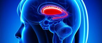
Depressed fractures of the skull bones are a violation of the integrity of the bony part of the head, which are characterized by pressing the bone into it. They are considered the most dangerous because they can cause damage to the dura mater, blood vessels, medulla and compression of intracranial structures.
The Neurology Department of CELT invites you to undergo diagnostics and treatment of depressed fractures of the skull bones in Moscow. Our multidisciplinary clinic is well known in the paid medical services market in the capital and region. We employ leading domestic specialists who have modern effective diagnostic and treatment methods. You can make an appointment with them online or by contacting our operators.
Signs and symptoms of a skull fracture
There are several reasons why a skull fracture can occur. The main ones are a powerful blow to the head with a heavy object, a traffic accident, a fall from a height, a bullet wound to the head, etc. Such damage is typical for middle-aged people who engage in active sports, work in heavy production, etc.
Traumatic brain injury causes severe headaches. Depending on the severity of the impact, signs of injury may vary.
In an adult
The main symptoms of a fracture and the presence of traumatic brain injury in an adult:
- loss of consciousness;
- short-term memory loss, numbness, inability to concentrate;
- local pain at the site of injury, severe headaches;
- breathing problems;
- visual deformation of the skull bones;
- vomiting, nausea;
- visual impairment: “fog”, “double vision”, “stars”, etc.;
- noise in ears.
With fractures, the patient can observe:
- bruises and tears at the site of injury;
- hemorrhage in the eyes, blood from the nose, ears (present in case of injury to the cranial fossa);
- heavy bleeding, swelling;
- impaired coordination of movements, balance;
- nervousness, anxiety, depression, etc.
If nerve endings are damaged as a result of the blow, the patient may experience loss of sensitivity and paralysis. If there are symptoms indicating traumatic brain injury, the victim must be urgently taken to the hospital.
The child has
Due to physiological development, the anatomy of a child’s skull differs from the mature skull of an adult, therefore the symptoms of traumatic brain injury in children are somewhat different. In infants, mild to moderate head trauma may be virtually asymptomatic. The patient may experience regurgitation during feeding, vomiting, causeless anxiety, poor sleep, and lack of appetite.
A preschooler who has suffered a head injury may experience the following symptoms:
- nausea, vomiting;
- headache;
- increased sweating;
- pale skin;
- unstable pulse.
After injury, the child's condition may be satisfactory. Over time, the victim will feel worse. Therefore, you need to see a doctor immediately.
Symptoms
Several general symptoms of a basal skull fracture are characteristic of any type of similar injury and do not depend on the specific location of the injury.
- Increasing headache.
- The pupils are of different sizes and do not constrict when exposed to bright light.
- Severe nausea, vomiting.
- Cardiac arrhythmia and unpredictable changes in blood pressure.
- Bradycardia.
- Due to the accumulation of blood in the orbit - protrusion of the eye (exophthalmos).
- Fainting or coma.
- Complete immobility or abnormal hyperactivity.
Fractures of the pyramid of the temporal bone
The most dangerous fracture in this case is the transverse type. Among the primary signs of such a fracture of the base of the skull is loss of consciousness, either for several hours or for several days. Due to damage to an area of dense bone, paralysis of the facial and outgoing nerves often occurs within an hour.
A typical manifestation is that the victim completely or partially loses his hearing, loses his sense of taste, and is unable to maintain balance. Liquor often leaks from the ears, and when it gets into the Eustachian tube, it begins to flow from the nose. All signs are observed against the background of rotational dizziness in combination with nausea and vomiting. A brain hematoma forms.
Anterior cranial fossa fractures
With fractures of the anterior cranial fossa, the patient experiences nasal liquorrhea and nosebleeds. 2-3 days after the injury, bruising appears around the eyes - a symptom of “glasses”. Due to cracks passing through the air sinuses, when the cells of the ethmoid bone are broken, subcutaneous emphysema develops in some cases.
Middle cranial fossa fractures
A characteristic sign of such an injury is unilateral auricular liquorrhea and bleeding. The victim's hearing completely disappears or partially decreases, and bruising appears behind the ear or in the area of the temporal muscle. The sense of taste is impaired and the function of the facial nerve is affected.
Posterior fossa fractures
In the case of a rupture or pinching of the caudal nerve, such fractures of the skull vault are distinguished by the fact that the victim’s larynx, palate and tongue are paralyzed. There are malfunctions in the functioning of all vital organs. Bruising appears behind one or both ears, and dysfunction of the auditory, abducens, and facial nerves occurs.
Diagnosis of damage
First of all, the doctor analyzes the patient’s complaints, finds out the circumstances due to which the injury occurred, and conducts an examination. If necessary, radiography, CT, MRI and other studies may be prescribed. Depending on the type of fracture and severity, the doctor prescribes treatment.
Computed tomography (CT) is the most effective method for diagnosing traumatic brain injuries, which allows a comprehensive examination of the damage and prescribing treatment.
X-rays and magnetic resonance imaging are used less frequently in diagnosing traumatic brain injuries, because the location of the fracture may not be clearly visible.
Damage classification
Traumatic brain injury can be simple (open) or complex (closed). The following forms are distinguished according to severity:
- mild (concussion);
- moderate severity;
- severe (fracture of the skull bones, severe brain contusions).
The following main types of traumatic brain injuries are distinguished:
- concussion (residual effects are possible; treatment by a neurologist is required);
- compression of the brain (reduction of intracranial space due to the formation of hematomas, immediate surgical intervention is required);
- brain contusion (possible mild, moderate and severe severity; diagnosed by CT scan);
- axonal damage (damage to nerve cells, accompanied by microscopic hemorrhages in the brain; the patient falls into a coma);
- intracranial hemorrhage (ruptures of blood vessels causing hemorrhage into the cranial cavity, possible formation of hematomas).
The following types of skull fractures are distinguished:
- linear skull fracture (a less dangerous type of fracture; it is thin lines that do not cause displacement of bone fragments);
- comminuted (the skull is broken into fragments that can damage the brain and cause the formation of hematomas, bruises, etc.; this type of fracture is life-threatening);
- depressed (during injury, the hard shell of the skull is pressed into the skull; the fracture can be severe);
- perforated (this type of fracture can occur as a result of a gunshot wound; it is considered severe and fatal).
Traumatic brain injury is life-threatening. Therefore, the victim should be hospitalized immediately.
Treatment methods
A person who has suffered a traumatic brain injury must be hospitalized immediately. If the injuries are not complicated (the patient has closed fractures, bruises, concussion, etc.), surgical intervention is not required; a conservative treatment method is used.
When treating linear fractures, doctors do not resort to emergency measures. Drug therapy consists of using painkillers, treating wounds, and constant monitoring by a specialist.
Severe traumatic brain injuries require surgical treatment. Doctors may resort to craniotomy, a surgical operation that removes fragments and other foreign bodies that have damaged brain tissue.
If the patient has lost a lot of blood, doctors will prescribe a transfusion. If brain tissue becomes infected, antibiotics are prescribed.
Patients are prescribed bed rest (its duration is determined by the attending physician). Depending on the severity and characteristics of the injury, the doctor may prescribe painkillers, drugs to improve blood circulation in the brain, antibiotics, anti-inflammatory drugs, etc.
Therapy
During the course of treatment, special attention is paid to preventing the development of purulent complications. For this purpose, the doctor prescribes broad-spectrum antibiotics. These drugs are used nasally and in the form of ear drops. They help prevent the occurrence of inflammatory processes. In the future, based on the type and nature of the injury, either conservative treatment or surgical measures are carried out. During the entire course of recovery, the patient must remain in bed with a slight elevation of the head. This is necessary in order to reduce the likelihood of leakage of cerebrospinal fluid.
If the fracture is accompanied by a concussion, the patient is prescribed additional nootropic and vasotropic medications. If there is serious brain damage, medications are prescribed that increase cerebral circulation.
Surgery is used only for severe fractures. These can be comminuted or depressed fractures. During the operation, the patient is given general anesthesia, after which a craniotomy is performed, and bone fragments of the skull and damaged soft tissue are eliminated. More often, operations are prescribed after hematomas are detected, which put strong pressure on the brain. In some cases, surgical intervention is required in the case of a developing purulent infection that cannot be eliminated with antibacterial drugs.
Complications and consequences of a fracture
During treatment, the patient may experience complications after injury. These include purulent formations, the development of intracranial hematomas, damage to the cranial nerves, etc. With non-operative intervention, complications can lead to death.
After treatment and discharge from the hospital, the patient can observe the consequences of a traumatic brain injury. These include:
- decreased performance, fatigue;
- disorders of speech, hearing, vision, memory;
- negative reaction of the body to drinking alcoholic beverages;
- painful reaction to changes in atmospheric pressure;
- disturbances in the functioning of the blood vessels of the brain, which is manifested by prolonged headaches, dizziness during physical activity, and changes in body position;
- epilepsy-like seizures (after severe traumatic brain injury).
The likelihood of consequences of traumatic brain injury is determined by the severity of the injuries suffered. In case of mild injuries, there is a possibility that the patient’s body will fully recover.
Care and rehabilitation
The following rehabilitation measures are taken to help a person return to normal life:
- neuropsychological correction (restoration of attention, memory);
- classes with a speech therapist;
- classes to restore motor activity;
- physiotherapy (restoration of brain activity);
- following a special diet;
- psychological recovery (conversations with a psychologist and family), etc.
The rehabilitation period lasts about 1.5-2 years. At this time, the patient has a chance to recover. After this time, recovery will be more difficult and less effective.
First aid
If you notice symptoms of a skull fracture, you should immediately seek medical help. Before doctors arrive, it is not advisable to give the victim painkillers, since some drugs can significantly increase bleeding or breathing difficulties, as well as provoke a comatose state of the patient.
Before the ambulance arrives, it is important to provide first aid to the patient:
- Place the patient on his back, on a hard surface, while fixing his head. You cannot put a pillow under your head. If the patient is unconscious, he must also be placed on his back, but in a half-turn, placing a roll of clothing on one side. The head should be slightly tilted so that the victim does not suffocate when vomiting.
- Treat the head wound with an antiseptic and apply a sterile bandage.
- Remove the victim's dentures, as well as all jewelry, watches and glasses.
- Unfasten clothing that may constrict the body, impede normal blood circulation and restrict breathing.
- Apply any cold object wrapped in a clean cloth to your head.
If the victim is not breathing, it is necessary to clear his mouth of vomit and perform mouth-to-mouth artificial respiration with chest compressions. To avoid direct contact with the victim’s mucous membranes, it is necessary to use a piece of clean fabric that is well breathable.
In the absence of breathing problems, it is allowed to give the victim Analgin with Diphenhydramine. In the process of transporting a patient to a medical facility, the following activities are performed:
- Glucose, diuretics and cardiac drugs are administered intravenously. However, in case of heavy bleeding, diuretics should not be used. Instead, Polyglucin or Getinol is administered. Lasix is most often used as a diuretic, and the main drugs that support the functioning of the heart muscle are Cordeamine and Sulfakamfocaine.
- If breathing problems occur, the patient is inhaled using an oxygen mask.
- Convulsions and increased motor activity are stopped with Suprastin.
In case of heavy bleeding, polyglucin is administered.
The use of narcotic painkillers is unacceptable. Such drugs are highly likely to cause respiratory arrest.