Structure
The division is part of the hindbrain. The structure and function of a bridge are very closely related, as in any other structure. It is located in front of the cerebellum, being a section between the midbrain and medulla oblongata.
It is separated from the first by the beginning of the 4th pair of cranial nerves, and from the second by a transverse groove. Outwardly, it resembles a roller with a groove, with nerves passing through it; they are responsible for the sensory abilities of the facial skin. In the groove there was also a place for the basilar arteries; their features include the fact that they supply blood to the back of the brain.
This section has a special diamond-shaped fossa located in the posterior part of the pons. The fossa is bordered at the top by the brain stripes, and above them are the facial colliculi.
Above them there is a median eminence, and next to it is the locus coeruleus, which is responsible for the feeling of anxiety; it includes many nerve endings of the norepinephrine type. The pathways look like thick fibers of nervous tissue that run from the pons to the cerebellum. Thus, they form the arms of the pons and the peduncles of the cerebellum.
Among other things, the structure of the bridge has a “tire”, which is an accumulation of gray matter. This gray matter is the centers of the cranial nerves and the parts that contain the pathways. That is, the upper part of the brain is reserved for the centers that have a connection with the cranial nerves (fifth, sixth, seventh and eighth pairs).
Useful to know: Brain stem: features and functions
Speaking of pathways, the medial lemniscus and lateral lemniscus pass through this part. The same tegmentum contains the reticular formation; it is part of six nuclei and contains structures that are responsible for auditory analyzers.
At the base there are paths that pass from the cerebral cortex to different parts:
- pons brain;
- medulla;
- spinal cord;
- cerebellum.
And the blood supply occurs through arteries that belong to the vertebrobasilar region.
to contents ^
Anatomy
Rice.
1. The cerebral bridge and its relationship with surrounding formations at the base of the brain: 1 - optic nerve; 2 - oculomotor nerve; 3 - mastoid body; 4 - trigeminal node; 5 - trochlear nerve; 6 - bridge; 7 - abducens nerve; 8 - facial nerve; 9 - vestibulocochlear nerve; 10 - pyramid; 11 - glossopharyngeal nerve; 12 - vagus nerve; 13 - accessory nerve; 14 - hypoglossal nerve; 15 - middle cerebellar peduncles. The bridge is located between the medulla oblongata and the cerebral peduncles, and on the sides it passes into the middle cerebellar peduncles (Fig. 1). From the base of the brain, the bridge is a dense white shaft measuring 30 X 36 X 25 mm. The anterior surface of the bridge is convex, facing forward and downward and is adjacent to the clivus at the base of the skull. The basilar groove (sulcus basilaris) runs in the middle of the anterior surface, in which lies the basilar artery (a. basilaris), which is the main source of blood supply to the cerebral pons.
Behind the pons of the brain, from the groove between the medulla oblongata, on the one hand, the pons and the middle cerebellar peduncle, on the other, the roots of the abducens, facial, intermediate and vestibulocochlear nerves emerge successively.
The posterior surface of the bridge faces upward and backward, into the cavity of the fourth ventricle, and is not visible from the outside, because it is covered by the cerebellum. It forms the upper half of the bottom of the diamond-shaped fossa.
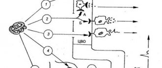
Rice. 2. Schematic representation of a cross section of the cerebral pons in its middle section: 1 - fourth ventricle; 2 - superior medullary velum; 3 - medial longitudinal fascicle; 4 - anterior spinocerebellar tract; 5 - superior cerebellar peduncle; b — descending root of the trigeminal nerve; 7 — central path of the tire; 8 - middle cerebellar peduncle; 9 - trigeminal nerve root; 10 - lateral loop; 11 - medial loop; 12 - pyramidal path; 13 - surface layer of transverse fibers of the bridge; 14—bridge cores; 15 - reticular formation; 16 - motor nucleus of the trigeminal nerve; 17 - superior sensory nucleus of the trigeminal nerve.
On transverse (frontal) sections of the pons (Fig. 2), a more massive anterior (ventral) part (pars ant. pontis), or base (basis pontis, BNA), and a small posterior (dorsal) part (pars post, pontis) are distinguished ), or tire (tegmentum, BNA). The boundary between them is the trapezoidal body (corpus trapezoideum), formed mainly by processes of cells of the anterior cochlear nucleus (nucleus cochlearis ant.). Clusters of nerve cells form the anterior and posterior nuclei of the trapezoid body (Gudden's nuclei). The front part of the bridge contains ch. arr. nerve fibers, between which numerous small accumulations of gray matter are scattered - the nuclei of the bridge (nuclei pontis). In the nuclei of the bridge, the fibers of the cortical-pontine tract (tractus corticopontini) and collaterals from the passing pyramidal tracts end. The processes of the cells of the pons nuclei form the cerebellopontine tract, the fibers of which pass predominantly to the opposite side and are the transverse fibers of the bridge (fibrae pontis transversae). The latter form the middle cerebellar peduncles (pedunculi cerebellares medii).
The rear part of the axle (tire) is much thinner. It contains the reticular formation (formatio reticularis) and the nuclei of the V, VI, VII, VIII pairs of cranial nerves. At the level of the middle of the bridge is the motor nucleus of the trigeminal nerve (nucleus motorius n. trigemini), and somewhat laterally there is the superior sensory nucleus (nucleus sensorius sup.). The latter is approached by fibers from the sensory cells of the trigeminal ganglion, which, as part of the sensory root, enter the substance of the bridge at its border with the middle cerebellar peduncle. Adjacent to the sensory root is the motor root, which is the processes of the cells of the motor nucleus of the trigeminal nerve.
At the level of the facial tubercle is the nucleus of the abducens nerve; nearby, in the reticular formation, is the motor nucleus of the facial nerve, the processes of the cells forming a knee that goes around the nucleus of the abducens nerve. Behind the motor nucleus of the facial nerve is the superior salivary nucleus (nucleus salivatorius sup.) and outward from the latter is the nucleus of the solitary tract (nucleus tractus solitarii). In the inferolateral part of the tegmentum of the bridge are located the nuclei of the vestibular-cochlear nerve (n. vestibulocochlearis). On the sides of the trapezoid body are the superior olives. The processes of the cells of the superior olive (oliva sup.) make up the lateral loop (lemniscus lat.), between the fibers of the latter is the nucleus of the lateral loop. (nucleus lemnisci lat.). The lateral lemniscus also includes processes of cells of the posterior nucleus of the cochlear nerve (nuci, cochlearis post.), the nuclei of the trapezoid body and the nucleus of the lateral lemniscus.
Inward from the superior olive above the trapezoid body there is a medial loop (lemniscus med.), which is a bundle of fibers of proprioceptive sensitivity, and a spinal loop (lemniscus spinalis) - a bundle of fibers of the path of pain and temperature sensitivity.
Motor and sensory functions
Speaking in more detail about motor and sensory function, let's talk about cranial nerves. When mentioning cranial nerves, it should be noted the ternary or mixed nerve (V pair). This pair of nerves is responsible for the movement of the masticatory muscles, as well as the muscles that are responsible for the tension of the eardrum and the palatine curtain.
To the sensory part of the trigeminal nerve there are afferent connections of nerve cells from receptors that are located in the skin of the human face, nasal mucosa, 60% of the tongue, eyeball and teeth. The sixth pair, or the so-called abducens nerve, is responsible for the movement of the eyeballs, namely for its rotation outward.
The 7th pair has one of the most important functions for human interaction; it is responsible for the innervation of muscles that allow the production of facial expressions. In addition, the facial nerve controls three glands: salivary, sublingual and submandibular. These glands provide reflexes such as salivation and swallowing.
The bridge also has a connection with the vestibulocochlear nerve. It is clear from the name that the cochlear part reaches the cochlear nuclei, but the vestibular part ends in the triangular nucleus. The eighth pair of nerves is responsible for analyzing vestibular stimuli; it determines the degree of their severity and where they are directed.
Useful to know: Gray matter of the brain, its structure, functions and properties
to contents ^
Formation in the prenatal period
Varolievye formation begins to form in the embryonic period from the rhomboid bladder. The bladder, in the process of its maturation and formation, is also divided into oblong and posterior. During the formation process, the hindbrain gives rise to the cerebellum, and the floor and its walls become components of the pons. The cavity of the diamond-shaped bladder will subsequently be common. At the stage of formation, the nuclei of the cranial nerves are located in the medulla oblongata and only over time do they move directly to the bridge.
Article on the topic: Urinary incontinence in men - causes, diagnosis and rules of personal hygiene, methods of therapy and prevention
When a baby is born, the bridge is located just above the back of the sella turcica. Only after 2-3 years does it begin to rise and thereby become fixed in its permanent place - the upper part of the skull.
At the age of 8, the child’s spinal fibers begin to become overgrown with a myelin sheath.
Integrating function
These pons functions connect parts of the brain called the cerebral hemispheres. Also, all other paths, both ascending and descending, pass along the bridge, connecting it with many departments of the central nervous system. Among them are the spinal cord, cerebellum and cerebral cortex.
Impulses passing through the pontocerebellar pathways of the cerebral cortex have an impact on the functioning of the cerebellum. The cortex cannot exert influence directly, so it uses the bridge as an intermediary for these purposes. The pons regulates the medulla oblongata, influencing the centers that are responsible for the respiratory process and its intensity.
to contents ^
What functions does the bridge perform?
The pons is responsible for several important forms of activity.
Among them:
- Reflexive automatic and voluntary movements of the eyes and eardrum in response to loud sounds, as well as tissues of the oral cavity (palate). Any violation ends in problems.
- The ability for purposeful motor activity. Since the pons in the brain ensures the functioning of the cerebellum, any damage causes problems with the ability to control the body.
- Perception of vestibular stimuli. In this case, we are talking about the ability to perceive one’s body as a whole, its orientation and location in space, to respond to any changes in environmental conditions, and also to dampen unnecessary movements (for example, during sudden braking in public transport, stumbling, etc.). When affected, there is a lack of coordination. Ability to navigate in space.
- Providing olfactory function. The bridge has this ability partially. Other subcortical clusters are also responsible for it.
- Normal innervation of the skin and mucous membranes of the face.
- The pons is involved in the formation of sleep. This is a complex and coordinated work of several cerebral formations at once. Any violations immediately lead to problems with night rest. The patient becomes lethargic, asthenic processes appear.
- The functions of the pons include the acts of chewing and swallowing. Vital for nutrition and breathing.
- In fact, the body’s ability to perform normal gas exchange depends on the functioning of this structure. In the absence of adequate conduction of impulses, problems begin, even lethal disorders.
Basic actions are performed by nervous tissues constantly. Even minor changes are noticeable immediately.
The pons is part of the brain stem, therefore deviations in its activity become an indirect cause of dysfunction of this entire formation.
Complications, including rapid and fatal ones, are likely. High-quality medical care is not always possible due to the complex localization of structures and complex structure.
CM pathologies
Like any organ in the human body, the VM can also stop functioning and this can be caused by the following diseases:
- stroke of the cerebral arteries;
- multiple sclerosis;
- head injuries. Can be obtained at any age, including during childbirth;
- tumors (malignant or benign) of parts of the brain.
In addition to the main reasons that can provoke brain pathologies, you need to know the symptoms of such a lesion:
- the process of swallowing and chewing is impaired;
- loss of skin sensitivity;
- nausea and vomiting;
- nystagmus is eye movements in one specific direction, as a result of such movements one can often feel dizzy, even to the point of loss of consciousness;
- may see double when turning the head sharply;
- disturbances in the functioning of the motor system, paralysis of certain parts of the body, muscles, or tremors in the hands;
- in case of disturbances in the functioning of the facial nerves, the patient may experience complete or partial anemia, lack of strength in the facial nerve;
- speech disorders;
- asthenia – decreased strength of muscle contraction, rapid muscle fatigue;
- dysmetria - incompatibility between the task of the movement being performed and muscle contraction, for example, when walking, a person may raise his legs much higher than necessary or, on the contrary, may stumble over small bumps;
- snoring, in cases where it has never been observed before.
Article on the topic: Ointment for sprains - list of drugs with instructions and composition