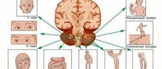
ALTERNATING SYNDROMES
(Latin alternus - alternating, changing; synonym:
alternating paralysis, cross paralysis
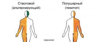
) - symptom complexes characterized by dysfunction of the cranial nerves on the side of the lesion (paralysis or paresis) and central paralysis or paresis of the limbs or sensory conduction disorders on the opposite side. Alternating syndromes include cross hemiplegia (see) - paralysis of one arm and the opposite leg and cross hemianesthesia - sensitivity disorder on one side of the face and hemianesthesia on the opposite half of the torso and limbs.
Alternating syndromes are distinguished: 1) according to the topic of the lesion - cerebral peduncles, pons, medulla oblongata, extracerebral localization is also possible; 2) according to clinical syndromes - motor, sensory, combined with dysfunction of cranial nerves, hyperkinesis and others; 3) according to the etiology of the disease - circulatory disorders, tumors, injuries, etc.; 4) along the flow - progredient, regredient.
Localization of the lesion in the brain stem is manifested by symptoms of damage to the cranial nerves (on the side of the lesion).
Paralysis or paresis of the limbs on the side opposite to the lesion develops due to damage to the corticospinal (pyramidal) tract. Cross hemianesthesia on the trunk and limbs opposite the pathological focus occurs when sensitive pathways are damaged (median lemniscus, spinothalamic tract). Hemiplegia or hemiparesis and hemianesthesia occur on the side opposite the lesion because the pyramidal tract and sensory pathways intersect in the lower part of the brain stem.
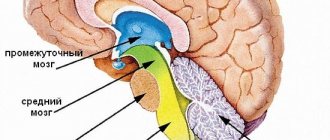
Alternating syndromes are divided according to the localization of the lesion in the brain stem: a) bulbar - with damage to the medulla oblongata; b) pontine - when the bridge is damaged; c) peduncular - with damage to the cerebral peduncle (colored Fig. 1-6).
- Localization of lesions in the brain stem that cause the appearance of alternating syndromes
- 1. Weber syndrome. (The lesions are indicated in yellow; the nuclei of the cranial nerves are in orange; the dotted line is the course of the cranial nerves.)
- 2. Benedict's syndrome. (The lesions are indicated in yellow; the nuclei of the cranial nerves are in orange; the dotted line is the course of the cranial nerves.)
- 3. Foville's syndrome. Rice. (The lesions are indicated in yellow; the nuclei of the cranial nerves are in orange; the dotted line is the course of the cranial nerves.)
- 4. Millard-Gubler syndrome. (The lesions are indicated in yellow; the nuclei of the cranial nerves are in orange; the dotted line is the course of the cranial nerves.)
- 5. Wallenberg-Zakharchenko syndrome. (The lesions are indicated in yellow; the nuclei of the cranial nerves are in orange; the dotted line is the course of the cranial nerves.)
- 6. Jackson syndrome. (The lesions are indicated in yellow; the nuclei of the cranial nerves are in orange; the dotted line is the course of the cranial nerves.)
Causes of the disease
There can be many reasons for this type of disease. Among the most common are:
- Blood supply disorders. This includes hemorrhagic and ischemic strokes, which are the most common causes of alternating syndromes.
- Neoplasms in the brain. If the brain stem is affected by a tumor, compression of the nerve endings occurs, which provokes the development of the disease.
- Inflammatory diseases that are localized in the trunk and act as an impetus for the development of pathology.
- Traumatic brain injury. Very often the disease occurs after damage to the skull.
Diagnostics
- Careful collection of anamnesis for previous infections and vaccinations.
- EMG is not always informative, as it is often within normal limits in the first weeks. Typical changes are:
- in acute inflammatory demyelinating polyneuropathy: decreased impulse conduction speed along motor nerves, proximal type conduction block, increased latency of F-waves.
— about acute sensorimotor axonal polyneuropathy: a decrease in the amplitude of the total action potentials of motor muscles and sensory nerve fibers, signs of demyelization in a maximum of 1 nerve.
- in acute motor axonal polyneuropathy: a decrease in the amplitude of the total action potentials of the motor muscles, sensory fibers remain intact, demyelination is also in a maximum of 1 nerve.
- in acute sensory polyneuropathy: motor fibers are not affected, there is a decrease in the amplitude of the total action potential of the sensory nerves.
- Study of cerebrospinal fluid: protein-cell dissociation. In the first days, however, it is not always possible to detect it, therefore the study of the cerebrospinal fluid should be carried out over time. It should be remembered that false-positive results are possible with intravenous administration of immunoglobulins.
- MRI of the brain and spine allows you to see changes in the roots, but to a greater extent this method is used in the differential diagnosis of tumors and stroke.
- Blood tests: detection of antibodies in the blood
- for acute inflammatory demyelinating neuropathy - antibodies unknown
— for acute motor axonal neuropathy – GM1+GM1b, GD1a
- for acute motor-sensory axonal neuropathy - GM1+GM1b, GD1a
- for acute sensory neuropathy - GD1b
- for Miller-Fisher syndrome - GQ1b.
Pathogenesis
The nuclei of the cranial nerves are located in the cerebral trunk. In the same part there is a pyramidal tract through which impulses are transmitted from the cortex to the neurons of the spinal cord. Sensory and motor nerve fibers intersect at the level of the spinal cord, so the innervation of one part of the body is carried out by nerve pathways that pass in another part of the trunk. If a brainstem lesion occurs involving the nuclei of the cranial nerves, then cross-symptoms appear. Such symptoms also appear with damage to the extra-stem part and motor cortex.
What else needs to be done?
Cardiac activity should be maintained, and also try to stabilize blood pressure at normal levels. After all, it is arterial hypertension that often leads to Wallenberg-Zakharchenko syndrome. The patient must take medications to lower blood pressure on an ongoing basis.
In addition, cardiac glycosides and nitrates are prescribed. It is necessary to normalize blood clotting and its thickness. Neuroprotectors protect brain cells. With the help of special medications, pain is relieved and muscles relax. Anticonvulsants and sedatives are also often prescribed.
Wallenberg-Zakharchenko syndrome is discussed in detail in this article.
Clinic of alternating syndromes
The signs of the disease in each patient are always obvious, which allows the neurologist to make the correct diagnosis after the initial examination. Patients show signs of dysfunction of the cranial nerves on the side of the body where they were affected, and there are various disorders: motor or sensory on the other side. Alternating symptoms can appear either gradually with an increase, or very abruptly and are accompanied by an increase in intracranial pressure and a deterioration in the general condition.
Description of the disease
A person rarely loses consciousness with Wallenberg-Zakharchenko syndrome, but there are attacks of severe dizziness, which provokes loss of balance because the cerebellar arteries are affected.
In addition, hiccups or retching occur, as well as a symptom of mild stupor.
Vascular atherosclerosis can lead to thrombosis. However, there are frequent cases of endarteritis of the vertebral artery, which is of the syphilitic type, or rheumovasculitis.
Slow thrombotic or non-thrombotic damage occurs. The most commonly affected area is the vertebral or basilar artery.
Wallenberg-Zakharchenko syndrome occurs with periods of improvement and deterioration. But at the same time, the disease progresses.
How can the syndrome manifest itself?
It can develop in five ways. This completely depends on where the pathology center is located and what clinical signs are present.
Let's look at the types of syndrome in more detail.
Bulbar group
Jackson syndrome occurs when the 12th nerve nucleus, as well as the pyramidal tracts, are affected. In such patients, partial paralysis of the tongue is observed: when protruded, it tilts towards the lesion, there are obvious signs of atrophy, and the articulation of individual sounds is impaired. Hemiparesis is observed on opposite limbs, and in some cases even sensitivity is lost.
Avellis syndrome is manifested by paresis of the laryngeal muscles, vocal cords, and pharynx due to damage to 10-11 nerve nuclei. Such patients very often have speech and voice disorders, and sensitivity in the opposite limbs decreases.
With Schmidt's syndrome, 9-12 nuclei of the cranial nerves are affected, which causes paresis of the trapezius and sternocleidomastoid muscles. In patients on the side that was affected, the shoulder drops significantly, and patients cannot raise their arm up.
Wallenberg (–Zakharchenko) syndrome
One of the alternating medullary syndromes of the brainstem. It was first described by the German physician Adolf Wallenberg in 1895. (Wallenberg A. Acute Bulbäraffection (Embolie der Art. cerebellar. post. inf. sinistr.?) // Archiv für Psychiatrie und Nervenkrankheiten. – 1895. – Bd.27. – S.504-540). Domestic neurologist M.A. Zakharchenko in 1911 described several clinical variants of the syndrome. Occurs with lesions of the posterolateral part of the tegmentum of the medulla oblongata. In this case, the process involves the double nucleus (IX, X nerves), the nucleus of the spinal tract (V nerve), sympathetic fibers from the ciliospinal center of Budge, the medial lemniscus, the inferior cerebellar peduncle (cordite body), the vestibular nuclei (VIII nerve) and other structures . The classic version of the syndrome is described in detail below.
Transverse section of the brain stem at the level of the medulla oblongata. The affected area in classic Wallenberg-Zakharchenko syndrome is shaded (source: www.medsch.wisc.edu/anatomy/bs97/text/bs/contents.htm, in its own modification)
Substrate of lesion and clinical symptoms:
| Structure involved | Ipsilateral | Contralateral |
| Nuclei of IX and X pairs of cranial nerves | Peripheral paresis of the muscles of the pharynx, soft palate and vocal cord (clinically – dysphagia, complete in the first hours; sometimes dysarthria, dysphonia). | |
| Nucleus of the spinal tract of the trigeminal nerve (V pair) | Dissociated hemianesthesia (analgesia, thermoanesthesia) on the face according to the “onion” type, most often in the outer Zölder zone* | |
| Connections between the Budge ciliospinal center and the hypothalamus | Bernard-Horner syndrome:
| |
| Inferior cerebellar peduncle (corpus filamentum) | Cerebellar symptoms: intention tremor, hemiatonia, hemiasynergia, lateropulsion, nystagmus. | |
| Medial loop | Dissociated hemihypesthesia of superficial sensitivity |
* Can be observed on both sides, less often - sensitivity on the face is not impaired.
The syndrome develops acutely, less often – subacutely. The first symptoms are vomiting, dizziness, nystagmus, staggering and even falling towards the affected side. As a rule, patients report severe headache on the affected side.
Among the variants of Wallenberg-Zakharchenko syndrome, the addition of disorders of vital functions (involvement of the reticular formation), damage to the VI and VII, XII nerves, cross paralysis, etc. is noted. Opalski's syndrome is considered separately when ischemia spreads below the level of the pyramidal decussation with the addition of ipsilateral hemiplegia to the lateral bulbar syndrome (Opalski A. Un nouveau syndrome sous-bulbaire: syndrome partiel de l'artère vertébro-spinale postérieur // Paris. Med., 1946. - Vol.1 – P.214-220).
The most common cause of the syndrome is thrombosis of the posterior inferior cerebellar artery (a branch of the vertebral artery) and circulatory failure in the vertebrobasilar region.
Pontine group
Millard-Gübler syndrome appears against the background of damage to the 7th nucleus of the cranial nerves and fibers of the pyramidal tract. Clinically manifested in the appearance of facial paresis and hemiparesis on the other side. In the Brissot-Sicard form, the lesion is located in a similar way, but in this case there is hemispasm on the face. Foville syndrome involves the presence of paresis of the 4th nerve, as a result of which patients develop strabismus. In Gasperini syndrome, nerves 5-8 are affected. Facial paresis, hearing loss, strabismus, and in some cases nystagmus are observed.
Complications
Alternating syndromes that appear simultaneously with spastic hemiparesis cause the appearance of joint contractures. This complication has a negative impact on already existing motor disorders. Paresis of the 7th pair of cranial nerves causes a distortion of the face to one side, which is why the patient not only experiences a lot of inconvenience, but also develops many complexes.
If the auditory nerve is affected, the patient will soon develop hearing loss, which can even lead to complete hearing loss over time. Paresis of the 3rd and 4th pairs causes a significant deterioration in vision and the appearance of double vision.
The most serious complication is damage to the brain stem, because It is here that all the most important centers for maintaining life are located. Such patients in most cases die.
Diagnostic methods for determining the disease
The type and presence of cross syndrome can be determined at an appointment with a neurologist. Already as a result of the initial examination, the doctor can judge the prevalence of the pathological process and its severity. If the root cause of this condition is a tumor, then the symptoms will gradually increase every month. If such a condition is caused by an infectious nature, the patient will complain of a change in health for the worse. During a stroke, alternating symptoms always appear suddenly and always increase rapidly.
To identify the root causes of this disease, the patient may be prescribed a series of examinations:
Magnetic resonance imaging. The study allows us to examine the lesion due to the fact that after its completion, specialists can evaluate three-dimensional images in high quality, due to which errors in making a diagnosis are practically eliminated.- Ultrasound diagnostics. One of the most common and accessible methods that provides detailed information about the condition of blood vessels and tissues.
- Neuroimaging of blood vessels. If doctors suspect that a patient has had a stroke, regardless of its type, magnetic resonance imaging of the blood vessels is prescribed. This diagnostic method allows you to identify the exact location of the pathological focus and its severity.
- Cerebrospinal fluid examination. A puncture is taken if an infectious disease is suspected.
Treatment
Treatment is aimed at eliminating the underlying pathology and includes many techniques:
- Conservative therapy. Patients are prescribed neuroprotective, decongestant and neuroprotective agents. For ischemic strokes, vascular therapy is carried out, for hemorrhagic strokes, calcium and aminocaproic acid preparations are prescribed.
- Neurosurgical treatment. In some particularly severe cases, it is performed for hemorrhagic stroke. The advisability of such an operation should be assessed by a neurosurgeon.
- Rehabilitation. After conservative therapy, patients must undergo a rehabilitation stage, which includes physical therapy, massage and other procedures.


