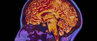
The midbrain is an ancient section of the brain included in the brain stem. It includes the ancient visual center. The midbrain is located below the cerebral cortex and above the hindbrain, being, as it were, in the very center of the brain. Caudally, the midbrain is adjacent to the hindbrain, and rostrally to the diencephalon. In the ventral part of the midbrain there are the so-called cerebral peduncles, most of which are occupied by the pyramidal tracts. In the midbrain, between the legs, there is an interpeduncular fossa, from which the third oculomotor nerve originates. Deep in the interpeduncular fossa there is a posterior perforated substance.
The midbrain includes: the roof of the midbrain (tectum), the lower tubercle (inferior colliculus), the colliculus (superior colliculi), the cerebral peduncle, the tegmentum of the midbrain (midbrain tegmentum), the black substance (substantia nigra), the cerebral peduncle (crus cerebri). It should be noted that there is no visible border with the diencephalon.
The midbrain is part of the brain stem. The substantia nigra of the midbrain is closely connected with the musculoskeletal system of the basal ganglia pathways. The substantia nigra and ventral tegmentum produce dopamine, which plays an important role in motivation and arousal. The midbrain transmits visual and auditory information.
Four Hills
The midbrain quadrigeminal consists of two pairs of inferior and superior colliculi. The upper pairs are visual, and the lower pairs are auditory. at the same time, the upper pairs of mounds are somewhat larger than the lower pairs. These hillocks are connected to structures of the diencephalon called the geniculate bodies. In this case, the upper colliculi are connected with the lateral ones, and the lower colliculi with the medial ones. The trochlear nerve emerges from the posterior surface of the midbrain. Four solid lobes help cross several optic fibers at right angles. The auditory nuclei are located inside the inferior colliculus.
Prevention of pathologies
The brain cannot function correctly without intellectual activity and physical activity. Typically, disruptions in the functioning of the central nervous system are observed in people over 70 years of age. But diseases of this group are diagnosed in those who, after retirement, no longer maintain their health and lead a healthy lifestyle. However, there are also congenital pathologies in the midbrain; you can get sick at any age.
Useful to know: Functions and structure of the cerebral pons, its description
To maintain your ability to think and work, it is enough to follow simple recommendations.
Exercise regularly to the best of your physical ability, walk in the fresh air, do gymnastics in the morning. Quit tobacco and alcoholic beverages. Switch to a healthy diet, eat as many fresh vegetables and fruits as possible. Do not eat products with preservatives and emulsifiers. Train your mind - for this you can read books, solve crosswords, play chess, gain new knowledge in an area of interest.
To get rid of vitamin deficiency - take vitamins and antioxidants. Since the brain is 60% fat, you cannot give up oil, but it must be natural. For example, olive oil is perfect. Avoid stressful situations. Do not engage in monotonous work too often, take breaks, switching to other activities. Monitor your blood pressure levels - hypertension can cause a stroke.
to contents ^
Brain legs
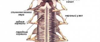
The cerebral peduncles are paired structures that are located on the ventral side of the cerebral aqueduct. They transfer the tegmentum to the dorsal side. The middle part of the brain contains the substantia nigra, which is a type of nucleus basalis. The substantia nigra is the only part of the brain that contains melanin. Between the legs is the interpeduncular fossa. which is filled with cerebrospinal fluid, is like a flush tank. The oculomotor nerve emerges between the crura, and the trochlear nerve prominently wraps around the outer sides of the crura. The oculomotor nerve (parasympathetically) is responsible for the constriction of the pupil and some eye movements.
Anatomical structure and functions
The midbrain is a very small formation, it contains several structures at once and is responsible for many functions, including reproduction. It would be a mistake to assume that the midbrain consists of the cerebral peduncles and the tegmentum. In addition to the base of the NM and the substantia nigra, it contains the quadrigeminal tract and the tegmentum. It is formed from a complex mesencephalic, ventral and dorsal part.
The main difficulty in considering the anatomical structure is the decision whether to include only the ridges from which the cerebral peduncles are formed, or whether to include in their composition the lid of the midbrain, the bottom and the specific substantia nigra:
- A traditional examination will not show anything other than the body of the legs itself, with a fossa located between them (tarina or interpeduncular). It is part of the interpeduncular cistern, located at the base of the human skull, from where the midbrain receives the cerebrospinal fluid necessary for life.
- In the interpeduncular area there is the exit of the nerve that moves the eyeball, eyelid and ensures the reaction of the pupil to a light stimulus, and on the outer surface there is the trochlear nerve, without which the movement of the oblique muscles and some characteristic turns of the eyes in the orbit are indispensable.
- The black mass, in one interpretation – a demarcating structure, and in another – an integral part of the operating system, develops from the mesencephalon and regulates the coordination of movements. Its remarkable feature is that this is the only area of the NCC in which the cells are stained with melanin.
- The base of the NM (base of the brain) is nothing more than the lowest part located in front. There are discrepancies in determining the identity of this part not only in old anatomy textbooks, where these terms were considered equivalent or were simply not distinguished as a separate entity. Now the ONM is precisely the lower part, in which there are axons stained with myelin.
- In contrast to the base, the tegmentum is located between the substantia nigra and the aqueduct of the brain, it forms both one and the other segments located nearby. Its structures include the red nucleus, its ventral and dorsal parts.
- It also contains gray (periaqueductal) matter, which is anatomically classified as the tegmentum, although it is located next to the cerebral aqueduct. OSV is considered the main center of regulation of the pain symptom, going along the descending line. It is capable of projecting to the nuclei of the thalamus and spinal cord, as well as to the raphe nuclei and locus coeruleus.
The importance of anatomical formation, regardless of its composition in the first or second case, is undeniable. After all, it is precisely this that provides orientation based on the visual image and the received stimulus, ensures the maintenance of a certain body posture and the regulation of muscle tone at the reflex level.
The connection of an anatomical formation with other areas of the brain is carried out in the most variable ways - from the fibers of the corticospinal tract going to the most important analyzers (ascending and descending), to information transmitted by neurons of all adjacent formations of gray, white and black.
Structure of the midbrain in sections
With a horizontal section of the midbrain at the level of the superior colliculus, the red nucleus, the nuclei of the oculomotor nerve and the associated Edinger-Westphal nuclei, the medullary peduncles, and the substantia nigra are observed.
With a horizontal section of the midbrain at the level of the inferior colliculus, the substantia nigra is also observed, the nuclei of the trochlear nerve and the cross of the superior cerebellar peduncles are also clearly visible.
In both cases, there is a cerebral aqueduct connecting the third and fourth ventricles and the periaqueductal gray matter.
Internal structure
If you have read my previous articles from the neuroanatomy series, then you probably know that any part of the central nervous system consists of gray matter (the body of neurons) and white matter (the processes of neurons). Gray matter is grouped into nuclei - clusters of neuron bodies that perform the same function. White matter is grouped into bundles, which are part of pathways in which bundles alternate with neurons and conduct impulses in a strictly defined direction.
As always, we study gray matter first and then white matter.
Midbrain gray matter
The gray matter of the midbrain is represented by the following groups of nuclei:
- Cranial nerve nuclei;
- Red kernels;
- Nuclei and neurons of the substantia nigra.
- Nuclei of the superior colliculus;
- Nuclei of the inferior colliculus;
- Nuclei and neurons of the reticular formation;
- Cajal and Darshkevich nuclei
Cranial nerve nuclei
The nuclei of 3 (oculomotor), 4 (trochlear) and 5 (trigeminal) pairs of cranial nerves are localized in the midbrain. These nuclei are localized around the opening for the circulation of cerebrospinal fluid, called the Sylvian aqueduct, or simply the aqueduct (aqueductus). In the lessons about the nuclei of the cranial nerves, we will definitely analyze their detailed localization, but for now we will note the formation called the gray central substance (substantia grisea centralis).
So, the central gray matter is a collection of cranial nerve nuclei that are located around the aqueduct. We mark it in gray:
By the way, the Sylvian aqueduct is an important topographical landmark, because it conditionally divides the midbrain into a tire and a roof. Everything above the conventional line passing through the aqueduct is the roof of the midbrain (tectum mesencephali), and the area from it to the substantia nigra is the tegmentum mesencephali. You can see these areas in the last illustration of this lesson.
Red kernels
As one cool anatomy professor said, red nuclei (nucleus ruber) are the calling card of the midbrain. They are very visible during any preparation. With any, even the most inept, incision, you will be able to see these large, really reddish, paired formations (the preparation here is quite high quality):
Now let’s mark the red nuclei in our diagram:
The red nuclei are one of the centers of the extrapyramidal system, that is, a system of movements that we do not think about and which we carry out without any comprehension. For example, this is moving the legs while walking or tying a shoelace, provided that we know how to tie it.
From the red nuclei begins the well-known rubro-spinal tract, also known as the red nuclear-spinal tract, also known as the Monakovsky bundle.
Black matter
Another very bright and noticeable anatomical formation of the midbrain is called the black substance (substantia nigra). The substantia nigra looks like a pair of black stripes. This strip is already present in my diagram, because in the study of anatomy it plays the role of a landmark - below it are the cerebral peduncles (which, let me remind you, are part of the midbrain, and not the greater bolus), and above it is the tegmentum of the midbrain.
But I suggest making it more contrasty and larger, because in reality it is very clearly visible to the naked eye:
The neurons of the substantia nigra, extremely rich in melanin, perform several important functions:
- Synthesis, accumulation and delivery of dopamine to the structures of the cerebral hemispheres, which are called the basal ganglia;
- Participation in coordinating the movements of the eyes, fingers, as well as the work of the chewing muscles and pharyngeal muscles;
- Participation in autonomic reactions, such as changes in blood pressure, pulse and respiratory rate.
It is with the damage and death of neurons in the substantia nigra that the symptoms of a terrible neurodegenerative disease - Parkinson's disease - are associated.
Consider these contrasting, clearly visible areas of the substantia nigra on the preparation:
Quadrigeminal nuclei
The nuclei of the superior and inferior colliculi produce subcortical processing of visual and auditory signals, respectively. How does cortical information processing differ from subcortical information processing? Cortical analysis involves longer and more meaningful work of neurons on information. Subcortical structures produce a primary, quick analysis and trigger any urgent actions, for example, turning the head after something quickly flashed before our eyes.
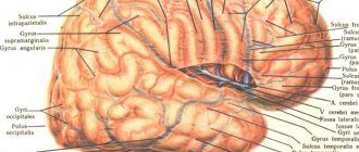
A horizontal section will not allow us to see both the superior and inferior colliculi at the same time, but we can look at the brainstem in the frontal plane and see them:
I hope you were able to identify the medulla oblongata with its thin and cuneate fasciculi, the pons and the massive middle cerebellar peduncles. Let's mark the nuclei of the superior colliculus (lilac) and inferior colliculus (light blue), keeping in mind that shades of blue represent sensory pathways and nuclei, and shades of red represent motor pathways:
By the way, pay special attention to the nuclei of the lower colliculi. There are the fourth neurons of the auditory pathway, to which axons go from the neurons of the trapezoid body. Read my articles about the Varoliev bridge and then you will have a great understanding of the auditory pathways. We haven't gone through the visual tracts yet, but we'll definitely look into it.
Nuclei of the reticular formation
The reticular formation is a set of nuclei and individual neurons that are located throughout the entire brain stem, including, of course, the midbrain. The reticular formation is formed primarily by interneurons, which provide interaction between other nuclei and pathways. Also, individual parts of the reticular formation ensure the transmission of pain sensations to the hypothalamus and the cerebral cortex.
Due to the fact that the reticular formation is located precisely as a diffuse formation, that is, it is distributed throughout the roof and tegmentum of the midbrain, I will not mark it in our drawing so as not to paint over the entire space
Cajal and Darshkevich nuclei
In the previous lesson about the pons nuclei, we studied the locus coeruleus as a special part of the reticular formation. The midbrain also contains nuclei of the reticular formation, which are sometimes isolated as independent formations. We are talking about two motor nuclei - the intermediate (nucleus interstitialis) and the nucleus of the posterior commissure (nucleus commisurae posteriois).
These nuclei are located between the red nuclei and the central gray matter, with the Darshkevich nucleus located much more lateral:
If you have taken my previous lessons, you, of course, remember the medial longitudinal fasciculus (fasciculus longitudinalis medialis), which transits through the pons and medulla oblongata. This bundle forms many anastomoses with the cranial nerves that control eye movements and the motor nuclei of the spinal cord, the processes of which innervate the neck muscles.
So, it is the nuclei of Cajal and Darshkevich that are the main, coordinating components of this conducting path. The medial longitudinal fasciculus begins from these nuclei.
PS - according to Prof. Gaivoronsky’s textbook, the nuclei of Cajal and Darshkevich are part of the central gray matter.
Midbrain Development
During embryonic development, the midbrain is formed from the second vesicle. It remains indivisible during further development, unlike the other two vesicles of the forebrain and hindbrain. Division into other areas of the brain does not occur during the development of the nervous system, unlike the forebrain, which is divided into the telencephalon and diencephalon.
During embryonic development, the midbrain undergoes continuous development of nerve cells, which are gradually compressed by the cerebral aqueduct. In some cases (with impaired development), partial or complete blockage of the cerebral aqueduct may occur, which leads to congenital hydrocephalus.
Functions
The midbrain, along with the pons, cerebellum and medulla oblongata, are classified as structures of the brain stem. Sometimes the diencephalon is also classified as a stem structure. The substantia nigra of the midbrain is involved in the motor pathways coming from the basal ganglia. The midbrain is archipallic in origin. Its general structure has remained virtually unchanged from the oldest and most primitive chordates to humans. Dopamine, produced in the substantia nigra and in the ventral tegmental area, plays a critical role in regulating the general level of central nervous system excitation, activity level, motivation, as well as in the development of addiction and addiction to a particular environment, food, type of activity, etc. including drug addiction, in all chordates, from humans to the most primitive. Moreover, areas structurally and functionally similar to the midbrain of chordates are also found in arthropods, such as insects, arachnids, and crustaceans. Laboratory mice that were selected to breed a line of mice with a predilection for “sports” (wheel running) have an increased size of the midbrain and, in particular, the ventral tegmental area [8]. The roof of the midbrain, namely the quadrigeminal, also plays an important role relay-relay and integrating station for visual and auditory information on its way to the thalamus.
Age characteristics and prevention
The brain is a complex structure. It operates with close interaction between all segments. The center that controls the middle section is the cerebral cortex. With age, connections become weaker, and reflex activity weakens. Since the area is responsible for motor function, even minor disruptions in this tiny segment lead to the loss of this important ability. It is more difficult for a person to move, and serious disorders lead to diseases of the nervous system and complete paralysis. How to prevent disorders in the functioning of the brain in order to remain healthy until old age?
First of all, you should avoid hitting your head. If this happens, it is necessary to begin treatment immediately after the injury. It is possible to preserve the functions of the midbrain and the entire organ until old age if you train it with regular exercises:
- The lifestyle a person leads is important for physical and mental health. Drinking alcohol and smoking destroy neurons, which gradually leads to a decrease in mental and reflex activity. Therefore, you should give up bad habits, and the sooner you do this, the better.
- Moderate physical activity and walks in nature supply the brain with oxygen, which has a beneficial effect on its activity.
- You shouldn’t give up reading, solving charades and puzzles: intellectual activity keeps the brain active.
- An important aspect of the functioning of brain structures is nutrition: fiber, protein, and greens must be present in the diet. The midbrain responds positively to intake of antioxidants and vitamin C.
- It is necessary to control blood pressure: the health of the vascular system affects the general condition of a person.
The brain is a flexible system that can be successfully developed. Therefore, by constantly improving your mind and body, you can maintain clarity of thoughts and motor activity until old age.
The midbrain, its structure and functions are determined by the location of the structure, providing movement, auditory and visual reactions. If you have difficulty maintaining balance or lethargy, you should consult a doctor and undergo an examination to find the cause of the disturbances and eliminate the problem.