Anatomy
Rice.
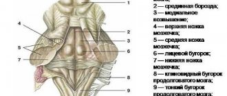
1. The cerebral bridge and its relationship with surrounding formations at the base of the brain: 1 - optic nerve; 2 - oculomotor nerve; 3 - mastoid body; 4 - trigeminal node; 5 - trochlear nerve; 6 - bridge; 7 - abducens nerve; 8 - facial nerve; 9 - vestibulocochlear nerve; 10 - pyramid; 11 - glossopharyngeal nerve; 12 - vagus nerve; 13 - accessory nerve; 14 - hypoglossal nerve; 15 - middle cerebellar peduncles. The bridge is located between the medulla oblongata and the cerebral peduncles, and on the sides it passes into the middle cerebellar peduncles (Fig. 1). From the base of the brain, the bridge is a dense white shaft measuring 30 X 36 X 25 mm. The anterior surface of the bridge is convex, facing forward and downward and is adjacent to the clivus at the base of the skull. The basilar groove (sulcus basilaris) runs in the middle of the anterior surface, in which lies the basilar artery (a. basilaris), which is the main source of blood supply to the cerebral pons.
Behind the pons of the brain, from the groove between the medulla oblongata, on the one hand, the pons and the middle cerebellar peduncle, on the other, the roots of the abducens, facial, intermediate and vestibulocochlear nerves emerge successively.
The posterior surface of the bridge faces upward and backward, into the cavity of the fourth ventricle, and is not visible from the outside, because it is covered by the cerebellum. It forms the upper half of the bottom of the diamond-shaped fossa.
Rice. 2. Schematic representation of a cross section of the cerebral pons in its middle section: 1 - fourth ventricle; 2 - superior medullary velum; 3 - medial longitudinal fascicle; 4 - anterior spinocerebellar tract; 5 - superior cerebellar peduncle; b — descending root of the trigeminal nerve; 7 — central path of the tire; 8 - middle cerebellar peduncle; 9 - trigeminal nerve root; 10 - lateral loop; 11 - medial loop; 12 - pyramidal path; 13 - surface layer of transverse fibers of the bridge; 14—bridge cores; 15 - reticular formation; 16 - motor nucleus of the trigeminal nerve; 17 - superior sensory nucleus of the trigeminal nerve.
On transverse (frontal) sections of the pons (Fig. 2), a more massive anterior (ventral) part (pars ant. pontis), or base (basis pontis, BNA), and a small posterior (dorsal) part (pars post, pontis) are distinguished ), or tire (tegmentum, BNA). The boundary between them is the trapezoidal body (corpus trapezoideum), formed mainly by processes of cells of the anterior cochlear nucleus (nucleus cochlearis ant.). Clusters of nerve cells form the anterior and posterior nuclei of the trapezoid body (Gudden's nuclei). The front part of the bridge contains ch. arr. nerve fibers, between which numerous small accumulations of gray matter are scattered - the nuclei of the bridge (nuclei pontis). In the nuclei of the bridge, the fibers of the cortical-pontine tract (tractus corticopontini) and collaterals from the passing pyramidal tracts end. The processes of the cells of the pons nuclei form the cerebellopontine tract, the fibers of which pass predominantly to the opposite side and are the transverse fibers of the bridge (fibrae pontis transversae). The latter form the middle cerebellar peduncles (pedunculi cerebellares medii).
The rear part of the axle (tire) is much thinner. It contains the reticular formation (formatio reticularis) and the nuclei of the V, VI, VII, VIII pairs of cranial nerves. At the level of the middle of the bridge is the motor nucleus of the trigeminal nerve (nucleus motorius n. trigemini), and somewhat laterally there is the superior sensory nucleus (nucleus sensorius sup.). The latter is approached by fibers from the sensory cells of the trigeminal ganglion, which, as part of the sensory root, enter the substance of the bridge at its border with the middle cerebellar peduncle. Adjacent to the sensory root is the motor root, which is the processes of the cells of the motor nucleus of the trigeminal nerve.
At the level of the facial tubercle is the nucleus of the abducens nerve; nearby, in the reticular formation, is the motor nucleus of the facial nerve, the processes of the cells forming a knee that goes around the nucleus of the abducens nerve. Behind the motor nucleus of the facial nerve is the superior salivary nucleus (nucleus salivatorius sup.) and outward from the latter is the nucleus of the solitary tract (nucleus tractus solitarii). In the inferolateral part of the tegmentum of the bridge are located the nuclei of the vestibular-cochlear nerve (n. vestibulocochlearis). On the sides of the trapezoid body are the superior olives. The processes of the cells of the superior olive (oliva sup.) make up the lateral loop (lemniscus lat.), between the fibers of the latter is the nucleus of the lateral loop. (nucleus lemnisci lat.). The lateral lemniscus also includes processes of cells of the posterior nucleus of the cochlear nerve (nuci, cochlearis post.), the nuclei of the trapezoid body and the nucleus of the lateral lemniscus.
Inward from the superior olive above the trapezoid body there is a medial loop (lemniscus med.), which is a bundle of fibers of proprioceptive sensitivity, and a spinal loop (lemniscus spinalis) - a bundle of fibers of the path of pain and temperature sensitivity.
The trapezoid body is composed of axons of the second auditory sensory neurons and their nuclei
1 - basilar groove 2 - trapezoid body 3 - base of the pons 4 - tegmentum 5 - transverse fibers of the pons 6 - longitudinal fibers of the pons 7 - rhomboid fossa 8 - superior cerebellar peduncle 9 - reticular formation 10 - medial lemniscus 11 - abducens nerve 12 - facial nerve 13 – vestibulocochlear nerve 14 – glossopharyngeal nerve 15 – vagus nerve 16 – accessory nerve
17 – hypoglossal nerve
Upper olive. superior olivary nucleus is localized in the tegmentum of the bridge in which part of the auditory fibers are switched, and the nuclei of the reticular formation are also located. In the superior olive, auditory information is interrupted for the purpose of processing it. From here descending fibers begin, heading into the inner ear to the receptors.
Vestibular nuclei. Balance system. The vestibular nuclei are an organ that records changes in body position in space and is located in the inner ear. Excitation of the vestibular nuclei occurs under the influence of an adequate stimulus acting on the vestibular apparatus.
Also, from the vestibular nuclei of the medulla oblongata there is a path to the so-called medial fasciculus, directed towards the spinal cord. This bundle performs an important function: it connects together all the nuclei of the nerves involved in regulating the activity of the muscles of the eyeball.
Signals coming from the vestibular nuclei enter the longitudinal medial fasciculus, due to which, when the vestibular apparatus is activated, the phenomenon of nystagmus occurs (involuntary oscillatory movements of the eyes with a high frequency (up to several hundred per minute)).
Thus, when the vestibular apparatus is irritated, a redistribution of muscle tone occurs and a change in the activity of the muscles of the eyeball occurs, as a result of which the animal is able to maintain balance and direct its gaze in the desired direction.
Neurons of the vestibular nuclei have the ability to respond to changes in the position of the limbs, turns of the body, signals from internal organs, i.e., to synthesize information coming from different sources. At the same time, they provide control and management of various motor reactions.
The most important of these reactions are vestibulospinal, vestibulo-vegetative and vestibuloculomotor. Vestibulospinal influences through the vestibulo-, reticulo- and rubrospinal tracts provide changes in the impulses of neurons at the segmental levels of the spinal cord.
In this way, a dynamic redistribution of skeletal muscle tone and reflex reactions necessary to maintain balance are carried out. The cerebellum is responsible for the phasic nature of these reactions: after its removal, vestibulospinal influences become predominantly tonic.
During voluntary movements, vestibular influences on the spinal cord are weakened.
The central axons of the primary sensory neurons of the vestibular ganglion terminate on the neurons of the vestibular nuclei.
These nuclei represent a single functional complex, which combines afferent information from the vestibular ganglia and from proprioceptors; this afferentation determines the nature of the activity of neurons in the vestibular nuclei.
The fibers of the vestibular nerve divide before approaching certain cell groups of the vestibular nuclei, where the second neurons begin. Some of its fibers transmit impulses directly, without switching to the cerebellum. The vestibular nuclei have a two-way connection with the cerebellum.
The vestibular nuclei complex includes:
– Superior vestibular nucleus (Bechterew’s nucleus) – Lateral vestibular nucleus (Deiters’ nucleus) – Medial vestibular nucleus (Schwalbe’s nucleus)
– Inferior vestibular nucleus (Roller’s nucleus)
Vestibular neurons send their fibers to: - spinal cord - brainstem - cerebellum
Projections above the cerebellum have not been proven, that is,
the representation of the vestibular system has not been proven either in the Thalamus or in the cerebral cortex (see first table 8 of lecture).
Own bridge cores. The gray matter of the base of the pons is represented by the pons' own nuclei. These nuclei play the role of intermediate centers of communication between the cerebral cortex and the cerebellum, i.e.
their neurons receive information and transfer it to the cerebellar cortex.
There are a lot of these nuclei (about 20 thousand), these are small inclusions of gray matter in the white matter (the base of the pons is represented by both white and gray matter, in contrast to the base of the medulla oblongata).
Pontine pathways: -corticospinal fibers -corticomopntine fibers
-corticobulbar fibers
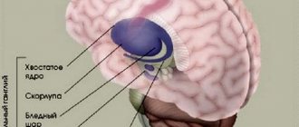
Red core. The red nucleus is a structure of the midbrain, located symmetrically in the thickness of the cerebral peduncles under the central gray matter.
It is a large accumulation of nerve cells, which, along with other formations, plays a key role in the extrapyramidal (automatic, without the participation of consciousness) system.
The red nucleus is closely connected with the structures of the cerebellum.
The neurons of this nucleus receive information from the cerebral cortex and cerebellum, that is, all information about the position of the body in space, the state of the muscular system, and skin. The influence on alpha motor neurons of the spinal cord is carried out using the rubrospinal tract.
The rubrospinal tract begins from the cells of the red nucleus located in the cortex of the cerebral peduncles. Activation of neurons in the red nucleus causes excitatory postsynaptic potentials in flexor motor neurons and inhibitory postsynaptic potentials in extensor motor neurons.
In this respect, the rubrospinal tract is similar to the corticospinal tract.
Tectal area: – reaction of a general startle (a sharp increase in the modality of any stimulus: there was nothing and suddenly a sharp impact on the sensory organ); – orienting reflex – orients towards the source of the stimulus, it is suppressed (when the stimulus is repeated, if it is insignificant, it is suppressed (we stop responding to it), suppression requires the participation of the cerebral cortex; it is based on the sentinel reflex;
Source: //studopedia.su/10_151637_trapetsievidnoe-telo-sostavleno-aksonami-vtorih-sluhovih-sensornih-neyronov-i-ih-yadrami.html
Functions
The important functional significance of the cerebral pons is due, on the one hand, to the location in it of the nuclei of the cranial nerves (V, VI, VII, VIII pairs), the reticular formation, and the pons nuclei; on the other hand, the passage of efferent pathways through the pons (corticospinal and corticonuclear, tegmental-spinal cord, red nucleus-spinal cord, reticular-spinal cord, etc.) and afferent pathways (spinothalamic, pathways of proprioceptive - deep - sensitivity, etc.), which are vital for the body and carry out two-way communication between the brain (see) and spinal cord (see).
The structure of the Varoliev formation
The formation is located on the surface of the brain. If we talk about the internal structure of the bridge, then it contains an accumulation of white matter, where the nuclei of gray matter are located. In the posterior part of the formation there are nuclei consisting of 5, 6, 7, and 8 pairs of nerves. One of the most important structures located on the bridge is the reticular formation. It performs a particularly important function; it is responsible for activating all the departments located above. The pathways are represented by thickened nerve fibers that connect the pons to the cerebellum, forming the streams of the formation itself and the cerebellar peduncles.
Saturates the Varoliev bridge artery of the vertebrobasilar region with blood. Outwardly, it looks like a roller that is attached to the brain stem. The cerebellum is attached to it on the posterior side. In its lower part there is a transition to the medulla oblongata, and from the upper part to the middle brain. The main characteristic feature of the Varoliev formation is that it contains a mass of pathways and nerve endings in the brain.
Four pairs of nerves diverge directly from the pons:
- ternary;
- abductor;
- facial;
- auditory.