7851 0
Intervertebral discs
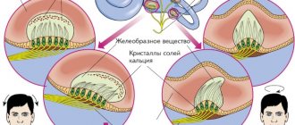
Intervertebral discs provide mobility to the spine, support it and distribute the load during movements. With age, the amount of proteoglycan in the disc core decreases, and the disc begins to dry out (loss of water). Mucoid degeneration and fibrous tissue ingrowth are observed, as well as ↓ disc space height and susceptibility to injury. Ring ruptures and nuclear protrusion occur as a result of intranuclear pressure under mechanical load.
Lumbar disc herniation
Clinical data
The posterior longitudinal ligament is strongest along the midline. The posterolateral portions of the annulus may be subject to greater stress. Therefore, most lumbar disc herniations (LDH) are observed posteriorly, somewhat to the side of the midline, compressing the nerve roots, causing characteristic radicular pain.
Various terms are used to describe the degree of disc protrusion based on sections or operating data: protrusion, sequestration, free fragment, etc.
Clinically, these differences are usually of little significance, the only difference being "limited prolapse" in which intradiscal interventions can be used. Characteristic data of the anamnesis:
1. complaints often begin with back pain, which after a few days or weeks gradually, and sometimes abruptly, turns into radicular pain with a decrease in back pain
2. it is rarely possible to identify factors that increase pain
3. pain decreases when bending the leg at the hip and knee
4. Patients usually avoid increased physical activity, however, prolonged stay in any position (sitting, standing or lying down) can increase pain, so a change in position is usually required every 10-20 minutes. This is in contrast to the severe pain observed, for example, with blockage of the ureter, when patients writhe from it
5. pain intensifies when coughing, sneezing or straining during defecation: in a series of observations66, a positive “cough shock symptom” was observed in 87% of cases
6. Urinary symptoms: The incidence of urinary disorders is 1-18%. In most cases, there is difficulty urinating or urinary retention. The earliest symptom may be decreased sensitivity of the bladder. Later, symptoms of “irritation” are often observed: increased urge, frequent urination (including nocturia), increased residual urine. Less common with radiculopathy are enuresis and urinary incontinence (note: true urinary retention may occur with cauda equina syndrome). Sometimes GPD may only present with urinary symptoms, which may improve after surgery. Laminectomy may improve bladder function, but this is not certain
Clinical manifestations of radiculopathy
Back pain as such is not a significant complaint (only 1% of patients with acute LBP have sciatica). If this is the only complaint, then other causes must be excluded. Sciatica is such a specific symptom of GPD that the likelihood of having clinically significant GPD in the absence of sciatica is only ≈1/1000. Exceptions are central hernias, which may cause symptoms of lumbar stenosis (eg, neurogenic claudication) or SCS.
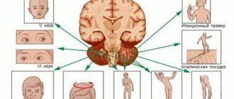
Compression of the nerve root leads to certain symptoms of varying severity. For the most commonly involved roots, characteristic syndromes have been described.
In a series of patients referred to a neurosurgeon for pain radiating to the leg, motor impairment was present in 28% (although only 12% reported muscle weakness as a complaint), sensory impairment in 45%, and reflex impairment in 51%.
The following symptoms may indicate compression of the nerve roots. The sensitivity and specificity of some symptoms in patients with sciatica are given in Table. 11-8.
Table 11-8. Sensitivity and specificity of clinical findings of lumbar disc herniation in patients with sciatica
1. complaints and symptoms of radiculopathy (see Table 11-9) A. pain radiating down the leg B. muscle weakness C. sensory disturbances along the dermatomes D. reflex disorders: mental factors can influence the symmetry of reflexes
2. positive symptoms of nerve root tension: including Lassègue's sign
Table 11-9. Lumbar disc syndromes
* the disturbance may be more noticeable when taking Jendrassik (during the test of the reflex, the patient pulls the clasped hands to the sides) † the reflex from the semimembranosus muscle is unreliable (it is not always only the L5 root), during testing the adductor muscles can also be stimulated ‡ sensory disturbances are more common observed along the lower border of the dermatome
Symptoms of nerve root tension
1. Lassegue's sign:
so-called straight leg raise test. Helps differentiate sciatica from hip pain. Methodology: the patient lies on his back, the sore leg is raised by the ankle joint until pain occurs (pain should appear at the angle
2. Crum test:
the patient lies on his back; lift the “sick” leg, slightly bending it at the knee, and then straighten it. The results are the same as when raising a straight leg
3. cross lift of a straight leg, so-called. Firestein test:
lifting the straightened “healthy” leg causes pain on the opposite side (this requires lifting to a greater angle than on the “sick” side). A more specific but less sensitive test than Lassegue's sign (97% of surgical patients who tested positive for this test had confirmed GPD). May correlate with a more central position of the hernia
4. hip stretch test or so-called. reverse straight leg raise test:
the patient lies on his stomach, the examiner's hand is on the popliteal fossa, the knee is maximally bent. The test is often positive for compression of the L2, L3 and L4 roots (ie, upper lumbar disc herniations) or for extremely lateral herniations (may also be positive for diabetic femoral neuropathy or psoas muscle hematoma). In these situations, the straight leg raise test (Lassegue's sign) is often negative (because the L5 and S1 roots are not involved)
5. bow string symptom:
After pain occurs when lifting your straightened leg, bend your knee and lower your foot onto the bed, continuing to keep your leg bent at the hip joint. With this maneuver, the pain caused by sciatica is relieved, but the pain in the hip joint remains
6. straightening the leg in a sitting position:
The patient is seated with both hips and knees flexed 90°. In this position, one knee is slowly extended. In this case, the same tension in the nerve roots occurs, as in moderate lifting of a straightened leg.
Other useful symptoms for radiculopathy
1. Patrick's test:
the ankle joint is placed on the knee of the other leg, and then the ipsilateral knee is moved towards the surface on which the patient is lying. This creates tension in the hip joint and usually does not increase true nerve root compression. The test is often positive in cases of hip involvement (eg, trochanteric bursitis) or mechanical LBP
2. Trendelenburg's sign:
the examiner looks at the pelvis from behind while the standing patient raises one leg. Normally, the pelvis should remain horizontal. If the symptom is positive, the pelvis tilts downwards on the side of the elevated leg, indicating weakness of the abductor muscles of the opposite thigh (main innervation of L5)
3. cross adductors:
When testing the knee reflex, the adductor muscles of the opposite thigh contract. With an increased ipsilateral knee reflex, this may indicate damage to the upper motor neuron (UMN), and with a decreased ipsilateral knee reflex, this may indicate pathological spread, indicating excitability of the nerve root
Nerve root syndromes
According to the data below, GPD usually does not affect the root emerging at the level of this space, but compresses the root that exits through the intervertebral foramen one level below (eg, L5-S1 GPD usually causes S1 radiculopathy). This leads to the formation of characteristic lumbar nerve root syndromes, shown in Table. 11-9.
Important anatomical facts about lumbar discs:
1. in the lumbar region, the nerve roots emerge below and close to the root of the arch corresponding to them by vertebral number
2. the intervertebral space is located below the level of the arch root
3. in the most typical structure of the human spine there are 24 presacral vertebrae, although in some cases there may be 23 or 25 A
. Therefore, the GPA of the last gap usually compresses the 25th nerve root (although, in accordance with the above individual characteristics, this may be the 24th or 26th root)
A
possible options: 11 or 13 vertebrae with ribs or lumbosacral transitional vertebra; the terms “S1 vertebral lumbarization” or “L5 vertebral sacralization” are imprecise and may be misleading
X-ray diagnostics
Conservative treatment
Surgery
Indications
Despite numerous attempts to determine which patients will improve on their own and which should be operated on more quickly, this has not yet been established.
1. ineffectiveness of conservative treatment: in more than 85% of patients with acute GPD, improvement will occur without surgery within an average of 6 weeks (in 70% within 4 weeks). Most experts recommend waiting about 5-8 weeks after the onset of radiculopathy before considering surgery (bearing in mind that there are no indications listed below)
2. “EMERGENCY INTERVENTION” (before 5-8 weeks have passed): indications: A. cauda equina syndrome (CAS) B. progressive movement disorders (eg foot drop): paresis of unknown duration is a questionable indication for surgery ( No studies have shown that patients in this situation have less severe motor deficits after surgery). However, acute onset or progression of movement disorders is considered an indication for urgent surgical decompression C. “emergency” surgery may be indicated for patients whose pain remains intolerable despite sufficient narcotic medications
3. ± patients who do not want to wait and try whether conservative treatment will help them, if there is still a question about surgery later
Greenberg. Neurosurgery
Lasègue's symptom occurs against the background of tension in the roots of the sciatic nerve, which occurs due to pinching of the nerve fiber in the spine or spasmodic contraction of the lumbar and gluteal muscles. This and some other symptoms make up a group of “tension symptoms” - pain that occurs when nerve fibers are stretched.
Pain in the projection of damaged nerve fibers occurs when lifting the leg up while the person is lying on his back. The pathogenetic mechanism for the occurrence of the symptom is due to the fact that when the lower extremities move, the sciatic nerve has to stretch, but when it is pinched, the length of the nerve fiber decreases. Against this background, when the limbs are slowly lifted upward, the sciatic nerve is “overstretched,” which is why pain occurs.
What it is
Physiologically, the sciatic nerve is normally capable of stretching to a length of up to 15 mm. It passes through the gluteal muscles and innervates the thigh and lower leg. Theoretically, its infringement can be localized throughout, but in practice compression is more common in the lumbosacral and gluteal region. Let's consider the mechanism of occurrence of Lasegue's symptom:
- In the supine position, the roots of the sciatic nerve are relaxed;
- Raising your leg up with a bent knee does not lead to pain, since the sciatic nerve is not “stretched”;
- Raising a straight leg upward in a supine position leads to pain in the lower back due to compression of the sciatic nerve.
False-positive Lasegue symptom
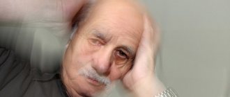
A false-positive Lasegue symptom is observed in people with reduced endurance of the muscle groups of the posterior thigh. If it is present, lifting the leg up leads to soreness of the femoral muscles. False-positive “triggering” is observed in older people. Its pathogenesis is not associated with compression of the sciatic nerve.
Neurological assessment of symptoms
Neurologists, when assessing the condition of the sciatic nerve in patients with suspected vertebrogenic lumbargia, note not only the presence of pain. What to look for when assessing Lasegue's symptom:
- The appearance of pain in the lower back when raising the lower limbs to an angle of 60 degrees raises doubts. The pain is associated with a psychological state rather than with compression of the sciatic nerve fiber;
- Pain syndrome when lifting legs above 60 degrees is reliable evidence of a decrease in the length of the sciatic nerve.
The lower limbs should not be raised above the achieved level if the patient experiences pain. The sciatic nerve can withstand loads of up to 3 kg. This pressure on it occurs when the leg is positioned above the horizontal plane at an angle of 60-65 degrees. With excessive loads, there is a risk of nerve fiber rupture!
Important rules for checking with Lasegue:
- You cannot quickly raise your lower limbs;
- The procedure is performed smoothly and stops after pain is achieved, even if the elevation angle is only 15 or 20 degrees;
- Pathological changes in the lower parts of the spinal column can be checked using magnetic resonance imaging (MRI);
- You should not check Lasegue's sm after anesthesia has been administered or painkillers have been taken. They distort the test results.
The importance of neurological diagnosis
- Almost all diseases of the spine cause muscle spasms of varying classes and degrees, and also provoke destruction of nerve roots and numbness of the damaged limb.
- In addition, quite often the symptom of tension can be an accompanying symptom of osteochondrosis and other degenerative diseases.
- In neurology, there are several common tension syndromes and each of them is tested in a certain way.
Lasègue's symptom
Almost always, with vertebrogenic pain syndrome, a muscle spasm occurs that blocks the nerve roots, and this, as a rule, causes a Lasegue tension symptom.
If, when assessing Lasègue syndrome, the patient made sudden movements, then additional examination is necessary for the presence of damaged nerve endings, since even moving the leg can break the fibers connecting the nerves to each other. Also, if the sciatic nerve is pinched (it is the largest in the entire body), bilateral Lasegue syndrome may begin to develop.
Symptoms of Lasègue:
- increased pain when raising the injured limb;
- pulsation in the leg.
Definition and treatment:
- first of all, the doctor determines the maximum angle of leg elevation;
- further, the leg bends at the knee joint as much as possible (in this case, the pain disappears or decreases significantly).
The doctor prescribes a treatment complex consisting of therapeutic massage and other physiological procedures.
Landing symptom
Planting syndrome allows you to fully study the nature of the pathological pain that has arisen in the spinal region and determine the condition of the nerve roots. A similar diagnosis occurs as follows:
- during the initial examination, the patient is asked to sit down and is in a semi-recumbent position (with legs fully extended);
- If a patient has tension pain syndrome, he will not be able to remain in this state for a long time due to exceeding the pain threshold and muscle contraction in the knee joint.
- The doctor probes possible affected areas until the patient experiences reflex spasms.
- The landing syndrome is of great importance for the diagnosis of neurological diseases of the peripheral nerves.
- For example, Lasegue syndrome often manifests itself as lumboischialgia (a disease whose symptoms are very similar to tension syndrome), but if the patient can sit quietly, then this disease should be excluded.
- When examining patients with complaints of verterobrogenic pain syndrome, practitioners very carefully study the characteristic clinical signs of the disease, which make it possible to distinguish back pain caused by damage to nervous, bone and muscle tissue from psychogenic pain caused by the characteristics of a person’s psycho-emotional state.
- One of the most specific signs that help distinguish between types of pain in the spine is the so-called tripod symptom.
- When examining a patient with true pain in the spinal column, an experienced vertebrologist pays special attention to:
- the position of the patient in bed - he can only sit, leaning with both hands behind his back and not allowing movement;
- getting out of bed - the patient tries not to strain the back muscles as much as possible;
- the position of the person on a chair - to reduce pain and unload the muscles, the patient tries to tilt the body back, leans on his hands.
The appearance of such a patient is quite characteristic, which is why this situation in patients with true vertebral syndrome is called the tripod symptom. Very often, this sign is detected in people who have undergone surgery on the spinal column with the installation of a metal structure.
Other tension syndromes
Symptoms of tension also include:
- Bonnet reaction . If a patient, during a preliminary neurological examination, cannot contract and spread his leg, this may mean that he has Bonnet’s symptom, which is characterized not only by an increase in the pain threshold, but also by numbness of the limbs. Painful sensations are localized only in the damaged area.
- Wasserman-Matskevich symptom . With strong muscle tension, the patient may experience Wasserman and Matskevich syndromes. They appear when there is damage to the peripheral nervous system or spinal nerves.
- Neri's symptom manifests itself when the dorsal roots of the spinal cord are irritated, developing as a result of osteochondrosis of the spine with bone osteophytes, disc herniations, tumors of the roots, which leads to unpleasant pain.
- Dejerine syndrome is a symptom of tension that is neuralgic in nature, and that is why when diagnosing the doctor pays attention to the smallest details. The pain is localized in the spinal region and appears only during physical activity. Manifests itself due to damage to the spinal cord column.
Provoking diseases
If, after carrying out all the necessary diagnostic measures, one or another tension syndrome was identified, then this may indicate the first stages of the following diseases:
- Spinal osteochondrosis is a fairly common disease that occurs due to pinched nerves between the intervertebral discs. This clogging is characterized by numbness of the damaged area, an increase in the pain threshold and shooting in the buttock area.
- Pinched sciatic nerve - pain spreads throughout the nerve (a similar syndrome is called sciatica in the medical literature).
- Ankylosing spondylitis is characterized by pain and deposits of salt and calcium throughout the spinal region.
- Radiculitis of the lumbosacral region is a pronounced disease that is manifested by the following symptoms: symptoms of tension in the nerve roots, sacral syndrome (when lifting a healthy limb, pain is radiated into it), pinched nerve and Neri syndrome, in which pain spreads throughout the body.
In addition, the symptom of tension is also an accompanying symptom of diseases such as intervertebral hernia, physiological deformation of muscle tissue, joint damage and tumors.
In order to prevent the development of severe pathologies, timely diagnosis and treatment are necessary. This can be done by a neurologist who, after carrying out special diagnostic measures, can identify a neurological disease.
Reasons for appearance
The most common cause of the pathological symptom of sciatic nerve tension is a herniated intervertebral disc in the lumbosacral spine of grades 2 and 3.
With severe prolapse of the disc into the spinal cord at the L5-S1 level, the patient experiences pain along the thigh and lower leg due to vertebrogenic lumbargia, so assessment of tension symptoms does not supplement the clinical status with important information. If such symptoms are present, an x-ray of the lumbar spine is required. If possible, an MRI of the lumbosacral spine is performed.
Other diseases in which tension of the sciatic nerve is observed:
- Piriformis syndrome - the nerve root is compressed not at the level of the spine, but in the gluteal region;
- Osteochondrosis of the lumbosacral spine;
- Bechterew's disease – calcification (deposition of calcium salts) of the ligamentous apparatus of the spinal column;
- Compression of the lower part of the spinal cord (cauda equina syndrome).
If pathological symptoms of tension are detected, the neurologist must refer the patient for additional examinations: radiography of the spine, magnetic resonance imaging (MRI) and computed tomography (CT). They are carried out to determine the cause of the disease and establish a reliable diagnosis.
Video showing Lasegue's symptom:
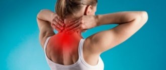
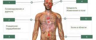
Pain in the lumbar region is a fairly common occurrence. It can be caused by quite a variety of conditions. Pain in the lumbar region is recorded in the following pathologies:
- vertebrogenic and discogenic lesions of nerve roots;
- renal colic;
- pancreatic diseases;
- pathological changes in vertebral bone tissue.
The symptoms described by various authors, indicating pathology of the spinal disc segment, help to establish the cause of the pain syndrome, diagnose damage to the nerve roots and clarify the severity of the disease.
The most frequently studied phenomenon, which has diagnostic significance and also plays a role in determining the patient’s ability to work, is Lasegue’s symptom.
Provoking diseases
If, after carrying out all the necessary diagnostic measures, one or another tension syndrome was identified, then this may indicate the first stages of the following diseases:
- Spinal osteochondrosis is a fairly common disease that occurs due to pinched nerves between the intervertebral discs. This clogging is characterized by numbness of the damaged area, an increase in the pain threshold and shooting in the buttock area.
- Pinched sciatic nerve - pain spreads throughout the nerve (a similar syndrome is called sciatica in the medical literature).
- Ankylosing spondylitis is characterized by pain and deposits of salt and calcium throughout the spinal region.
- Radiculitis of the lumbosacral region is a pronounced disease that is manifested by the following symptoms: symptoms of tension in the nerve roots, sacral syndrome (when lifting a healthy limb, pain is radiated into it), pinched nerve and Neri syndrome, in which pain spreads throughout the body.
In addition, the symptom of tension is also an accompanying symptom of diseases such as intervertebral hernia, physiological deformation of muscle tissue, joint damage and tumors.
In order to prevent the development of severe pathologies, timely diagnosis and treatment are necessary. This can be done by a neurologist who, after carrying out special diagnostic measures, can identify a neurological disease.
Mechanism of occurrence
In a normal state, in a healthy person, the nerve root is located freely in the intervertebral foramen, without tension and without causing pain when flexing the hip (except for some discomfort at extreme degrees of flexion).
Stretching of a nerve root becomes painful to one degree or another when it is pinched in the intervertebral foramen, as well as when it is stretched due to the convexity of a herniated intervertebral disc. In these situations, any strain while slowly lifting the lower limb will be painful.
A positive Lasègue symptom occurs when pain occurs when raising the hip less than 60 degrees. The appearance of discomfort or pain at an angle of more than 60 degrees may be a false-positive Lasegue symptom. This situation can arise even when examining a healthy person who leads a sedentary lifestyle or has reduced flexibility.
How is Lasegue's symptom caused?
The symptom manifests itself when the lower leg is extended in a patient lying on his back with the hip flexed. The examination should be carried out carefully, without making sudden movements, since pain with a positive Lasègue symptom can be of high intensity. Lasègue's symptom is positive if pain occurs in the lumbar region, along the back of the thigh and lower leg of the corresponding side. Soreness is usually localized on the side of the affected nerve root. For example, Lasegue's sign on the right is caused by flexion of the right leg.
- The first phase of Lasegue's symptom: the occurrence or appearance of pain in a patient lying on his back when trying to bend or flex an extended leg at the hip joint.
- The second phase of Lasegue's symptom: if you subsequently bend the leg at the knee without straightening the hip joint, then the pain syndrome either disappears or its intensity sharply decreases.
An important point: it is not allowed to cause the Lasegue tension symptom after the use of anesthesia, since the protective pain reflex in such conditions is significantly reduced; manipulation in this case can lead to tearing of axons and cause motor disorders (up to paresis).
Symptoms of tension in cervical osteochondrosis
Osteochondrosis of the cervical spine also gives symptoms of tension, which manifest themselves in the collar area, upper chest, and shoulders. Determining the symptoms of tension in cervical osteochondrosis is quite simple - for this, the person needs to be asked to raise or move their arm to the side, and tilt their head.
For example, with positive Bonnet tension syndrome, the patient cannot move the arm raised to shoulder level to the side and bring it back. In this case, the upper limb on the side where there is no damage to the radicular nerve moves freely in different directions. In Bonnet syndrome, sensory disturbances are also present. Local areas of decreased skin sensitivity are identified along the back surface of the shoulder and forearm. When palpating the cervical spine, shoulder, collar area and upper limb, pain is detected only at the site of localization of degenerative dystrophic destruction of the intervertebral disc.
The Wasserman-Matskevich symptom is a combination of two clinical signs that appear with severe muscle tension in the neck and collar area. To determine them, the patient is asked to lie on his back on a hard surface. Then he should take turns raising his arms up in front of him and abducting them in different directions at different angles. With positive tension syndrome, raising your arm up in front of you will be accompanied by severe pain in the area of the shoulder blade and forearm.
Neri's tension syndrome may indicate inflammation or compression of the dorsal roots of the spinal cord. To determine it, the patient must stand up straight with his legs straight. Then the doctor asks him to tilt his head as far as possible towards his chest so that his chin touches his chest. If it is impossible to do this on their own, the doctor helps the patient until the chin touches the sternum or until the patient says that he has severe pain in the lumbar or collar area. This tension syndrome is present with radiculitis, myelitis of the roots, herniated intervertebral disc in the cervical spine.
Neri's tension syndrome can be positive when osteophytes grow in the area of the cervical and cervicothoracic spine, osteochondrosis with radicular syndrome, displacement of vertebral bodies, destruction of intervertebral joints with compression of the roots, etc.
Also, with cervical osteochondrosis, Dejerine's syndrome is of great diagnostic importance. It indicates damage to the membranes of the spinal cord. If a person experiences pain only during a certain type of physical activity, for example, when turning the head to the side, then this indicates a positive symptom of Dejerine’s tension.
Interpretation of Lasegue's symptom
In the presence of a pathological process localized in the area of the nerve root, pain may appear when the hip is flexed at different angles (10, 15, 20, 30 degrees). The level at which Lasegue's symptom occurs allows us to judge the severity of the patient's condition and is of great importance for diagnosing the stage of the disease.
In order to track the dynamics of the clinical manifestations of a radicular lesion, it is advisable, when recording the examination results, to indicate the approximate angle at which the patient experiences pain (an example of such a recording: Lasegue’s symptom on the right is positive, 30 degrees). This will make it possible in the future, during repeated examinations, to compare the current result with the previous one and, based on the comparison results, make a judgment about the dynamics of the patient’s condition. In particular, an increase in the angle between the plane on which the patient lies and his thigh will indicate a gradual improvement in the patient’s condition.
Other diagnostic signs accompanying Lasegue's symptom
- Neri - the occurrence of pain in the lumbar region when the head is flexed.
- Matskevich's symptom is pain in the groin area and the anterior surface of the thigh when the patient bends the lower leg while lying on his stomach.
- Dejerine's symptom is increased pain when coughing or sneezing.
- Bekhterev's symptom (in a number of sources referred to as Lasegue's cross symptom) - when a lying patient sits down, his leg bends at the knee joint on the side of the nerve root lesion. If you straighten this leg, the healthy one will bend. In cases where a positive Lasègue symptom is recorded, ankylosing spondylitis' symptom is usually also positive.
- Lerrey's phenomenon - a pronounced increase in pain in the lumbar region in the event of a sharp transition from a lying position to a sitting position - is almost always a symptom accompanying the Lasègue sign.
Tactics for identifying a positive Lasègue phenomenon
The symptom indicates an acute course of the disease. A patient whose examination reveals this sign is disabled (for at least 10 days).
- The first step is to relieve pain (the use of NSAIDs is optimal; in case of severe pain, drug blockades can be used).
The patient must be provided with bed rest for the duration of the acute pain syndrome (this is especially important in case of violations of the integrity of the fibrous ring).
An integrated approach is required: the treatment regimen includes NSAIDs, muscle relaxants, physiotherapy, and, as the exacerbation progresses, exercise therapy.
To avoid relapses, a patient with discogenic disorders may be advised to wear a lumbosacral corset in situations involving stress on the lumbar spine.
Lasegue's symptom is one of the syndromes of tension of the iliac muscles, used in neurology to assess the condition of the spinal nerves in pathology of the spinal column.
Spastic contractions of the skeletal muscles of the back appear when the nerve roots between the vertebrae or muscle “clamps” are pinched. They are important when assessing the prognosis for intervertebral hernias, which can lead to disability.
Lasègue's sign is assessed by lifting the legs of a person who is lying on their back. The procedure is performed slowly, until the first pain appears.
The symptom is positive if pain occurs when raising the leg to an angle of 30 degrees. Further continuation of the manipulation will lead to a sharp increase in pain, since the nerve located in the thickness of the spasmodic iliacus muscle is strongly compressed. It is impossible to bring a patient to such a state! At the first sign of pain, you should stop performing the test.
The specificity of Lasegue is the cessation of pain when bending the knee or hip joint. This symptom appears due to relaxation of the L5 or S1 roots, pinched by the paravertebral muscles.
Due to the existence of several specific manifestations when assessing the condition of spastic muscles, some doctors separately distinguish Lasegue syndrome, which includes the following symptoms:
- Pain when raising the leg;
- Cessation of pain when flexing the hip and knee joints;
- Numbness of the skin of the anterior thigh when performing the test;
- If pain appears when lifting the leg more than 70 degrees, it is formed against the background of compression of the root, and not the nerve fiber;
- Radicular pain syndrome is confirmed by raising the leg to the maximum limit and then quickly flexing the ankle joint. In this case, pain should spread throughout the entire leg;
- When you raise your healthy leg while lying on your back, pain should appear in the affected limb - Lasegue's cross symptom.
For a qualified assessment of the condition of the spinal nerves, it is not enough to know what Lasegue tension syndrome is, because there are additional confirmatory tests: Neri (), Wasserman (). They help more accurately confirm the doctor’s assumption, since there is a subjective assessment of pain by each patient and individual thresholds of nerve sensitivity.
How do we treat ankylosing spondylitis?
Non-steroidal anti-inflammatory drugs are used as basic therapy for ankylosing spondylitis. To significantly slow down the development of the disease, it is recommended to use them in long courses.
We examine the patient for various infections and, if detected, treat them. This helps prevent exacerbations of the disease.
Hirudotherapy (leech treatment) is also successfully used in the treatment of this disease. The enzymes contained in the saliva of leeches can have a complex effect on the disease: they “soften” and make the spine more flexible, increase immunity and have an anti-inflammatory effect.
Kinesiotherapy, a unique set of exercises for the spine and joints, also helps prevent ankylosis of the spine. This complex was developed by our specialists. It is therapeutic and educational. During individual or group sessions, patients learn to perform exercises on their own so that they can practice at home.
We also use massage, acupuncture, bee therapy and other methods, which can be found here.