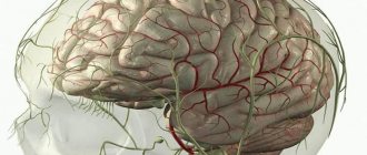
Whatever this phenomenon is called: symptoms of irritation of the meninges (meningeal symptom complex) or meningeal signs, we are always talking about the same process in the brain or spinal cord - the process of intoxication with a disorder in the course of biochemical reactions in the tissues of the higher structures of the nervous system .
And it doesn’t matter at all what agent the intoxication is caused by:
- whether the activity of one’s own chronic microbial infection, “dormant”, but suddenly “awakened” from many years of hibernation at the right moment;
- the “import” of some super-new virus;
- whether a toxic chemical (technical or household) product or allergen has entered the bloodstream.
Because the symptoms will be approximately the same in all cases.
And even in the case of mechanical pressure on the meninges, during hemorrhages in one or another part of the brain, toxic products are formed from the destruction of one’s own tissues, leading to a disorder in the fine tuning of nervous processes. To diagnose neurological pathology, there are many research methods - both clinical, instrumental and laboratory. In turn, among the clinical ones, there are several of the most reliable symptoms of irritation of the meninges, “tested” to brilliance by centuries of medical practice.
The classic syndrome-reflexes of the symptom complex bear the names of the “fathers” of medical science and neurology who discovered them for the first time many years ago: Kernig, Brudzinsky, Gordon and others.
General principles and mechanisms
The essence of the process is that in response to a chemical attack on the nerve structures located in the meninges, a reflex arc closes, accompanied by a muscle reaction from both smooth muscles and skeletal muscles.
The response from the latter is especially strong and rudely noticeable, and therefore simply cannot be ignored. Against the background of the obligatory headaches, excessive sensitivity to sound and light, nausea and vomiting, general weakness and muscle pain, as well as fever and confusion (symptoms that are not at all necessary for meningitis) - phenomena aggravated by tapping on the skull, spine, as well as touching the body, opisthotonus is also inevitably formed.
This is the name of the classic pose, found and adopted by the patient to relieve suffering caused by edematous and inflammatory changes in the affected meninges.
Opisthotonus is a body lying on its side with the head thrown back sharply to the limit, with the legs folded like a penknife and drawn to the stomach and chest, with the arms folded and pressed to the body in a similar way.
Its appearance means that the time has come to identify pathological reflexes.
Diagnostics
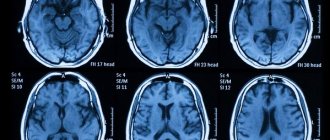
Computed tomography of the brain is one of the ways to diagnose meningeal syndrome
Meningeal syndrome is only a preliminary diagnosis. Further actions of the doctor are reduced to identifying the root cause. For this purpose, instrumental and laboratory techniques are used.
First of all, the patient is sent for a lumbar puncture to obtain cerebrospinal fluid for testing. Analysis of cerebrospinal fluid can reveal signs of inflammation, as well as the causative agent of infection. If there are no changes in the cerebrospinal fluid (bacteria are not detected, and the composition is within normal limits), the patient is diagnosed with meningism, and further diagnosis is reduced to the search for conditions that can provoke the development of such symptoms.
To detect an inflammatory process in the body, a routine clinical blood test can be used. In this case, an increase in ESR and an increase in the level of leukocytes indicates the presence of inflammation.
To perform differential diagnosis, the following techniques are used:
- Magnetic resonance imaging.
- CT scan.
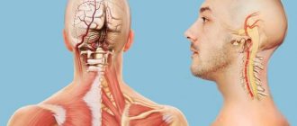
Brudzinski's signs and neck stiffness
To assess the degree of rigidity of the nuchal muscles, the diagnostic neurologist's hand is placed under the back of the patient's head and the patient's neck is flexed in order to reach the surface of the chest with his chin; the test result is assessed by the distance “traveled” by the head to the specified body level.
This test may not be reliable enough when performed on old people - they have stiff neck muscles due to age, as well as in children.
In addition to neck rigidity - reflexive resistance exerted by tension-bound cervical and occipital muscles to an attempt to bend the patient's head to the chest, the meningeal symptom complex is manifested by Brudzinski's syndromes.
There are four Brudzinski symptoms:
- A positive upper occipital symptom is manifested by flexion of the lower extremities at the knee and hip joints with their adduction to the body with an effort to bend the neck and tilt the patient’s head forward.
- Brudzinski's symptom, middle pubis , is manifested by adduction to the body and flexion of both lower extremities in both the hip and knee joints when pressing on the pubic area.
- The contralateral (literal translation: on the other side) or lower version of the symptom is a reflex of flexion of the second limb when the thigh of his leg is brought to the patient’s stomach, bent by the diagnostician at the knee joint at a right angle (not completely).
- With the buccal version of the symptom, pressing on the cheek under the zygomatic arch causes a reflex lifting of both shoulders and folding and bending of both the patient’s arms at the elbow joints. In the version with tapping on the zygomatic arch, a reflex involuntary bending of the legs at the knees occurs.
Meningitis. Reasons for development. Symptoms. Diagnostics. Treatment.
Meningitis is an inflammation of the lining of the brain and spinal cord. Inflammation of the dura mater is called pachymeningitis
, and inflammation of the soft and arachnoid meninges -
leptomeningitis
.
In the clinic, inflammation of the soft meninges is most common and the term “meningitis” is used. Its causative agents can be various pathogenic microorganisms: viruses, bacteria, protozoa.
Classification
Based on the nature of the inflammatory process in the membranes and changes in the cerebrospinal fluid, serous and purulent meningitis are distinguished. In serous meningitis, lymphocytes predominate in the cerebrospinal fluid; in purulent meningitis, neutrophils predominate. According to pathogenesis, meningitis is divided into primary and secondary. Primary meningitis develops without a previous general infection or infectious disease of any organ, and secondary meningitis is a complication of an infectious disease (general or local). When the process is widespread in the meninges, generalized and limited meningitis are distinguished (for example, at the base of the brain - basal meningitis, on the convex surface of the cerebral hemispheres - convexital meningitis). Depending on the rate of onset and course of the disease, the following are distinguished: fulminant, acute (sluggish) meningitis, subacute and chronic; and according to the severity of the clinical picture - mild, moderate, severe and extremely severe forms. According to the etiology, there are bacterial (meningo-, pneumo-, staphylococcal, tuberculosis, etc.), viral (acute lymphocytic choriomeningitis caused by enteroviruses Coxsackie and ECHO, mumps, etc.), fungal (candidiasis, torulosis, etc.) and protozoal meningitis (toxoplasmosis, malaria, etc.).
Pathogenesis
There are several ways of infection of the meninges:
- hematogenous, lymphogenous, perineural, transplacental;
- contact - the spread of pathogens to the meninges with an existing purulent infection of the paranasal sinuses (sinus meningitis), the middle ear or mastoid process (otogenic meningitis), the upper jaw with pathology of the teeth (odontogenic meningitis), the eyeball, etc.
- with open craniocerebral and spinal cord injuries, with fractures and cracks of the base of the skull, accompanied by liquorrhea.
Meningitis may be the main or only manifestation of bacteremia. The entrance gates for infectious agents during meningitis are the mucous membrane of the nasopharynx (with the occurrence of nasopharyngitis, sore throat), bronchi (with bronchitis), and the gastrointestinal tract with disorders of its function and subsequent hematogenous or lymphogenous spread of the pathogen to the meninges. The pathogenetic mechanisms of clinical manifestations of meningitis include inflammation and swelling of the meninges, as well as adjacent brain tissue, microcirculation disorders in the meningeal and cerebral vessels, hypersecretion of cerebrospinal fluid and slower resorption, which leads to the development of cerebral hydrocele and increased intracranial pressure. Irritation of the sensitive receptors of the meninges and the perforating membranes of the roots of the cranial and spinal nerves also have their own characteristics of clinical manifestations.
Pathomorphology
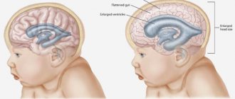
Pathological changes in acute purulent meningitis do not depend on the pathogen. When a microorganism penetrates the meninges through the blood or lymph flow, their inflammation quickly and diffusely spreads to the entire subarachnoid space of the brain and spinal cord. For example, with pneumococcal meningitis, the subarachnoid space is filled with green-yellow purulent exudate. In a localized area of infection, purulent inflammation may be more limited. Swelling of the membranes and substance of the brain is observed. The cortical veins are filled with blood. The cerebral convolutions are sometimes flattened due to internal hydrocephalus. Microscopically, inflammatory infiltration is detected in the pia mater, in the early stages consisting of polynuclear cells, and then lymphocytes and plasma cells are also detected. Intrinsic hydrocephalus is most often caused by inflammatory adhesion of the cerebellomedullary cistern, which impedes the flow of cerebrospinal fluid. With serous viral meningitis, there is swelling of the membranes and substance of the brain, expansion of the cerebrospinal fluid spaces.
Clinical picture and diagnosis
The symptoms of all forms of acute meningitis are very similar, regardless of etiology. The diagnosis of meningitis is made based on a combination of three syndromes:
- general infectious disease;
- meningeal (meningeal);
- inflammatory changes in the cerebrospinal fluid.
The presence of one of them does not allow reliably diagnosing meningitis. For example, meningeal symptoms can be caused by irritation of the membranes without inflammation (meningism). An increase in the number of cells in the cerebrospinal fluid may be due to a reaction of the membranes to a tumor or bleeding. The diagnosis is clarified on the basis of a visual examination of the cerebrospinal fluid, as well as bacteriological, virological and other methods for diagnosing infectious diseases, taking into account the epidemiological situation and the characteristics of the clinical picture.
General infectious symptoms include chills, fever, usually increased temperature, inflammatory changes in the peripheral blood (leukocytosis, increased ESR, etc.), and sometimes skin rashes. The heart rate may be slow early on, but as the disease progresses, tachycardia appears. Breathing becomes faster and its rhythm is disrupted.
Meningeal syndrome includes headache, nausea, vomiting, general hyperesthesia of the skin, photophobia, meningeal posture, rigidity of the neck muscles, Kernig's symptoms, Brudzinski's, zygomatic Bechterew's symptom, etc. The initial symptom is headache, which increases in intensity. It is caused by irritation of pain receptors in the meninges and their vessels due to the inflammatory process, the action of a toxin and irritation of baroreceptors as a result of increased intracranial pressure. The headache is intense and has a bursting, tearing character. It can be diffuse or localized more in the frontal and occipital regions, radiating to the neck and along the spine, sometimes spreading to the limbs. Already at an early stage, nausea and vomiting may be observed, not associated with food intake, occurring against the background of increased headaches. Children often, and less often adults, develop seizures. Psychomotor agitation, delirium and hallucinations are possible, but as the disease progresses, drowsiness and stupor develop, which can then progress to coma.
Meningeal symptoms are manifested by reflex muscle tension due to irritation of the meninges. The most common symptoms are neck stiffness and Kernig's sign. In severe cases of meningitis, the head is thrown back, the stomach is retracted, the anterior abdominal wall is tense, the legs are brought to the stomach, and opisthotonus is detected (meningeal posture of the patient). Trismus, zygomatic ankylosing spondylitis (local pain when tapping along the zygomatic arch), pain in the eyeballs when pressing and eye movements, skin hyperesthesia, increased sensitivity to noise, loud conversation, odors, Brudzinski's symptom (upper and lower) are often observed. Patients prefer to lie motionless with their eyes closed in a darkened room.
In infants, tension and protrusion of the fontanel are observed, a symptom of Lesage’s “suspension”.
Venous hyperemia and papilledema may be detected in the fundus. In severe cases of the disease, the pupils are usually dilated, and sometimes there is strabismus and diplopia. Difficulty in swallowing, paresis and paralysis of the limbs with muscle hypotonia, Babinski's sign, incoordination of movements and tremor indicate damage not only to the membranes, but also to the substance of the brain, which is observed in the final stage of the disease. Control of the pelvic sphincters is impaired late, but severe mental disorders can contribute to the development of urinary retention or incontinence.
Lumbar puncture should be performed in all patients with signs of meningeal irritation. With meningitis, cerebrospinal fluid pressure is often increased. Low pressure occurs when there is obstruction of the cerebrospinal fluid pathways, usually in the area of the base of the skull.
Kernig reflexes
Increased irritation of the meninges also occurs when checking Kernigie's sign, which is performed in two stages.
After the patient is laid on his back, in the first stage, the neurologist researcher bends the patient's leg at a right angle at both the hip and knee joints.
Then the patient is asked to attempt to straighten the leg at the knee, which causes a reaction of sharp resistance from the muscles that flex the lower leg. A sharp exacerbation of pain in a patient is noticeable to the researcher even when the patient is in an unconscious state - in this case, the second phase of the test is performed by the examining doctor.
In addition to irritation of the meninges due to meningitis, the Kernig sign can be positive and:
- with excessive intracranial pressure;
- with a skull injury;
- in the presence of a hematoma in the brain tissue.
At the same time, it can also be negative, as in the case of hemiparesis with an increase or decrease in muscle tone, or in another neurological pathology, Parkinson's disease, for example.
Features of treatment
Therapy is carried out in a hospital setting. Depending on the clinical manifestations and etiology of the disease, the following treatment directions are possible:
- Etiotropic. If the disease is bacterial in nature, then the patient is prescribed antibiotic therapy using a wide range of medications. If the cause of the disease is a virus, then antiviral drugs are used. In case of fungal infection, antimycotics are used.
- Symptomatic. Involves relief of symptoms. In the presence of hyperthermia, the patient may be prescribed antipyretics, and in case of severe vomiting, antiemetic drugs. If the patient has arterial hypertension, then he is prescribed antihypertensive medications. In case of development of epileptic paroxysm, the patient is administered anticonvulsants.
- Decongestant therapy. It is carried out to prevent cerebral edema. Involves the use of glucocorticosteroids and diuretics.
Other symptoms of meningeal irritation
If necessary, also take:
- meningeal test is a reaction in the form of an involuntary contraction of the quadriceps femoris muscle (m. guadriceps femoris) of the second leg, as a response to compression of the body of the quadriceps muscle of the opposite leg. It is pathognomonic for meningitis; a healthy person does not have this reflex.
- Diagnostically indicative and valuable is also checking Gordon's symptom , manifested by extension - or dorsal flexion-extension of the big (I) toe when the examining doctor squeezes the muscles of the patient's lower leg.
- In addition to reflex muscle - flexor and extensor reactions, increased painful pain when the patient is conscious is also achieved by pressing on Kehrer's points . In the meningeal condition, both trigeminal points (based on the name of the points of exit under the skin of the branches of the trigeminal nerve, these are supraorbital, infraorbital and mental, respectively) and occipital - occipital points have diagnostic significance. With massive, bilateral diffuse damage to the meninges, the pain will intensify when pressing on the indicated points on both sides of the body, with unilateral damage - only on one side.
Other milestones on the path to diagnosis
In addition to the classic ones, there are other signs and symptoms of irritated meninges, such as:
- Lobzin's test - increased pain when pressure is applied to the wall of the external auditory canal;
- Flatau phenomenon - when the head is tilted forward, the patient’s pupils dilate;
- Bekhterev's test : with percussion of the zygomatic arch, the headache intensifies and a reflex contraction of the facial muscles occurs;
- Pulatovsky, or craniofacial reflex - the appearance of a grimace of pain even with the most gentle percussion of the skull.
As for children, the most significant are:
- Lesage's symptom (or hanging symptom) - a child with meningitis, raised by the armpits, pulls his legs to his stomach, holding them in this position until he is lowered;
- a symptom called “the sound of a cracked pot ,” which occurs when a large fontanelle is percussed in infants, as well as its painful tension and causeless bulging.