Principles of conservative treatment of traumatic brain injury
Conservative treatment of the acute period of traumatic brain injury is pathogenetic. There are two stages in the treatment of closed craniocerebral injury.
At the first stage, in case of impaired consciousness, especially for persons in a state of alcoholic intoxication, it is necessary to administer analeptic mixtures: 2 ml of 20% caffeine and 25% cordiamine subcutaneously or 10% sulfocamphocaine 2 ml subcutaneously (intramuscular or slow intravenous).
In cases of development of intracranial hypotension, manifested by an increase in stupor, severity of neurological focal symptoms, tachycardia, decreased arterial and cerebrospinal pressure, 500-1000 ml of 5% glucose, distilled water in a dose of 10 ml 2 times a day, hydrocortisone 100 mg per 500 should be administered intravenously. ml of physiological solution 2-3 times a day intravenously. Up to 40 ml of polyglucin or rheopolyglucin can be administered intravenously. Additionally, use 1 ml of 1% mesatone, 1% fethanol or subcutaneous 5% ephedrine. It is also advisable to administer a mixture of 40% glucose (100 ml), 10 units of insulin, 100 mg of cocarboxylase, 0.06% corglucon (0.5 ml), 5% ascorbic acid (6 ml).
For high blood pressure, ganglion blockers are used: 5% pentamine or 2.5% benzohexonium is injected intravenously, 0.5-1 ml per 50 ml of saline until blood pressure decreases by 20-30%. This can be supplemented with intravenous administration of 5-10 ml of 2.4% aminophylline.
In the fight against increasing cerebral edema, diuretics and glucocorticoid hormones are administered. Already at the prehospital stage, 2 ml of 1% Lasix in 20 ml of 40% glucose is used intravenously or 50 mg of Uregit in 100 ml of 5% glucose. It is recommended to use 15% mannitol (mannitol) at a dose of 1-1.5 g per 1 kg of patient body weight. In severe cases, glucocorticoid hormones should be administered intravenously: 8-12 mg of dexazone or 40-80 mg of methylprednisolone in 200 ml of 5% glucose. After 6-8 hours, they switch to intramuscular administration of one of the drugs in smaller doses (4 mg of dexazone or 40 mg of methylprednisolone).
If there is psychomotor agitation or convulsive syndrome, it is necessary to inject 2-4 ml of seduxen intravenously; if there is no effect, repeat the injection after 20 minutes. For the same purpose, use an intramuscular mixture of 2 ml of 2.5% aminazine, 1% diphenhydramine, 0.5% seduxen and 50% analgin or 2 ml of droperidol with fentacyl. In the case of convulsive syndrome during a traumatic illness or registration of epileptic activity on the EEG, longer anticonvulsant therapy is indicated. Depending on the form and frequency of paroxysms, phenobarbital, diphenine, benzonal, finlepsin, chloracon, etc. are used. A control EEG is carried out after 6 months. treatment.
Diagnosis of focal brain injury
- Blood pressure measurement.
- Lumbar puncture (increased cerebrospinal fluid pressure up to 200 mm water column).
- X-ray of the skull (linear fracture of the bones of the calvarium).
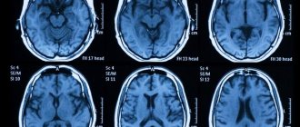
- Computer and magnetic resonance imaging of the brain (presence of a hypodense lesion corresponding to local edema; linear fractures of the bones of the calvarium).
Differential diagnosis: other traumatic brain injuries.
Treatment of mild cervical trauma
The basis of therapy for mild brain injury is desensitizing drugs (diphenhydramine, tavegil, pipolfen, calcium preparations) and vasoregulating drugs. Of the vasoregulators, Cavinton 2 ml (10 mg) intravenously 1-2 times a day per 200 ml of saline has a good therapeutic effect. You can also use aminophylline, halidor, papaverine. Drugs that improve microcirculation are used (curantil 0.05 mg, 1 tablet 3 times a day, trental OD mg, 1 tablet 3 times a day, prodectin 0.25 mg, 1 tablet 3 times a day), venotonic agents (anavenol 20 drops 3 times a day, aescusan 15 drops 3 times a day orally), as well as diuretics (diacarb, triampur, veroshpiron) in average therapeutic doses. According to appropriate indications, symptomatic therapy is carried out with analgesics (acetylsalicylic acid, amidopyrine, baralgin, analgin, pentalgin, etc.), tranquilizers (seduxen, tazepam, mebicar, elenium, eunoctin). Increased excitability of the autonomic nervous system is reduced with bellataminal, belloid, phenibut, butyroxane. Vitamin therapy, glutamic acid, nootropil, aminalon, encephabol are prescribed.
Treatment of mild brain contusion
Treatment of severe brain contusion is aimed at correcting vascular and metabolic disorders, combating increasing hypoxia, cerebral edema, hemorrhagic syndrome and preventing complications. At the earliest stage, means of protecting the brain from hypoxia are used. 20% sodium hydroxybutyrate is administered - 20 ml in 200 ml of 5% glucose, to prevent hypokalemia, also 10% potassium chloride - 10 ml or panangin (asparkam) 10 ml intravenously. In parallel, a neurovegetative blockade is carried out, which includes: 2.5% aminazine, 0.5% seduxen solution, 1 ml intramuscularly after 4 hours. In case of arterial hypertension, ganglion blockers are included in the mixture or 100 ml of 0.25% novocaine is administered intravenously. The initial period of treatment can be carried out under light barbiturate anesthesia (sodium thiopental, hexenal, etc.). This increases the brain's resistance to hypoxia, reduces its energy needs and delays lipolysis processes, preventing metabolic disorders. Against the background of dehydration therapy, 400 ml of a glucose-insulin-potassium mixture of reopolyglucin, reogluman or hemodez can be administered.
Treatment of hemorrhagic syndrome
Hemorrhagic syndrome is relieved by the following means: 10% calcium chloride - 10 ml intravenously, 1% vikasol - 1 ml intramuscularly, ascorbic acid - 2 ml intravenously or intramuscularly. For the same purpose, proteinase inhibitors are used - trasylol (or contrical) 25 thousand units drip in saline after 12 hours, or 5% aminocaproic acid - 100 ml intravenously, drip after 6 hours. In case of massive subarachnoid hemorrhages, repeated lumbar examinations are performed together with neurosurgeons punctures with active washing of the cerebrospinal fluid spaces with saline solution or installing cerebrospinal fluid drainage with the removal of 200-300 ml of cerebrospinal fluid during the day. This speeds up its sanitation and serves to prevent the development of aseptic arachnoiditis.
To improve microcirculation and prevent thrombosis, in the absence of hemorrhagic syndrome, heparin is administered subcutaneously - 2-3 thousand units every 8 hours. In the acute period (up to 1 month) to prevent infectious complications (pneumonia, pyelonephritis) in average therapeutic doses, broad-spectrum antibiotics are used actions: erythromycin, oletethrin, ceporin, etc. If swallowing is impaired in a comatose state, one should not forget about parenteral nutrition. The loss of protein is compensated by the introduction of hydrolysine or aminopeptide through a probe up to 1.5-2 l / day, anabolic hormones (nerobol, retabolil).
Causes and consequences
Knowledge of causative factors. which can lead to cerebral hemorrhage, will prevent their effect on the body. The main factors are:
- arterial hypertension (chronic increase in blood pressure, especially when there is no treatment for this pathology);
- diabetes mellitus (a condition that develops with a constant increase in blood glucose levels, against which various complications develop);
- vascular aneurysms (expansion of an artery along any length), which can be either congenital or acquired during life;
- traumatic damage to cerebral vessels, especially after injury from a sharp object;
- hemorrhagic diathesis (conditions that are accompanied by increased fragility of the vascular wall);
- uncontrolled use of anticoagulants (drugs that prevent blood clotting). They are usually prescribed after thromboembolic conditions;
- damage to blood vessels by amyloid (a substance that is deposited in the vascular wall and leads to a change in the strength of the vessel);
- tumor lesion;
- infectious and inflammatory process in brain tissue (encephalitis) and others.
Regardless of the primary cause affecting the nervous tissue, a change in vascular permeability occurs towards its increase. Ultimately, this leads to rupture of the vascular wall.
This is accompanied by the appearance of hemorrhage in the brain. However, very rarely, diapedetic intracerebral hemorrhage may appear, which is characterized by the release of formed elements of blood and plasma through an intact vascular wall.
This becomes possible as a result of the expansion of the spaces between endothelial cells (choroid cells). This is how damage to the brain or spinal cord occurs.
Most often, cerebral hemorrhages affect the cerebral hemispheres. Less commonly, hemorrhages may develop in the brainstem or cerebellum.
A hemorrhagic stroke that affects the brain stem leads to disruption of vital functions, since the respiratory center, the center of the cardiovascular system, etc. are located in the medulla oblongata. The consequences in this case are very serious.
The symptoms in this case are vivid and progress quickly. Therefore, signs of such hemorrhage are easily diagnosed.
In this case, surgery is the only method of salvation, but it is not always effective.
Drug therapy for traumatic brain injury
On the 3-5th day of traumatic brain injury, medications are prescribed that stimulate metabolic processes in the brain. This is aminalon (0.25 g, 2 tablets 3 times a day), glutamic acid (0.5 g, 1-2 tablets 3 times a day), cocarboxylase (200 mg intramuscularly), vitamins 5% B6, B12 (200-500 mcg), ATP (1 ml intramuscularly). A course of treatment is carried out with nootropic and GABAergic drugs - cerebrolysin, nootropil (piracetam), encephabol (pyriditol), etc. Desensitizing therapy is also recommended (gluconate and calcium chloride, ascorutin, tavegil, diphenhydramine, diazolin). Vasoregulatory drugs (cavinton, halidor, papaverine, aminophylline) and drugs that improve the condition of the venous wall (anavenol, aescusan, troxevasin) are used. According to indications, dehydration therapy is continued (diacarb, veroshpiron, triampur).
Differentiated treatment of the acute period of severe head injury can be schematically presented in the following form. The first five days of treatment are carried out in the intensive care unit. On the day of admission, a skull x-ray and a lumbar puncture are required. This allows you to exclude or confirm a skull fracture, pneumocephalus, intracranial hematoma, as well as clarify the massiveness of the subarachnoid hemorrhage and the presence of liquor hyper- or hypotension. Attention should be paid to the displacement of the pineal gland. In cases of an increase or appearance of focal neurological symptoms, the patient’s stunned state, or the development of a convulsive syndrome, an urgent consultation with a neurosurgeon is necessary. EEG, Echo-EG, carotid angiography or the application of diagnostic burr holes are performed to exclude intracranial hematoma.
Surgical treatment for intracranial hematoma of any location is practically performed without taking into account contraindications. Explorator burr holes are applied even in the terminal stage.
Treatment of hematomas
In case of a head injury, the patient needs an external examination. X-rays and CT scans are then performed to identify skull injuries and intracranial hemorrhages. Based on their results, the doctor prescribes treatment. Conservative therapy is often carried out, but in certain situations surgical treatment methods cannot be avoided.
Conservative therapy
Conservative treatment involves the use of medications that eliminate symptoms and prevent complications.
The course begins with the use of drugs that help stop bleeding, after which drugs are added that accelerate the resorption of blood clots. In addition, diuretics (required to reduce ICP) and painkillers are additionally prescribed.
When a hematoma on the head after a blow is small in size, then first of all cold is applied to it. When treated at home, the lump is lubricated with ointments that accelerate the resorption of blood clots. Among traditional medicines, the use of compresses using vinegar or ethyl alcohol is effective for eliminating bumps from impact.
Hematoma in children, which is obtained during childbirth, is unpleasant, but not in all cases dangerous. When it is small, therapy is not needed. With proper care, the neoplasm resolves in 2-4 weeks. It is necessary to monitor the condition of the sick baby to avoid complications.
A hematoma on a child’s head as a result of a fall can vary in size. When prescribing treatment, it is necessary to take into account the patient’s age, height of the fall, and severity.
Surgical intervention
Only after a final diagnosis has been established, the patient is prescribed appropriate treatment for a cerebral hematoma.
It can be conservative (daily regimen, medications) and surgical (an operation is performed). The treatment method for cerebral hematoma directly depends on the nature and severity of the injury, and on the patient’s well-being.
Treatment of subdural and epidural hematomas of the brain differs, but the main ones are the same.
Conservative techniques
For brain contusions that are small in size and do not pose a threat to the patient’s life, as well as after surgery, conservative treatment is carried out. It consists of the following: adherence to bed rest.
Symptomatic treatment
These include:
- if headaches are present, analgesics (ketan, analgin) are prescribed;
- for vomiting, use cerucal (metoclopramide);
- if the patient is overly excited, antipsychotics and tranquilizers are prescribed;
- artificial ventilation will help cope with depressed breathing;
- for cerebral edema, the doctor prescribes mannitol;
- To prevent vasospasm, calcium channel blockers are taken;
- to improve blood microcirculation, heparin or pentoxifylline is administered;
- To restore the patient’s condition, nootropic drugs are used, as well as B vitamins and multivitamins.
Also, for minor subdural and epidural hematomas, cold and pressure bandages are usually applied. Within a month, the small hematoma resolves.
Surgical removal
For large intracranial hematomas, craniotomy and removal of the hematoma are performed. The consequences after removal of a brain hematoma can be the most severe and such an operation requires long-term rehabilitation.
For subdural hematoma, osteoplastic or resection trepanation is performed. In this case, a hole is made in the skull, the dura mater of the brain is opened, blood and clots are removed, after which drainage is installed for a day and the shell is sutured.
Endoscopic removal of the hematoma using a small hole may also be performed.
Epidural hemorrhage, which is severe, requires urgent surgery. In this case, a milling hole is made through which part of the hematoma is removed. After this, osteoplastic trepanation is performed, the hematoma is completely removed, and the bleeding stops.
If there is a cerebral hemorrhage, treatment should begin immediately. The time of first aid is a factor that directly affects the prognosis.
If left untreated, stroke leads to the development of severe complications, which are considered the main causes of death. When selecting a treatment regimen, it is necessary to take into account the localization of the lesion and the reasons for its formation.
First of all, measures should be taken aimed at restoring and maintaining vital functions, especially in case of hemorrhage in the brain stem. It is necessary to begin providing first aid before the doctors arrive; the patient should be transported to a medical facility as early as possible.
Conservative therapy can eliminate signs of cerebral hemorrhage and prevent the development of complications. Normalization of blood pressure levels is facilitated by the administration of antihypertensive drugs in combination with sedatives.
The use of diuretics is aimed at eliminating swelling of brain tissue. The most effective are osmotic diuretics, as well as Dexamethasone, which reduces the permeability of vascular walls.
Hemostatic therapy is aimed at strengthening them and increasing blood clotting.
Surgery if there is a cerebral hemorrhage is indicated in almost all cases of hemorrhagic stroke. This is due to the fact that subsequent pathological processes develop as a result of changes in the shed blood. When is surgery necessary? The main indications for neurosurgical intervention are:
- localization of the hematoma in the area of the brain that is accessible for surgical removal (for example, the cerebellum, temporal lobes, brain stem, etc.);
- rupture of a vascular aneurysm, especially if new symptoms appear that indicate herniation of the brain into the foramen magnum.
Surgical treatment should be performed as soon as possible. It is most optimal to perform it during the first two days. Performing it in the following days will not lead to a pronounced therapeutic result, but can only worsen the general condition of the patient.
Treatment of cerebral hemorrhages depends on the volume of blood released into the brain, the degree of compression of the brain, the presence or absence of edema, and concomitant disorders. If the volume of blood released is less than 50 ml, and no serious abnormalities are detected, then doctors resort to conservative treatment.
For large volumes of hematomas, emergency surgery is necessary, since with each passing hour the hematoma can grow and put more and more pressure on the brain. Signs of cerebral edema are increasing.
The surgery is performed in the neurosurgery department.
If a patient is in a coma, surgery can only be performed in an emergency, since many vital functions of the brain are affected during coma. Such an intervention is extremely undesirable, but may be the only chance to save the patient’s life.
After the patient is brought out of the coma and the hematoma is removed, restoration of health largely does not depend on further rehabilitation measures, but is determined by the volume of the lesions that were received.
Therefore, with mild lesions in patients, it is possible to restore motor and speech functions, regain memory, and improve consciousness. However, severe injuries threaten long-term recovery, mental disorders, lack of certain reflexes, motor impairment, and paralysis.
Maximum rehabilitation of such patients should take place in the first year after the incident, otherwise it will be more difficult to restore all functions later.
In comatose patients, a positive prognosis for recovery is given only if the coma lasts less than two weeks, and in the future doctors do not provide guarantees for a positive outcome. A significant proportion of patients who have been in a coma for more than three months die, the rest remain in a state of deep disability.
Work ability examination: MSEC after traumatic brain injury.
For mild closed traumatic brain injury (concussion), the period of inpatient treatment is 2-3 weeks. The total duration of temporary disability is 1-1.5 months. In some cases, if you continue to feel unwell, the period of temporary disability can be extended to 2 months. Employment through MSEC is indicated; disability group III can be determined.
In case of moderate trauma (mild to moderate brain contusions), the duration of inpatient treatment is from 3-4 weeks to 1.5 months. The duration of temporary disability is on average 2-4 months and depends on the immediate work forecast. If the prognosis is favorable, sick leave through MSEC can be extended for up to 6 months. If signs of persistent disability are detected, then patients are referred to MSEC after 2-3 months. after being injured.
If the head injury is severe (severe contusion, compression of the brain), the treatment period in the hospital is 2-3 months. The clinical prognosis is often either unclear or unfavorable, so decide on temporary disability for up to 4 months. inappropriate, excluding operated hematomas. Depending on the severity of the motor defect, psychopathological, convulsive and other syndromes, it is possible to establish (with the participation of a psychiatrist) disability group II or I. The duration of temporary disability and disability group after removal of surgical hematomas are determined individually, taking into account the immediate prognosis and the nature of the work performed.
© Doctor of Medical Sciences, Leonovich Antonina Lavrentievna, Minsk, 1990 (edited by MP Krasgmu.net)
Symptoms of hematoma
Signs of a hematoma in the head may appear immediately after the injury or after 4-8 hours. Therefore, the victim requires 2 examinations: immediately after the impact and 3-5 hours later. The formation of blood clots under the skin can be noticed through a simple external examination. The area of the skin where blood has accumulated rises above the rest.
It looks like a red swelling, the color of which changes over time due to the conversion of bilirubin. In the process of pressing on the formation, the patient feels significant discomfort. The lump is quite dense and does not move when palpated.
Hemorrhages often occur spontaneously. In case of a head injury or a sharp deterioration in the patient’s condition, he may experience the following symptoms of cerebral hemorrhage:
- sudden severe headache;
- confusion;
- convulsions, episyndrome;
- vomiting, nausea.
If the hematoma has formed in the deep parts of the brain, then the signs of hemorrhage in the brain may worsen: patients experience loss of consciousness, abnormal heart rhythm, impaired respiratory function, lack of pupillary response to light, and the onset of a coma.