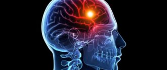
A stroke is a dangerous acute disorder of the blood circulation of the brain as a result of blockage or narrowing of the main veins and arteries that supply cerebral structures. An inevitable consequence of such a formidable condition is swelling. This is an extremely dangerous phenomenon, which is characterized by an increase in the amount of fluid in the peri-cerebral area. As a result, intracranial pressure increases.
Cerebral edema during a stroke is deadly, since there is a high probability that the organ will move and cerebral structures will become wedged into the foramen magnum.
What is cerebral edema and what is worth knowing about such a dangerous condition?
Immediate causes of edema and features of the condition
There is only one direct cause of swelling of the cerebral structures: an increase in the concentration of cerebrospinal fluid and intercellular fluid in the skull. Why does cerebral edema form during a stroke?
There are several likely factors:
- Disturbance of energy metabolism in brain cells. Lead to congestion and local inflammation. As a result, a large amount of excess fluid is formed, the cells increase in volume.
- Circulatory disorders in the main veins and arteries. Lead to cerebral swelling. Considering that the blood supply during a stroke is disrupted in all cases, blood circulation changes in 100% of clinical situations.
- Lack of oxygen and nutrients. Transportation of substances is insufficient. As a result, swelling develops.
- Changes in blood composition. It becomes many times more alkaline. This factor reduces blood fluidity and increases the formation of intercellular fluid.
- Changes in blood plasma pressure. The higher it is, the more intensely the intercellular fluid enters the space of the cerebral structures.
Understanding a stroke
In order to understand how and why cerebral edema occurs after a stroke, it is necessary to have an understanding of the disease itself.
The word stroke in medical practice means an acute disturbance of blood circulation in the vessels of the brain.
Hemorrhagic stroke is a violation of the integrity of a vessel, its rupture with subsequent hemorrhage in the brain.
Blood flow can be disrupted due to obstruction of blood vessels or their rupture, hence the division of strokes into two separate types:
- Ischemic is the most common type of pathological process; in this case we are talking about partial or complete obstruction of blood vessels due to their blockage. The danger is that the tissues that were fed from the blocked vessel stop receiving nutrients and oxygen from the blood. As a result, tissue damage occurs in a certain area of the brain and necrosis may begin.
- Hemorrhagic is a rarer and more dangerous type of pathology, which is characterized by a violation of the integrity of the vessel, that is, its rupture with subsequent hemorrhage into the brain. It is after a hemorrhagic stroke that the likelihood of edema is greatest.
In other articles we talk in more detail about the ischemic and hemorrhagic forms.
Features of the course of the condition
There are several schemes for the development of cerebral edema after a stroke.
- As already mentioned, a cerebral stroke always leads to disruption of the blood supply to nerve cells. The result is blood stagnation. Normal blood flow promotes the evacuation of toxic substances from organic matter into intercellular structures, and then they are completely eliminated from the body in the urine. In addition, due to a critical lack of oxygen, the concentration of toxins increases exponentially. The result is the development of cytotoxic damage. The intercellular substance, together with toxins, enters the space of nerve cells.
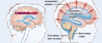
- Brain edema after a stroke can also develop due to banal cellular stagnation. As a result of an increase in the osmotic pressure of the blood, the intercellular substance begins to enter the brain space and increases intracranial pressure and the volume of cerebral fluid (CSF).
- Finally, when it comes to hemorrhagic stroke, another factor in the development of edema is a hematoma (an accumulation of blood in the subarachnoid space). Often it is hematomas in combination with increased cerebral fluid pressure that increase the likelihood of death.
A new remedy for the rehabilitation and prevention of stroke, which is surprisingly highly effective, is the Monastic Collection. The monastic collection really helps fight the consequences of a stroke. Among other things, tea keeps blood pressure normal.
Life forecast
Cerebral edema is a serious condition with a very high risk of death. Predicting survival depends entirely on how timely assistance is provided to the patient. To avoid irreversible consequences, medical intervention should be carried out in the first few hours.
No doctor can give an accurate prognosis. Each patient is individual. If a person manages to survive cerebral edema, he will still have to face the severe consequences of this disease. At the same time, it is much more difficult for older people to cope than for young people. Few people succeed in restoring impaired body functions.
Cerebral edema is a dangerous pathological condition that requires immediate medical intervention. The complications of this disease are quite serious, so the sooner help is provided, the greater the likelihood of achieving positive dynamics.
Clinical picture
Among the typical symptoms of the development of cerebral edema, general and focal manifestations are observed.
The more extensive the lesion and the more pronounced the swelling, the more intense the symptoms.
Characteristic manifestations include:
- Cramps. They are provoked by the pressure of a hematoma or cellular fluid on the temporal lobe of the brain. The so-called temporal lobe epilepsy. Characterized by the appearance of typical symptoms of epileptic seizures. Occurs in 15% of cases.
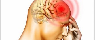
- Intense headaches. The brain itself is incapable of experiencing pain: there are no nerve endings responsible for pain. However, the blood supply structures are richly innervated. With their stenosis, which inevitably occurs during a stroke, the vessels begin to hurt. An additional factor is compression of veins and arteries by cellular fluid.
- Feeling of nausea, vomiting of an indomitable nature, which is not related to diet. The manifestation is due to the development of compression of the vomiting center.
- Disorders of the sensory organs. Depending on the location of the lesion, damage to vision, hearing, and touch is distinguished. According to statistics, it is vision that suffers most often. Fog develops before the eyes, partial or complete loss of vision, hemianopsia (loss of visual fields), photopsia (lightning flashes), spots before the eyes. The manifestation occurs in 60% of cases and is explained by damage to the occipital lobe of the brain. Hearing impairment is less common, in no more than 25% of cases. Manifested by hearing loss in one or both ears.
- Feeling of derealization, disorientation. A person cannot understand where he is, who he is and what time of day it is. Mental disorders are one of the most severe manifestations of swelling.
- With deep damage to the brain structures, problems with breathing and heart rhythm are observed. These are deadly symptoms of destruction of the nuclei of the cerebral trunk (including the pons, thalamus, etc.). Careful treatment is required to prevent such consequences of cerebral edema.
- Memory impairment. Partial or complete amnesia is possible. A person does not remember anything after the onset of a stroke. In rare cases, complete loss of memory is possible, when a person does not remember who he is or where he is.
- Coma and comatose states.
- Aphasia. Full or partial. It is a disorder of speech and its perception by the patient.
Read also: Ischemic stroke recommendations
The most dangerous manifestations associated with worsening the condition:
- Sluggish reaction of the pupils to a light stimulus.
- Hyperthermia at levels above 40 degrees.
- Deep coma or disturbances of consciousness.
Symptoms of cerebral edema are not characteristic enough. For the most part, they aggravate the clinical picture of stroke. But the damage does not always affect the same parts of the brain where the acute lack of blood circulation occurred. Compression of several hemispheres and even the entire brain is possible.
Brain edema: symptoms, causes, consequences
Brain edema is a consequence of interrelated physical and biochemical processes occurring in the body as a result of diseases or pathological conditions.
This complication, depending on the severity, may remain unnoticed and pass without a trace, for example, with a mild concussion (symptoms). Much more often, the consequences of cerebral edema are further severe complications in the form of:
- changes in mental and mental activity
- visual impairment
- auditory
- motor
- coordination functions of the body that are the cause of disability
- cerebral edema often ends in death
What is cerebral edema
The essence of the accepted definition of this condition is a nonspecific reaction of the whole organism in response to the influence of severe damaging factors. The latter are the reason:
- disorders of blood microcirculation in brain tissue;
- lack of oxygen transport to the brain, especially in combination with excessive accumulation of carbon dioxide in the blood;
- disturbances of water-electrolyte, protein and energy metabolism with accumulation of lactic acid in nerve cells;
- violations of the acid-base state of the blood;
- changes in osmotic (electrolyte) and oncotic (protein) pressure of plasma.
All these reasons lead to swelling and swelling of the brain. With edema, the permeability of the capillary walls is impaired and the liquid part of the blood leaks into the surrounding tissues. During swelling, due to the difference in oncotic pressure, water molecules enter directly into the nerve cells of the brain through their membrane. Here they are bound by intracellular proteins and the cells increase in volume.
However, most authors of scientific articles consider swelling as one of the stages of edema, leading to a volumetric increase in the brain. This leads to its compression and displacement (dislocation) around its axis inside a closed space limited by the bones of the skull.
The spread of cerebral edema causes compression of the underlying structures (medulla oblongata) in the foramen magnum. It contains vital centers - breathing regulation, cardiovascular activity and a thermoregulation center.
Signs of cerebral edema are clinically manifested in disruption of the functioning of nerve cells and brain centers even before complete damage to their structures occurs, which can already be determined using modern research methods.
Types and causes of edema
There are two types of cerebral edema:
- Local or regional edema, that is, limited to a certain area surrounding a pathological formation in the brain tissue - an abscess, tumor, hematoma, cyst.
- Generalized. spread throughout the entire brain. It develops with traumatic brain injury, suffocation, drowning, intoxication, loss of large amounts of protein in the urine due to various diseases or poisoning, with hypertensive encephalopathy resulting from severe forms of high blood pressure, and other disorders.
In many cases, with the exception of traumatic brain injury or asphyxia (suffocation), identifying cerebral edema can be difficult against the background of symptoms of other diseases and pathological conditions. The onset of the development of edema can be assumed when the signs of the underlying disease decrease or do not progress, but neurological symptoms, on the contrary, appear and increase.
The main causes of cerebral edema:
- traumatic brain injury, concussion and contusion of the brain, asphyxia with vomit during an alcoholic coma or after hanging, laryngeal stenosis in children with acute respiratory infection (see treatment of laryngitis in a child);
- subdural hematoma, formed under the dura mater as a result of mechanical impact without violating the integrity of the bones of the skull;
- brain tumors, subarachnoid (under the arachnoid mater)
- hemorrhage, which often occurs as a result of a stroke with high blood pressure (see first signs of stroke. consequences of ischemic stroke);
- acute infectious diseases - influenza, meningitis, encephalitis, including severe childhood infections - mumps, measles, scarlet fever, chicken pox;
- gestosis in the second half of pregnancy - severe nephropathy, preeclampsia and eclampsia;
- diseases accompanied by convulsive syndrome - hyperthermia in children (high temperature) with infectious diseases, heat stroke, epilepsy;
- severe diabetes mellitus, especially occurring with episodes of hypoglycemic state, acute and chronic renal, hepatic or renal-hepatic failure;
- severe allergic reactions and anaphylactic shock;
- poisoning with medications, chemical poisons and gases;
- swelling of the brain in newborns as a result of entanglement in the umbilical cord, protracted labor, severe gestosis in the mother (see gestosis during pregnancy), birth injury to the child’s brain.
In addition, swelling of the brain almost always occurs after cranial surgery. Sometimes - after operations performed under spinal or epidural anesthesia or accompanied by large blood loss, due to a pronounced and prolonged decrease in blood pressure, with excessive intravenous administration of saline or hypotonic solutions during surgery, due to the difficulty of tracheal intubation for the purpose of artificial ventilation or inadequacy of ventilation itself and anesthesia.
Symptoms of cerebral edema
Depending on the duration of the disease, the localization of the lesion, the prevalence and rate of growth of the process, the symptoms of cerebral edema may be different. Local, limited edema is manifested by general cerebral symptoms or single signs characteristic of a given part of the brain. With increasing or initially generalized edema, but slowly increasing, there is also a gradual increase in the number of symptoms, indicating damage to several parts of the brain. All symptoms are divided into three groups:
The appearance of diffuse (scattered) neurological symptoms
This is a reflection of the increase in the pathological process, which carries the risk of developing coma due to cerebral edema. This is caused by the involvement of the cerebral cortex first in the edema, and then the subcortical structures. In addition to impaired consciousness and transition to a coma, the following occur:
- generalized (widespread) repeated seizures
- psychomotor agitation between attacks of epileptic seizures, occurring with a predominance of increased muscle tone
- pathological defensive and grasping reflexes
Group of the most dangerous symptoms
They are associated with a further increase in cerebral edema, dislocation (displacement) of its structures, with their wedging and pinching in the foramen magnum. These signs include:
- Coma of varying degrees.
- Hyperthermia (up to 40 degrees or more), which cannot be reduced with the use of antipyretic and vasodilating drugs. Sometimes it is possible to slightly reduce the temperature only by using cold in the area of large vessels or general hypothermia.
- There are different sizes of pupils and their lack of reaction to light, strabismus, “floating” eyeballs, unilateral paresis and unilateral convulsive contractions of the extensor muscles, heart rhythm disturbances with a tendency to decrease the heart rate, and the absence of pain and tendon reflexes.
- If the patient is not given artificial ventilation, the frequency and depth of breathing initially increases, the breathing rhythm is disturbed, followed by a stop and cessation of cardiac activity.
Diagnostics
In an outpatient setting, diagnosing cerebral edema is quite difficult, since this condition does not have any special, specific neurological symptoms. In the early stages, cerebral edema may be mild or asymptomatic. The diagnosis is made based on the symptoms of the underlying disease or injury that caused the swelling, as well as the results of a fundus examination.
If the development of cerebral edema is suspected, the patient should be taken to the intensive care unit or neurosurgical department. In a hospital setting, the issue of performing a lumbar puncture and angiography is decided. MRI and CT are informative, which help to identify edema, assess the degree of its severity and prevalence.
Consequences of cerebral edema in adults and children
The sooner such a pathology is detected and intensive and adequate medical care is provided, the higher the chances of recovery. In a hospital setting, the blood supply to the brain, cerebrospinal fluid dynamics, and dehydration therapy are restored; the prognosis is largely determined by the severity of the disease.
Because with minor perifocal edema, complete recovery is possible, but with the development of cystic-atrophic processes in the brain tissue, only partial restoration of functions can be achieved. When only the underlying disease, accompanied by cerebral edema, is treated, recovery is not possible in all cases and the risk of death is high.
The success of treatment and consequences depend on the severity of the disease that caused cerebral edema and the degree of development of the edema itself, which can resolve with complete recovery. In more severe cases, there are:
- When edema develops in the medulla oblongata, where the main life support centers of the body are located, the consequences of cerebral edema can be respiratory failure, convulsions, epilepsy, and impaired blood supply.
- Even after treatment, the patient may continue to have increased intracranial pressure (symptoms). which greatly worsens the patient’s quality of life, as it is accompanied by headaches, disturbances of consciousness, loss of a person’s orientation in time, social communication skills decrease, lethargy and drowsiness appear.
- Infringement of the brain stem, as well as its displacement, is very dangerous; this threatens respiratory arrest and the development of paralysis.
- After treatment and a course of rehabilitation, many patients are left with adhesions between the membranes of the brain, in the ventricles of the brain or in the cerebrospinal fluid spaces, which is also accompanied by headaches, disorders of neuropsychic activity and depressive states.
- With prolonged cerebral edema without treatment, brain dysfunction may subsequently appear and a person’s mental abilities may decrease.
In children, complete recovery is also possible or:
- development of cerebral palsy and hydrocephalus (see increased intracranial pressure in infants)
- epilepsy (see symptoms and treatment of epilepsy) and dysfunction of internal organs
- speech and motor coordination disorders
- neuropsychic instability and mental retardation
Cerebral edema is a serious, often very severe pathology that requires further observation and treatment of adults by a neurologist, psychoneurologist, and children by a neurologist together with a pediatrician. The duration of observation and treatment after cerebral edema depends on the severity of residual effects.
Our readers write
At the age of 45, pressure surges began, I suddenly became ill, with constant apathy and weakness. When I turned 63, I already understood that I didn’t have long to live, everything was very bad. An ambulance was called almost every week, I always thought that this time would be the last.
Everything changed when my daughter gave me an article on the Internet. You can’t imagine how grateful I am to her for this. This article literally pulled me out of the other world. Over the last 2 years I have started to move more; in the spring and summer I go to the dacha every day, grow tomatoes and sell them at the market. My aunts are surprised how I manage to do everything, where so much strength and energy comes from, they still can’t believe that I’m 66 years old.
Who wants to live a long and energetic life without strokes, heart attacks and blood pressure surges, take 5 minutes and read this article.
Stroke due to stage 4 stomach cancer
Good afternoon. I really need your advice or consultation. I hope I can formulate the question correctly. Man, 63 years old, stage 4 stomach cancer. I had a stroke. The left side of my face was paralyzed and my left hand functions weaker than my right. Speech is impaired. Does cancer contribute to strokes? How is recovery going and is it possible? what procedures are required?
According to the doctors' diagnosis, I recently had a micro stroke. During chemotherapy I lost consciousness several times. One course completed.
Good afternoon. I'll try to answer your questions. I will post three files of extracts, translation from Lithuanian. Unfortunately, there are no originals in Russian. I tried to translate the latest extract into Russian, the primary ones in Lithuanian and English, and they are just too difficult for me to translate. If English doesn’t help you, I’ll try to find a doctor-translator.
Very thin, getting weaker, I won’t tell you the weight (not yet possible), conscious, sane, no activity, mostly gets up with help from others (very weak), dizzy, holding the right side of his head (possibly hurts, doesn’t complain) , right side aches, acute pain, after morphine and platyra, no, there is a dull pain - not always;
I hope I was able to answer as much as possible.
DETAILS: Treatment of lymph node cancer
EPICRISIS Patient case description No. 10-27979 Issued day: 2010-November-10 State institution Vilnius University Hospital Santariskes clinics VLADIMIR MAGIT Date of birth: 1947-12-21 Home address: Geroves Str. 19-11, Vilnius
Abdominal surgery center The first abdominal surgery department Date of hospitalization: 2010-November-04 12:25:00 Date of leaving: 2010-November-10 Diagnosis at the moment of hospitalization: STOMACH NEOPLASM
Clinical Diagnosis: Ventricular adenocarcinoma T3N2M1. Hepatic metastases. Carcinomatosis. Chronic pulmonary embolism. Bilateral thrombosis of lower calf veins.
Patient complaints and disease history: general malaise, constant pain in the epigastrium, loss in body weight. Symptoms appeared three weeks ago. After performing Esophagogastroscopy (EGD) the stomach neoplasm is diagnosed, the biopsy results showed the adenocarcinoma. The computed tomography (CT) showed metastases in the liver and omentum. The purpose of hospitalization was to perform the liver biopsy for verification of the extent of the disease.
Patient status at presentation: The general condition is satisfactory. Blood pressure (BP) 130/80 mg/mm H, Pulse rate (P): 74/min. Lungs: breathing is vesicular without crackles. Palpated abdomen is soft, tender in the epigastrium, without signs of peritoneal irritation.
Conducted diagnostic and laboratory tests: Laboratory tests Blood test (2010-November-04): WBC (*10e9/l)-12.07, NEU (%) – 72.2, LYM (%) -16.5, MON (%) 9.9 EOS (% ) -0.8, Lazdeliniai (%)3.6, Segmintuoti (%) – 83.7, BAS (%) – 0.2, Monocytes (%) – 3.6, IG (%) – 0.4, NRBC (%) – 0.0, Basophils (%) - 0.9, NEU (*10e9/l) – 8.
72, LYM (*10e9/l) – 1.99, MON (*10e9/l) 1.19, EOS (*10e9/l) -01, BAS (*10e9/l) -0.02, IG (*10e9/l) – 0.05 , NRCB (*10e9/l) -0.00, RCB (*10e9/l) – 4.12, Hgb (g/l) – 104, Hct (l/l) -0.329, MCV (fl) – 79.9, MCH (pg) – 25.5, POLY polychromatophilia – 0, HYPO hypochromia – 1, MCHC (g/l) – 316, RDW-SD (fl) – 41.
OTHER DIAGNOSTIC TESTS: Esophago-gastro-duodenoscopy and biopsy (2010-November-04): Endoscope was easily introduced into esophagus. Lumen of esophagus is unobstructed, esophagus mucosa is unchanged. Entrance into cardia is tight. There is some bilious fluid and mucus. There is approx. 6 cm neoplastic ulceration with surrounded tissue infiltration in the stomach angular region, the greater curvature.
X-RAY OF THE LUNGS (2010-November-04): There are no visible foci or dim-outs in x-ray picture. Lung branches are unexpanded, structured. The fluid is not visible in the pleural cavity. Diaphragm is partly relaxed from the right side. Heart shadow is unexpanded. Aorta is normal. Conclusion: Changes in the lungs are not seen.
Types of edema
There are two main types of edema. With a stroke, as a rule, both develop at once:
- Local or regional swelling of cerebral structures. It is formed due to the formation of edema or a partial increase in the volume of cerebral fluid in the brain area.
- Generalized edema. Formed in response to the factors described above.
Generalized edema is fraught with the development of massive damage to nerve cells. Extensive neurological symptoms are observed, represented by the symptoms described above.
Diagnostics
Diagnosis of this problem is carried out by a specialist neurologist or neurosurgeon. Since edema does not have pathognomonic manifestations, the examination is carried out only in a hospital setting.
The first priority is a neurological diagnosis with examination of reflexes, etc. This is only possible if the patient is conscious.
Intraocular pressure measurement is required. A spinal tap (lumbar puncture) is prescribed. It is necessary to measure pressure in the spinal cord, as well as determine the presence of blood in the cerebral and cerebrospinal structures.
Computer or magnetic resonance imaging is mandatory. These studies make it possible to assess the condition of the brain and fluid volume.
Additionally, it is worth conducting a number of laboratory tests, including a coagulogram, blood biochemistry, and a general blood test.
These measures are sufficient to make an accurate diagnosis.
Treatment
Treatment at first is conservative, medicinal.
The following pharmaceutical groups are prescribed:
- Antiplatelet agents. Prevents the formation of blood clots.
- Anticoagulants. They increase blood fluidity and normalize its composition.
- Nootropic drugs for rapid restoration of cerebral cells.
- Antihypertensive drugs. Intended for the treatment of hypertension and blood pressure reduction in secondary hypertension of other origins. It is advisable to carry out such treatment with high tonometer readings, over 180 to 100.
- Diuretics. They help quickly evacuate excess fluid from the brain.
In exceptional cases, craniotomy and drainage of the peri-cerebral space are indicated.
Consequences
Among the most common consequences of cerebral edema are:
- Cramps.
- Lethargy, drowsiness.
- Impaired coordination, breathing, heart rhythm (tachycardia, bradycardia, irregular rhythm).
- Lethargy.
- Impaired consciousness, stupor, coma.
- Paralysis and paresis.
- Mental disorders (induced psychoses).
However, there is a chance for recovery. It is important to show the person to the doctor in a timely manner and not delay the start of treatment.
Swelling of cerebral structures is a serious condition, fraught with life-threatening and health-threatening consequences. Timely diagnosis and adequate therapy are the key to complete recovery.
Drawing conclusions
Strokes are the cause of almost 70% of all deaths in the world. Seven out of ten people die due to blocked arteries in the brain. And the very first and main sign of vascular blockage is a headache!
Blockage of blood vessels results in a disease under the well-known name “hypertension”, here are just some of its symptoms:
- Headache
- Increased heart rate
- Black dots before the eyes (floaters)
- Apathy, irritability, drowsiness
- Blurred vision
- Sweating
- Chronic fatigue
- Swelling of the face
- Numbness and chills in fingers
- Pressure surges
Read also: Examination for stroke
Attention! If you notice at least 2 symptoms, this is a serious reason to think about it!
The only remedy that gave significant results. READ MORE. >>>
My sister was diagnosed with a tumor in her brain and then had a stroke - what is the time frame for resuscitation?
Lyudmila
: Hello. My sister was diagnosed with a brain tumor at the age of 47, and had a stroke within 2 days after the tumor was discovered. I would like to know the timing of resuscitation of such patients and the outcome of this disease.
Doctor's answer:
Hello, Lyudmila. The period of resuscitation for these pathologies can be long, since the diseases are very serious. Both ischemic and hemorrhagic stroke can occur against the background of a brain tumor. Since the tumor puts pressure on the brain tissue, compression of the blood vessels occurs, which can lead to disruption of blood supply, resulting in an acute condition - ischemic stroke. Also, this situation can lead to the formation of blood clots and aneurysms, which, due to a small blow or a sharp rise in blood pressure, rupture, causing a hemorrhagic stroke; the spilled blood from the ruptured vessel accumulates and causes even greater compression of the brain structures and disruption of their function.
The condition for both types of stroke is severe. But hemorrhagic stroke is more difficult to tolerate and often leads to death, especially against the background of a brain tumor. To predict the condition, it is important to know what kind of tumor, in what area of the brain it is located and what type of stroke occurred.
In any case, with such a brain pathology, a person remains disabled, since irreversible consequences occur. The situation is worsened by the fact that stroke therapy involves vascular and nootropic drugs, which are contraindicated for brain tumors. With ischemic stroke, the prognosis is more positive.
At the first stage, the priority task is to normalize blood pressure and stabilize the general condition; treatment in serious condition is carried out in intensive care, where the patient is kept until the main indicators stabilize. If necessary, in case of hemorrhagic stroke, surgical treatment is performed in the first hours to remove hemorrhagic contents.
Further treatment depends on the type of tumor: if it is benign, then you can start using metabolic drugs (Mexidol, Mexiprim, Actovegin) for a short course. In case of a highly malignant tumor, drugs that improve the nutrition and restoration of brain cells cannot be used. When the condition is stabilized after a stroke, it is possible to perform a craniotomy and tumor biopsy, followed by surgery, if necessary, or radiation. Health to you and your loved ones!
Stroke may indicate cancer
A group of Spanish scientists from the Medical Oncology Center recently made a discovery - people who have suffered a stroke may suffer from hidden oncological processes.
Previously, it has been suggested that patients who have been given a similar diagnosis constitute the main risk group for the formation of malignant neoplasms. However, scientists could not determine the extent of the possibility of developing cancer to which patients were exposed. In their new scientific study, experts tried to establish a relationship between stroke and oncology, as well as find out what factors indicate the formation of a malignant tumor and determine the outcome of the disease.
Scientists decided to study the records of approximately 1 thousand patients who went to the emergency department regarding the manifestation of stroke symptoms. Almost 400 people met the necessary criteria, whom scientists observed for a year and a half after the diagnosis of this disease.
As it turned out, cancerous tumors were found in 7.6% of individuals. It is noteworthy that the most common sites of neoplasms were the colon, lungs and prostate gland, in addition to which metastases were present. On average, cancer developed six months after the stroke.
Such information helped to draw the conclusion that people suffer from cancer after a stroke 2 times more often than was previously thought. In addition, advanced stages of cancer indicate that patients suffered from cancer even before they suffered a stroke. Thus, it was found that such a disease acts as a symptom of cancer.
Sources used: simptomer.ru
The concept of cerebral edema and its causes
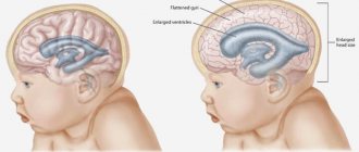
Brain edema after a stroke is a pathological process in which too much fluid accumulates in the brain cells and intercellular space. As a result, the brain increases in size. This increase reaches its peak after 2–5 days, then the swelling begins to subside if the patient survives.
Doctors identify several reasons that can cause swelling. These include:
- Failure of energy metabolism in tissues, which causes stagnation and local inflammation. All this leads to the formation of a lot of excess fluid, and the cells become larger.
- Impaired blood flow in the vessels.
- Lack of oxygen and cell nutrition.
- Increasing the alkalinity of the blood, which reduces its fluidity, increases the likelihood of excess fluid forming between the cells.
- Changes in blood plasma pressure. High pressure promotes the removal of intercellular fluid into the space of cerebral structures.
Causes
Cerebral edema can develop not only as a result of a stroke, but also due to reasons such as:
- mechanical damage to the skull;
- surgical intervention in the brain area;
- oxygen deficiency experienced by the brain and other internal organs;
- infectious processes that have spread to the meninges and tissues (meningitis, encephalitis);
- epilepsy;
- tumor growths in the skull.
The pathological process can occur not only in adults, but also in children, including newborns. In the latter case, edema develops as a result of injuries during childbirth, infectious diseases of the mother during gestation.
Swelling of the brain as a result of a stroke occurs as a result of:
- sweating of fluid into the tissue, which is caused by increased intracranial pressure;
- hypoxia, in which tissue blood flow is disrupted and congestion occurs;
- violations of the permeability of the walls of blood vessels and cells.
The prognosis is complicated by the fact that cerebral edema requires emergency care, but this pathology is not always accompanied by obvious symptoms that would help differentiate it.
Symptoms of cerebral edema
When a person's upper or lower extremities swell, the swelling is immediately visible. But cerebral edema cannot be noticed visually, so it is necessary to focus on certain signs. You can suspect the development of swelling in the brain based on the following symptoms:
- sudden headache;
- nausea and vomiting;
- blurred vision.
- loss of sensation;
- violation of orientation in space;
- failure of the respiratory system;
- stupor;
- memory impairment;
- convulsive conditions;
- fainting;
- in some cases, patients fall into a coma, from which not everyone recovers.
Overview of Brain Tumors
The brain is a complex structure. It consists of approximately ten billion neurons, which make about 13 trillion connections among themselves. It is much more complex than any modern supercomputer. Despite its complexity, most of the brain is made up of supporting cells rather than neurons. The main part of these cells are astrocytes. They seem to serve the main cells of the brain - neurons. Oligodendrocytes are another type of brain cell that are much smaller in number. Their function is to protect nerve fibers in the brain - myelin synthesis. There are the fewest ependymal cells in the brain. They simply cover the inner surface of the ventricles of the brain.
Which stroke most often causes swelling?
With any type of stroke, the risk of developing cerebral edema is very high. The differences are only in the mechanism of its occurrence. In the case of a hemorrhagic stroke, blood vessels rupture, blood and plasma escape into the brain and provoke a tumor. In such a situation, edema develops very quickly and can lead to serious consequences due to compression of brain structures.
If an ischemic stroke occurs, the supply of oxygen to cells is disrupted, which leads to their starvation. As a result of this, the metabolic process in the cells is disrupted, as a result of which a lot of fluid comes out of them.
Read also: 4th degree stroke
First aid
If there is a suspicion of cerebral edema, it is necessary to urgently call an ambulance; this may be the only way to save the patient’s life.
On this topic
A person who has suffered a stroke must be constantly monitored, so if a sharp increase in pressure or signs of swelling are noticed, first aid should be provided while awaiting the arrival of a specialist. Swelling can spread to large areas very quickly, in about thirty minutes, which is very dangerous.
Having called the emergency room, it is necessary to give the patient medications that will reduce blood pressure and sedatives. Also, in some cases, a cold compress is applied to the head.
Treatment of cerebral edema
Before prescribing treatment for cerebral edema after a stroke, the doctor conducts an examination. To provide emergency assistance, which is required in the first hours after the onset of symptoms of blood flow disturbance, the presence of external signs of pathology, high blood pressure, and increased pulse rate is sufficient.
After the attack has stopped, a more detailed diagnosis is carried out to assess the state of the brain, determine the extent of the lesion, identify tissue swelling, as well as the pathologies that caused the development of the stroke.
Methods such as magnetic resonance and computed tomography, angiography, and laboratory blood tests can help doctors with this. The condition of the heart is also checked using an electrocardiogram and ultrasound.
Emergency care and medication administration
In the process of providing emergency care for cerebral edema, the patient’s body is provided with oxygen, since swelling leads to a lack of cell nutrition and their subsequent necrosis. Oxygen is supplied to the respiratory tract using inhalers and special equipment.
Then the patient is given medications that can reduce the amount of fluid in the body. They are called diuretics. These medications are taken under strict control of water and electrolyte balance. If it is impaired, blood pressure will drop and blood clotting will worsen.
As soon as the patient’s condition stabilizes and the pressure drops, doctors move on to eliminating the cause that caused the cerebral edema and the resulting consequences. To normalize blood circulation, patients are given anticoagulants and antiplatelet agents. It is also recommended to take drugs that improve metabolism and regulate blood pressure.
Surgical intervention
Sometimes doctors resort to surgery to treat cerebral edema after a stroke. The purpose of the operation is to remove excess water in the tissues. To solve this problem, one of the following types of intervention is used:
- Trepanation of the skull. It is used if the patient has extensive brain swelling or the formation of a large blood clot.
- Catheterization, where fluid is removed using a catheter.
If a person falls into a coma after a stroke, maintenance therapy is prescribed. If the functioning of the respiratory organs is impaired, artificial ventilation is used.
Forecasting
Understanding the mechanisms and causes of the development of swelling after a stroke, as well as knowing the main symptoms, we can talk about prognosis. It is important to understand that when it comes to cerebral edema during a stroke, the prognosis assumes the presence of other abnormalities (speech, consciousness, memory, motor functions, paralysis of the limbs, etc.). Also, much depends on the type of stroke, its location, the extent of the damage, and the age of the patient.
However, with favorable factors, in the first stages of edema the prognosis is very optimistic. If, when the first symptoms appear, the patient is provided with appropriate assistance, the development of swelling is stopped, and the swelling itself is eliminated, we can talk about recovery and restoration of lost functions.
But in cases where the development of edema has reached the fifth point presented above or the person has fallen into a coma, the prognosis for life is sharply reduced. According to statistics, only 40% of all patients whose swelling after a stroke has developed so severely survive.
Consequences of the disease
Acute cerebrovascular accident with swelling of brain cells cannot pass without a trace. The pathological process always entails life-threatening consequences. After all, cells that are responsible for important body functions die in the head.
Frequent complications are the following:
- Impaired coordination of movements and orientation in space.
- Complete or partial paralysis of the body.
- Loss of sensation.
- Deterioration of vision.
- Speech dysfunction.
The most terrible consequences of cerebral edema are the development of coma, recurrent stroke and death.
ATTENTION.
It should be clearly understood that prolonging life after a stroke largely depends not only on the timeliness of treatment, but also on compliance with all the instructions of the attending physician.
Stroke lung cancer
Registration: 08/24/2009 Messages: 7
Good afternoon A huge request for help with advice. My mother is 70 years old, on June 16 she was admitted to hospital No. 15 with a diagnosis of stroke, then she was diagnosed with lung cancer. They did an MRI on him and said that there was no metastasis in the head, they also did scentigraphy and said that the entire spine was affected. He was completely paralyzed, we were peeing with a catheter.
Registration: 05/02/2006 Messages: 3,025
Answer the questions in the “Important” topic of this section in DETAIL and ACCURATELY.
These are extracts from hospital 15. Age 70 years old, fully conscious and memory, completely paralyzed, can only move her neck a little, stroke, no allergic reactions to drugs, blood pressure 100/73, in the evening it happens 150/90, pulse 70 Pain in the neck and arms, radiates to the back, pain constantly aching. but it can also be acute. The stool is disturbing, it happens once every 4-5 days, and then only with our help and after laxatives, senode, regulax, urination was with a catheter, but it’s been 2 days since the catheter was removed, difficulty breathing, yesterday I couldn’t clear my throat for a long time , my left hand has been very swollen for more than a week, and since yesterday my right hand has also started to swell.
was Ketanov - 4 times a day, now he drinks Pentalgin-M and Novigan 400 mg in turn - 4 times a day, enough for 5 hours. He takes a trace of medication: Furasimide 40 mg - every other day one tablet Detralex 500 mg - every day 1 tablet Sirdalud 2 mg - every day morning, evening, 2 tablets Neuromultivit - 2 tablets Thrombo ACC 50 mg - 1 tablet per day THIOGAMMA 600 mg - 1 tablet per day
The 2nd file does not open. You do not answer all the questions (concomitant diseases, pain therapy) Read the questions carefully. There should be stool at least once every 3 days. Write down the exact dosage of laxatives and the regimen for taking them. Pain therapy is carried out categorically incorrectly
Rehabilitation activities
Returning a person to their usual way of life is very difficult, and this process takes a lot of time. Not all patients manage to be rehabilitated; most remain disabled. The recovery period becomes significantly more complicated if coma develops after a stroke. The vast majority of patients do not come out of it.
Rehabilitation of a stroke patient is aimed at restoring his physical and mental condition. To get rid of paralysis and impaired motor activity, it is recommended to engage in therapeutic exercises. First, classes are conducted with a specialist, then the patient performs them at home independently.
In addition to physical therapy, the patient has to attend various physiotherapeutic procedures, work with speech therapists and psychologists. Diet and giving up bad habits are also important. A favorable family environment and comprehensive support from relatives are of great importance for successful recovery.
Prevention measures
The best way to prevent the development of cerebral edema is to prevent stroke. If this does happen, you must strictly follow the doctor’s recommendations and follow the prescribed regimen.
In addition, prevention methods include:
- giving up bad habits - smoking, drinking alcoholic beverages;
- following a diet that allows you to lower the level of cholesterol in the blood, which worsens the condition of blood vessels;
- weight control and blood pressure levels;
- feasible sports activities.
Cerebral edema is a likely complication after an ischemic or hemorrhagic stroke. It occurs due to severe vascular disorders. The surest way to prevent is to prevent the development of strokes.