The parasympathetic nervous system is a component of the autonomic system. The latter is responsible for human growth, controls the energy received from the lungs and intestines, and regulates blood circulation. It is completely interconnected with heart rate.
The autonomic system works with impulses coming from the brain, due to which the tone of the vessels through which blood and lymph move is regulated. If a person has suffered a head injury, this functionality may be impaired.
What is the parasympathetic nervous system
The department is responsible for the functionality of the body without its participation. For example, parasympathetic fibers provide respiratory function, regulate heartbeat, dilate blood vessels, control the natural process of digestion and protective functions, and provide other important mechanisms. The parasympathetic system is necessary for a person to help the body relax after physical activity. With its participation, muscle tone decreases, the pulse returns to normal, the pupil and vascular walls narrow. This happens without human participation - arbitrarily, at the level of reflexes
The main centers of this autonomous structure are the brain and spinal cord, where nerve fibers are concentrated, ensuring the fastest possible transmission of impulses for the functioning of internal organs and systems. With their help, you can control blood pressure, vascular permeability, cardiac activity, and the internal secretion of individual glands. Each nerve impulse is responsible for a specific part of the body, which, when excited, begins to react.
It all depends on the localization of the characteristic plexuses: if the nerve fibers are located in the pelvic area, they are responsible for physical activity, and in the organs of the digestive system - for the secretion of gastric juice and intestinal motility. The structure of the autonomic nervous system has the following structural sections with unique functions for the whole organism. This:
- pituitary;
- hypothalamus;
- nervus vagus;
- pineal gland
This is how the main elements of the parasympathetic centers are designated, and the following are considered additional structures:
- nerve nuclei of the occipital zone;
- sacral nuclei;
- cardiac plexuses to provide myocardial impulses;
- hypogastric plexus;
- lumbar, celiac and thoracic nerve plexuses.
The influence of age on autonomic tone
In newborns, the influence of the sympathetic department predominates against the background of a general immaturity of nervous regulation. Therefore, their pulse is significantly accelerated. Then both parts of the autonomic system develop very quickly, reaching a maximum during adolescence. At this time, the highest concentration of nerve plexuses in the myocardium is noted, which explains the rapid change in pressure and contraction speed under external influences.
Until the age of 40, parasympathetic tone predominates, which affects the slowing of the heart rate at rest and its rapid return to normal after exercise. And then age-related changes begin - the number of adrenergic receptors decreases while the parasympathetic ganglia are preserved. This leads to the following processes:
- the excitability of muscle fibers worsens;
- the processes of impulse formation are disrupted;
- the sensitivity of the vascular wall and myocardium to the action of stress hormones increases.
Under the influence of ischemia, cells become even more responsive to sympathetic impulses and respond to even the slightest signals by spasming the arteries and accelerating the pulse. At the same time, the electrical instability of the myocardium increases, which explains the frequent occurrence of arrhythmia during angina pectoris, and especially during a heart attack.
It has been proven that disturbances in sympathetic innervation are many times greater than the zone of destruction in acute coronary circulatory disorders.
Sympathetic and parasympathetic nervous system
Comparing the two departments, the main difference is obvious. The sympathetic department is responsible for activity and reacts in moments of stress and emotional arousal. As for the parasympathetic nervous system, it “connects” in the stage of physical and emotional relaxation. Another difference is the mediators that carry out the transition of nerve impulses at synapses: in sympathetic nerve endings it is norepinephrine, in parasympathetic nerve endings it is acetylcholine.
Features of interaction between departments
The parasympathetic division of the autonomic nervous system is responsible for the smooth functioning of the cardiovascular, genitourinary and digestive systems, while parasympathetic innervation of the liver, thyroid gland, kidneys, and pancreas takes place. The functions are different, but the impact on the organic resource is complex. If the sympathetic department provides stimulation of internal organs, then the parasympathetic department helps restore the general condition of the body. If there is an imbalance between the two systems, the patient needs treatment.
- Birch sap - homemade recipes
- Reproductive health of men, women and adolescents. Factors influencing and preventing reproductive health
- Pork stew: recipes with photos
The influence of two systems on the heart
Although sympathetic and parasympathetic stimulation have opposing effects on the cardiovascular system, this is not always so clear-cut. And the mechanisms of their mutual influence do not have a mathematical pattern; not all of them have been sufficiently studied, but it has been established:
- the more the sympathetic tone increases, the stronger the suppressive effect of the parasympathetic department will be - accentuated opposition;
- when the desired result is achieved (for example, acceleration of the rhythm during exercise), the sympathetic and parasympathetic influence is inhibited - functional synergism (unidirectional action);
- the higher the initial level of activation, the less the possibility of its increase during irritation - the law of the initial level.
Watch the video about the effect of the sympathetic and parasympathetic systems on the heart:
Where are the centers of the parasympathetic nervous system located?
The sympathetic nervous system is structurally represented by the sympathetic trunk in two rows of nodes on both sides of the spine. Externally, the structure is represented by a chain of nerve lumps. If we touch on the element of so-called relaxation, the parasympathetic part of the autonomic nervous system is localized in the spinal cord and brain. So, from the central parts of the brain, impulses that arise in the nuclei go as part of the cranial nerves, from the sacral parts - as part of the pelvic splanchnic nerves, and reach the pelvic organs.
Intuition failed
Since the classic work of the British physiologist Walter Gaskell, it was believed that parasympathetic innervation is carried out by long cranial nerves (oculomotor, facial, glossopharyngeal and - main - vagus), originating in the nuclei of the midbrain and diencephalon and regulating the functioning of the eyes and nasal mucosa , glands and internal organs to the lower parts of the colon, as well as the sacral splanchnic nerves, which begin in the nuclei of the lateral horns of the sacral spinal cord and regulate the functioning of the pelvic organs.
The reason for this was some features of the sacral nerves. Anatomically, they are less branched than the sympathetic nerves of the thoracic and lumbar regions, their ganglia are located further from the spine, and they innervate internal organs that are not reached by the branches of the vagus nerve. Physiologically, the sacral nerves act on some organs in the opposite way to the thoracic and lumbar. And, finally, the organs pharmacologically innervated by them are sensitive to blockers of postganglionic acetylcholine receptors.
Classical idea of the structure of the sympathetic (red) and parasympathetic (blue) nervous systems
Anatomy of the Human Body, Henry Gray, 1918 / Wikimedia Commons
Share
The validity of classifying the sacral splanchnic nerves as a parasympathetic system has already been questioned, since the fibers of the cranial nerves extend from the central nervous system dorsally (from the back), and the sacral splanchnic nerves - ventrally (from the chest and abdomen), like sympathetic fibers. This, in turn, indicates different sources of their development in the embryonic period. However, this did not lead to the rewriting of textbooks.
Functions of the parasympathetic nervous system
Parasympathetic nerves are responsible for the natural recovery of the body, normal myocardial contraction, muscle tone and productive relaxation of smooth muscles. Parasympathetic fibers differ in local action, but ultimately act together - in plexuses. When one of the centers is locally damaged, the autonomic nervous system as a whole suffers. The effect on the body is complex, and doctors highlight the following useful functions:
- relaxation of the oculomotor nerve, constriction of the pupil;
- normalization of blood circulation, systemic blood flow;
- restoration of normal breathing, narrowing of the bronchi;
- decreased blood pressure;
- control of an important indicator of blood glucose;
- reduction in heart rate;
- slowing down the passage of nerve impulses;
- decreased eye pressure;
- regulation of the functioning of the glands of the digestive system.
In addition, the parasympathetic system helps the blood vessels of the brain and genital organs dilate, and smooth muscles become toned. With its help, natural cleansing of the body occurs due to phenomena such as sneezing, coughing, vomiting, and going to the toilet. In addition, if symptoms of arterial hypertension begin to appear, it is important to understand that the nervous system described above is responsible for cardiac activity. If one of the structures - the sympathetic or parasympathetic - fails, measures must be taken, since they are closely related.
Central department
This part of the autonomic nervous system represents various structures of the brain. It turns out that it is scattered throughout the entire brain. In the central section, segmental and suprasegmental structures are distinguished. All formations belonging to the suprasegmental department are united under the name hypothalamic-limbic-reticular complex.
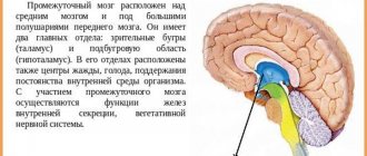
Hypothalamus
The hypothalamus is a structure of the brain located in the lower part, at the base. This cannot be said to be an area with clear anatomical boundaries. The hypothalamus smoothly passes into the brain tissue of other parts of the brain.
In general, the hypothalamus consists of a cluster of groups of nerve cells, nuclei. A total of 32 pairs of nuclei were studied. Nerve impulses are formed in the hypothalamus, which reach other brain structures through various pathways. These impulses control blood circulation, breathing, and digestion. The hypothalamus contains centers for regulating water-salt metabolism, body temperature, sweating, hunger and satiety, emotions, and sexual desire.
In addition to nerve impulses, substances with a hormone-like structure are formed in the hypothalamus: releasing factors. With the help of these substances, the activity of the mammary glands (lactation), adrenal glands, gonads, uterus, thyroid gland, growth, fat breakdown, and the degree of skin color (pigmentation) is regulated. All this is possible thanks to the close connection of the hypothalamus with the pituitary gland, the main endocrine organ of the human body.
Thus, the hypothalamus is functionally connected with all parts of the nervous and endocrine systems.
Conventionally, two zones are distinguished in the hypothalamus: trophotropic and ergotropic. The activity of the trophotropic zone is aimed at maintaining the constancy of the internal environment. It is associated with a period of rest, supports the processes of synthesis and utilization of metabolic products. It exerts its main influences through the parasympathetic division of the autonomic nervous system. Stimulation of this area of the hypothalamus is accompanied by increased sweating, salivation, slowing of heart rate, decreased blood pressure, vasodilation, and increased intestinal motility. The trophotropic zone is located in the anterior parts of the hypothalamus. The ergotropic zone is responsible for the body’s adaptability to changing conditions, ensures adaptation and is realized through the sympathetic division of the autonomic nervous system. At the same time, blood pressure increases, heartbeat and breathing accelerate, pupils dilate, blood sugar increases, intestinal motility decreases, and urination and bowel movements are inhibited. The ergotropic zone occupies the posterior parts of the hypothalamus.
Limbic system
This structure includes part of the temporal lobe cortex, hippocampus, amygdala, olfactory bulb, olfactory tract, olfactory tubercle, reticular formation, cingulate gyrus, fornix, and papillary bodies. The limbic system is involved in the formation of emotions, memory, thinking, ensures eating and sexual behavior, and regulates the sleep-wake cycle.
To realize all these influences, the participation of many nerve cells is necessary. The functioning system is very complex. In order for a certain model of human behavior to be formed, it is necessary to integrate many sensations from the periphery, transmitting excitation simultaneously to various structures of the brain, as if circulating nerve impulses. For example, in order for a child to remember the names of the seasons, repeated activation of structures such as the hippocampus, fornix, and papillary bodies is necessary.
Reticular formation
This part of the autonomic nervous system is called the reticular system because, like a network, it interweaves all the structures of the brain. This diffuse location allows it to participate in the regulation of all processes in the body. The reticular formation keeps the cerebral cortex in good shape, in constant readiness. This ensures instant activation of the desired areas of the cerebral cortex. This is especially important for the processes of perception, memory, attention and learning.
Individual structures of the reticular formation are responsible for specific functions in the body. For example, there is a respiratory center, which is located in the medulla oblongata. If it is affected for any reason, then independent breathing becomes impossible. By analogy, there are centers of cardiac activity, swallowing, vomiting, coughing, and so on. The functioning of the reticular formation is also based on the presence of numerous connections between nerve cells.
In general, all structures of the central part of the autonomic nervous system are interconnected through multineuron connections. Only their coordinated activity allows the vital functions of the autonomic nervous system to be realized.
Segmental structures
This part of the central part of the visceral nervous system has a clear division into sympathetic and parasympathetic structures. Sympathetic structures are located in the thoracolumbar spinal cord, and parasympathetic structures are located in the brain and sacral spinal cord.
Sympathetic department
Sympathetic centers are localized in the lateral horns in the following segments of the spinal cord: C8, all thoracic (12), L1, L2. Neurons in this area are involved in the innervation of smooth muscles of internal organs, internal muscles of the eye (regulation of pupil size), glands (lacrimal, salivary, sweat, bronchial, digestive), blood and lymphatic vessels.
Parasympathetic Division
Contains the following structures in the brain:
- accessory nucleus of the oculomotor nerve (nucleus of Yakubovich and Perlia): control of pupil size;
- lacrimal nucleus: accordingly, regulates tear secretion;
- superior and inferior salivary nuclei: provide saliva production;
- dorsal nucleus of the vagus nerve: provides parasympathetic influences on internal organs (bronchi, heart, stomach, intestines, liver, pancreas).
The sacral section is represented by neurons of the lateral horns of segments S2-S4: they regulate urination and defecation, blood flow to the vessels of the genital organs.
Diseases
Before using any medications or doing research, it is important to correctly diagnose diseases associated with impaired functioning of the parasympathetic structure of the brain and spinal cord. A health problem manifests itself spontaneously, it can affect internal organs and affect habitual reflexes. The following disorders of the body of any age may be the basis:
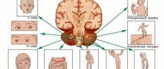
- Cyclic paralysis. The disease is triggered by cyclical spasms and severe damage to the oculomotor nerve. The disease occurs in patients of all ages and is accompanied by nerve degeneration.
- Oculomotor nerve syndrome. In such a difficult situation, the pupil can dilate without exposure to a stream of light, which is preceded by damage to the afferent portion of the arc of the pupillary reflex.
- Trochlear nerve syndrome. A characteristic disease manifests itself in the patient with a slight strabismus, invisible to the average person, with the eyeball directed inward or upward.
- Injured abducens nerves. In the pathological process, strabismus, double vision, and pronounced Foville syndrome are simultaneously combined in one clinical picture. The pathology affects not only the eyes, but also the facial nerves.
- Trinity nerve syndrome. Among the main causes of pathology, doctors identify increased activity of pathogenic infections, disruption of systemic blood flow, damage to the corticonuclear tract, malignant tumors, and previous traumatic brain injury.
- Facial nerve syndrome. There is an obvious distortion of the face when a person voluntarily has to smile, while experiencing painful sensations. More often this is a complication of a previous illness.