Excitability
Excitability is the ability of a tissue to respond to irritation with a specialized reaction - excitation . Excitability is a form of irritability .
Excitable tissues are those tissues whose cells are capable of generating a specific reaction—excitation—in response to irritation.
Excitation is a specialized response of a living object to the action of a stimulus , manifested in changes in its certain parameters.
Excitable tissues include:
- nervous,
- muscular,
- glandular.
Signs of excitement:
- are common,
- specific.
General signs of excitation (inherent in all excitable tissues):
- Changes in the level of metabolic processes in tissues;
- Release of various types of energy - thermal, electrical.
Specific signs of excitation (characteristic of a certain type of tissue):
- Muscle tissue - contraction,
- Glandular - secretion of secretion,
- Nervous - generation and conduction of nerve impulses.
Non-excitable are:
- epithelial,
- connective tissue.
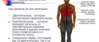
Muscle and nerve tissue
Muscle tissue
are heterogeneous in their origin in embryogenesis, their structure also differs, but there is one property that unites them into a group - the ability to
contract
. Their cells have an elongated, elongated shape, they perceive irritating nerve impulses well and contract in response. Without muscle tissue, the body would not be able to move in space, and organs would not be able to function - the heart would pump blood, the tongue would “hang out,” the intestines would move food, the fingers would press keyboard buttons…
So, the main properties of this type of tissue: excitability
(the ability to respond to irritation),
contractility
(the ability of cells to shorten and stretch),
conductivity
(the ability of the cell membrane to “drive” a wave of excitation, to transmit an impulse). There are two types of muscle tissue - smooth and striated. Let's look at their features in more detail.
1. Smooth muscle tissue
present in internal organs.
Its cells resemble spindles and have one rod-shaped nucleus. Contraction of smooth muscles occurs involuntarily, proceeds slowly, while the muscles contract strongly, but become tired little. For example, the intestines contract up to 12 times in one minute, moving food along. The structural unit is a muscle cell, myocyte
, containing glycogen and myofilaments (myofibrils), and externally covered with a basement membrane. It is interesting that the myocytes of this tissue can divide throughout their lives, unlike, say, cardiomyocytes (cells of cardiac tissue), which divide during the development of the embryo, but then almost lose this ability.
2. Striated muscle tissue
It is distinguished by transverse striation of fibers and high elasticity.
The striation is caused by the special distribution in the cytoplasm of the fibers of many filaments - myofibrils
(consisting of protein sarcomeres), which are combined into bundles. As a result, the muscle fiber along its entire length is densely filled with myofibrils. They are the contractile element of the muscle cell. There are two types of striated muscle tissue: skeletal and cardiac.
1) Skeletal tissue
forms skeletal muscles, it can be controlled voluntarily by directing movements.
Its structural unit is muscle fiber
.
It consists of myosymplast
(a multinuclear structure in which sarcoblast cells merge; myofibrils are located in the center) and
myosatellite cells
(mononuclear stem cells). From the outside, these formations are enveloped by a basement membrane. The muscle fibers are thin, but their length can reach several centimeters. Several muscle fibers form a bundle and have a common sheath, the sarcolemma. Several bundles also have their own membrane - this is how muscle is formed. Skeletal muscles are attached to bones or to each other using tendons.
2) Heart tissue
characterized by good conductivity.
Its cells usually contain one nucleus, less often two. This tissue forms the heart muscle - myocardium
.
The structural unit is a cardiomyocyte
with multiple mitochondria. The heart tissue contracts involuntarily; this process cannot be controlled from the outside.
Nervous tissue
Nervous tissue
creates the basis of the nervous system.
Its main properties are excitability and conductivity; it perceives a nerve impulse and transmits it. Thanks to nervous tissue, all organs interact. This tissue is found in the nerves, brain and spinal cord. Its base is made up of nerve cells - neurons
, and a specific substance called
neuroglia
(auxiliary cells), which provides nutrition and protection to neurons.
Neurons are perhaps the most beautiful of all cells. Many of them have the shape of a star or a tree, others look like pears, spindles, pyramids... They consist of a body and processes - dendrites and axons
.
Dendrites (short, multiple, branched) perceive irritation, axons (long, single) transmit a signal to other cells. A synapse is where axons make contact with other nerve cells. Do you want to pass the exam with flying colors? Click here - OGE courses in biology
Physiological properties of excitable tissues
The main properties of all excitable tissues are:
- excitability,
- conductivity,
- lability.
Lability (functional mobility) is the ability of a tissue to respond to various influences at a certain speed, that is, the ability to reproduce a certain frequency of stimulation .
A measure of lability is the largest number of responses with which excitable tissue is capable of reacting per unit of time in accordance with the frequency of stimulation applied to it .
Conductivity is the ability of tissue and cells to transmit excitation.
Irritation is the process of exposure of living tissue to agents external to this tissue.
A stimulus is a cause that can cause a response.
Excitability and conductivity are the main properties of which tissue
1. Basic physiological properties of excitable tissues
- · Excitability
–
the ability of a tissue to respond to irritation with excitation.
The excitability of envy depends on the level of metabolic processes and the charge of the cell membrane .
The excitability indicator the irritation threshold
, is the minimum strength of the stimulus that causes the first visible tissue response.
Stimuli are: subthreshold, threshold, suprathreshold. Excitability and irritation threshold are inversely proportional values. - · Conductivity – the ability of tissue to conduct excitation along its entire length
.
The conductivity indicator is the speed of excitation
. The speed of excitation through skeletal tissue is 6-13 m/s, through nervous tissue up to 120 m/s. Conductivity depends on the intensity of metabolic processes, on excitability (directly proportional). - · Refractoriness (non-excitability) – the ability of a tissue to sharply reduce its excitability when excited.
At the moment of the most active response, the tissue becomes inexcitable.
There are: - · Lability (functional mobility) – the ability of tissue to reproduce a certain number of excitation waves per unit of time in exact accordance with the rhythm of applied stimulation.
This property characterizes
the rate at which excitation occurs
. Lability indicator: the maximum number of excitation waves in a given tissue: nerve fibers - 500-1000 impulses per second, muscle tissue - 200-250 impulses per second, synapse - 100-125 impulses per second. Lability depends on the level of metabolic processes in the tissue, excitability, and refractoriness. - ·
For muscle tissue, to the four listed properties, a fifth is added -
contractility.
absolutely refractory period
– the time during which the tissue does not respond to absolutely any pathogens;
relative refractory period
- the tissue is relatively inexcitable - excitability is restored to its original level.
Refractoriness index
is
the duration of the refractory period (t).
The duration of the refractory period for skeletal muscle is 35-50 ms, and for nervous tissue – 5-5 ms.
Tissue refractoriness depends on the level of metabolic processes and functional activity (inverse relationship).
2. The concept of a state of relative physiological rest and activity.
A state of rest
is observed in the absence of the action of a stimulus.
Characterized by a relatively constant level of metabolic processes
(since this level is still constantly changing - a state of relative rest);
lack of functional manifestations
of this tissue.
The state of activity
occurs under the influence of stimuli.
It is characterized by a pronounced change in the level of metabolic processes
,
manifestations of the functional functions
of a given tissue.
According to A. A. Ukhtomsky: “Rest and activity are two different levels of metabolic processes.” 3. Forms of the active state of excitable tissues There are 2 forms of the active state of excitable tissues
:
excitation; braking
_
Excitation
is
an active process - a tissue response to irritation
.
Characterized by the manifestation of functional functions
.
Any excitement has a number of signs. 1. Nonspecific signs
: present in all tissues - changes in the permeability of the cell membrane, changes in the movement of ions through the cell membrane, changes in the charge of the cell membrane, changes in the level of metabolic processes, changes in oxygen consumption and carbon dioxide emissions, changes in tissue temperature.
Changes in viscosity, etc. The easiest way to register is a change in the charge of the cell membrane. 2. Specific signs
(
function
) - characteristic of a certain type of tissue (for example: muscle tissue - contraction, nervous tissue - generation of nerve impulses).
Inhibition
-
occurs in tissue in response to irritation and is characterized by inhibition of the functional functions of a given tissue
.
Inhibition occurs with the expenditure and release of energy, but they are less than during excitation. Conclusion:
when irritation is applied to tissue, either excitation or inhibition occurs; these processes are closely interrelated and (according to Pavlov) are two sides of one process.
4. Types of excitation Excitation can be of 2 types: local
(local response);
propagating
(pulse).
Local excitation
is the most ancient type (lower forms of organisms and low-excitable tissues - for example, connective tissue).
Local excitation also occurs in highly organized tissues under the influence of a subthreshold stimulus
or as a component
of an action potential
.
With local excitation there is no visible response. Features of local excitation
: no latent (hidden) period -
occurs immediately upon action of the stimulus
;
no irritation threshold;
local excitation
is gradual
- the change in the charge of the cell membrane is proportional to the strength of the subthreshold stimulus;
there is no refractory period
; on the contrary,
a slight increase in excitability
;
propagates with decrement (attenuation
).
Impulse (spreading) excitation
is inherent in highly organismal tissues and occurs under the influence
of threshold and superthreshold stimuli
.
Features of impulse excitation: has a latent period
- some time passes between the moment of application of irritation and the visible response;
has a threshold of irritation; not gradual
- the change in the charge of the cell membrane does not depend on the strength of the stimulus;
presence of a refractory period; impulse excitation does not fade
.
Conclusion:
local and impulse excitation is observed in the animal and human body.
The occurrence of one or another type of excitation depends on the degree of tissue development and the strength of the stimulus. 5. Laws of interaction of stimulus with excitable tissue There is a certain dependence of the response on
the stimulus parameter .
Laws:
law of stimulus strength;
law of stimulus duration; law of stimulus gradient. Law of stimulus strength
.
The tissue response is proportional to the strength of the applied irritation up to a certain limit
.
The increase in response is the result of excitation of an increasing number of tissue fibers. When the maximum stimulus is applied, the greatest response occurs, since all excitation fibers and a further increase in the response are impossible. Law of stimulus duration
.
The tissue response depends on the duration of the stimulus, but up to a certain limit.
The nature of the response depends on the strength of the stimulus and the time of action.
The Hoffweg-Weiss
-
Lanin
force-time curve reflects this dependence: P – rheobase, a.p.
- useful time. Explanations: under the influence of weak stimuli over time there is no visible reaction.
When the threshold is reached, a visible response appears.
This threshold value is called rheobase
- the minimum electrical current that causes the minimum tissue response.
The time during which a current equal to the rheobase causes a response is useful time.
Since the irritation threshold is not a constant value, in clinical studies an irritant equal in strength to two rheobases is used.
The time during which a stimulus equal to two rheobases causes a response is called chronoxia.
Chronoxia is determined to judge the functional activity of tissue (nervous and muscle).
Chronoxia
is
one of the indicators of excitability
; the greater the excitability, the less chronoxia.
Law of stimulus gradient
.
Gradient
-
the steepness of the increase in stimulus strength
.
The tissue response depends on the stimulus gradient
up to certain limits.
Accommodation
is the adaptation of tissue to a stimulus that slowly increases in strength.
With a slow increase in the strength of the stimulus, there may be no response. Accommodation mechanism: under the influence of a stimulus slowly increasing in strength, sodium inactivation
and, as a consequence, a constant increase in the threshold of irritation.
Conclusion :
1) depending on the strength, duration and gradient of the stimulus, different tissue responses are observed; 2) this dependence is not unlimited.
Classification of stimuli
By nature:
- physical,
- chemical,
- physico-chemical,
- biological.
According to their biological significance, irritants are divided into 2 groups:
- Adequate - irritants to the effects of which tissues in the process of evolution are adapted to the greatest extent.
- Inadequate - irritants to which excitable tissues are not specifically adapted .
Plasma membrane composition
- Lipids (mainly phospholipids),
- Proteins (glycoproteins),
- Carbohydrates (mucopolysaccharides).
Lipids are very tightly packed in the membrane, there are practically no distances between them, so the membrane does not allow water to pass through and is practically impermeable to ions and other large molecules.
Protein molecules can be immersed in the lipid layer from the extracellular or cytoplasmic side, or they can completely penetrate the membrane.
If proteins are attached to the surface of the membrane, they are called peripheral . On the inside there will be enzyme proteins , and on the outside there will be receptor proteins .
If proteins penetrate the entire thickness of the cell membrane, then they are called integral or transmembrane .
Such proteins form structures that allow the movement of ions through the membrane.
If proteins form the walls of a pore through which ions pass by simple diffusion, then these are ion channels .
If transmembrane proteins pump ions against concentration and electrical gradients, then they are ion pumps .
All channels present in living tissues can be divided into 2 types:
- the first type is rest channels, which spontaneously open and close without any external influences ;
- the second type - gate channels (gate channels) - at rest they are closed and open under the influence of stimuli .
Ion channels:
- nonspecific (leakage channels are always open),
- specific (selective), having the ability to allow only certain ions to pass through when the charge on the membrane changes or the action of chemicals occurs.
Transport of substances
Particle transport through channels is a vital process for cells.
Usually, the transport of substances is divided into passive (without energy consumption), i.e. transport of substances along concentration, osmotic and electrochemical gradients and active (with energy consumption).
Did you like the site? Support us by subscribing on social networks!
- Site group in VK
- Website profile on Twitter
- Site community on Facebook
There are primary and secondary active transport
Primary active transport of ions is provided by special ion pumps, carried out with the expenditure of energy ATP , against the concentration gradient , i.e. the transfer of substances occurs from a lower concentration through the membrane to a higher concentration .
Secondary active transport:
- This is a type of transport for the transfer of substances (glucose, amino acids, etc.) across a membrane also against a gradient, but without energy consumption.
- These substances pass through the membrane with the help of special carriers (for example, Na ions), the transport of which requires energy, and these substances move as if in parallel.
Membrane potential or resting potential
At rest, charge or potential difference , which was later called the membrane potential (MP) or resting potential (RP).
Positive charges are concentrated on the outer surface of the membrane, and negative charges are concentrated on the inner surface.
The membrane potential is measured in negative values , because the inner surface of the membrane is negatively . Its value ranges from -60 to -90 mV in different cells.
Methods for measuring membrane potential
Depending on the location of application of the electrodes:
- extracellular using macroelectrodes ,
- intracellular using microelectrodes .
1. The study of the PP using macroelectrodes is carried out by applying one of them to an undamaged area of tissue, and the other to a damaged area of tissue.
2. Microelectrode method
The microelectrode is a micropipette with a diameter of 0.5 - 1 µm, filled with a concentrated saline solution (KCl). It may also contain a non-polarizing electrode - made of silver, gold or platinum .
The second electrode is placed in the extracellular fluid.
Both electrodes are connected to an amplifier and an oscilloscope to record the potential. At the moment the membrane is pierced, the oscilloscope records the appearance of a negative potential corresponding to the PP.
The first reason is ionic asymmetry:
- There are 30-50 times more K ions in the cell than outside;
- There are 8-10 times more Na ions outside the cell than inside it;
- There are many times more Ca ions outside the cell;
- There are also 50 times more Cl ions in the extracellular fluid than inside the cell;
- There are more organic anions inside the cell compared to the outside surface.
Thus, for these ions the direction of the concentration gradient is different!
- For K from the cell (from higher to lower concentration);
- For Na, Ca and Cl into the cell.
2nd reason causing membrane polarization:
- different permeability of the membrane for different ions. At rest, the membrane is 25 times more permeable to K ions than to Na, because the number of potassium channels per unit membrane area is much greater than sodium channels.
- Since the concentration of K ions in the cytoplasm is much higher than outside the cell, they begin to move through the channel and out of the cell.
- K ions carry positive charges, so the outside of the membrane becomes positively charged.
The negative charge on the inner surface of the membrane is due to the presence of organic anions - large molecular compounds that are negatively charged and for which the membrane is impermeable (glutamate, aspartate, organic phosphates, sulfates, etc.)
Thus, a potassium-equilibrium potential is formed on the membrane, because the forces of diffusion (the release of K from the cell along the concentration gradient) and electrostatic interaction (repulsion of the outgoing K ions by the positive charge on the outer surface of the membrane) are balanced.
Potassium is the main ion ensuring the formation of MP (PP), which is confirmed by the Nernst formula. Using it, you can, knowing the concentration of potassium ions inside and outside the cell, calculate the PP value.
General physiology of excitable tissues
Biological reactions . Living organisms and all their cells have irritability, i.e. the ability to respond to environmental influences or disturbances in their condition by changing their structure or function, which is inextricably linked with quantitative and qualitative changes in metabolism and energy. Changes in the structure and functions of the body and its cells in response to various influences are called biological reactions , and the influences themselves that cause them are called irritants or stimuli .
The concept of a biological reaction includes all types of response activities of the body, its cells and organs to various influences. Cell reactions are manifested in changes in their shape, structure, growth and division process, in the formation of various chemical compounds in them, the transformation of potential energy into kinetic (electrical, mechanical, thermal, light), the performance of one or another work (moving in space, excreting certain substances, work on concentrating certain electrolytes in the cell, etc.). The reactions of the entire organism are even more diverse, especially complex forms of behavior. In the process of their implementation, the activity of many organs and countless cells changes, because the body always reacts to various influences as a single whole, as a single complex system.
Irritants . Any change in the external environment or internal state of the organism can be a stimulus to a living cell or organism as a whole, if it is large enough, occurs quickly enough, and lasts long enough.
The entire infinite variety of possible stimuli can be divided into 3 groups: physical, physicochemical and chemical. Physical stimuli include temperature, mechanical (impact, injection, pressure, movement, acceleration, etc.), electrical, light. Physico-chemical stimuli are represented by changes in osmotic pressure, active reaction of the environment, electrolyte composition, and colloidal state. Chemical irritants include many substances that have different compositions and properties and can change cell metabolism (food substances, drugs, poisons, hormones, enzymes, metabolites, etc.).
The stimuli to cells that cause their activity and are of particular importance in life processes are nerve impulses. Being natural, i.e. arising in the body itself, electrochemical stimuli to cells, nerve impulses, traveling along nerve fibers from nerve endings to the central nervous system or coming from it to peripheral organs, cause directed changes in their condition and activity.
All stimuli are divided into external (extero-) and internal (intero-) stimuli according to their place of origin, and according to physiological significance - into adequate and inadequate. Adequate are those stimuli that act on a given biological structure in natural conditions, to the perception of which it is specially adapted by evolution and the sensitivity to which it is usually extremely high (eye - light, ear - sound, etc.). Inadequate are those stimuli for which a given cell or organ is not specifically adapted to perceive, but which under certain conditions can cause changes in structure or function (muscle - can contract upon impact, rapid warming, exposure to electric current, sudden stretching, exposure to acid, etc. .).
Excitability. Cells of nervous, muscle and glandular tissues are specially adapted to carry out rapid reactions to irritation (get excited). The cells of these tissues are called excitable, and their ability to respond to various stimuli with excitation is called excitability. Excitability is the property of a cell membrane to respond to the action of an irritating (stimulating) factor by changing its permeability and its electrical state. This phenomenon is called excitation. Excitation is a complex biological reaction, manifested in a combination of physical, physicochemical and functional changes. A mandatory sign of excitation is a change in the electrical state of the surface cell membrane (a change in its membrane potential, MP, and the generation of a propagating action potential, AP). Having arisen in one cell or in one part of it, excitation spreads to other parts of the same cell or to other cells.
The response of a living cell to stimulation, whether in the form of excitation and the associated electrical reaction, or in the form of contraction or secretion, always occurs after a certain hidden or latent period. This is the name given to the period of time between the onset of the stimulus and the tissue response to its action. During the latent period, the changes in tissue condition necessary for a reaction to occur must occur. The latent period of excitable tissues is shorter than that of non-excitable tissues, and the latent period of the electrical reaction of the tissue is shorter than that of muscle contraction and, especially, the secretory reaction.
History of the discovery of electrical phenomena in tissues.
In 1786, the Italian doctor and physiologist Galvani, hanging frog legs on the balcony to dry, noticed that when the leg swayed by the wind comes into contact with the metal grating of the balcony, its contraction occurs. Galvani concluded that if a short circuit is established between a nerve and a muscle through a metal conductor, and at the same time the muscle contracts, then this is evidence of the manifestation of “animal electricity”. He believed that nerve and muscle are charged oppositely.
However, the physicist Volta showed the fallacy of Galvani's conclusion by conducting the following experiment: he noticed that the balcony railings were copper, and the hooks on which the paws hung were iron. Having tried to apply tweezers to the paw, one leg of which was made of copper, and the other of zinc or iron, Volta received a contraction of the muscle. Consequently, he concluded, the muscles contract not because “animal electricity” is released, but because a current flows between the two metals in contact with the electrolyte, which irritates the nerves of the frog’s leg.
Disagreeing with Volta, Galvani set up a second experiment. It consisted of a non-metallic contraction of the muscle. The contraction was achieved by draping the nerve over the prepared muscle using glass instruments. However, it turned out that contraction could be obtained only when the muscle was damaged, and if the muscle was prepared carefully, without damaging its surface, then contraction did not occur during such an experiment. Later, the German physiologist Hermann showed that if galvanometer electrodes are applied to an intact muscle, no potential difference can be seen. But if damage is made to a muscle or nerve, an incision is made, and one of the electrodes is immersed in this incision, then the galvanometer needle deviates, which shows that an electric current arises between the damaged and undamaged areas of the living muscle, and the damaged area carries a negative charge. This current was called fault current, or quiescent current .
In 1837, Matteuci showed that the resting current of skeletal muscle decreases when it contracts. Matteuci did another experiment. He took two neuromuscular drugs and threw the nerve of the 2nd onto the muscle of the 1st. At the same time, it irritated the nerve of the first drug, causing the muscle to contract. It turned out that the second muscle also began to contract. This cannot be explained by the influence of a resting current on the nerve, since the contraction of the second muscle occurred only when the first was excited. This experiment is even more demonstrative if, instead of the first muscle, we take the working heart of a frog. When a glass hook of a nerve of a neuromuscular drug is thrown onto the heart of a frog, the leg muscle begins to contract in the rhythm of the beating heart. The reason for this phenomenon was discovered later.
In 1850, the famous French researcher Dubois-Reymond, irritating the sciatic nerve of a frog, discovered that following the irritation a wave of electric current runs through the nerve. In 1868, Herman showed that the reason for this is that the electric current arising during irritation reaches the neighboring area, excites it, then reaches the next area and through such contacts the excitation wave runs along the nerve, like fire along a fuse-ford cord.
If a section of a nerve is irritated with single blows of direct current, and the current is diverted from the next section with two electrodes to a galvanometer or to the tube of a cathode oscilloscope, then initially, at the moment of application of irritation, no deviations are recorded. since there is the same potential under both output electrodes. After some time, spreading. the excitation reaches the first output electrode and then the galvanometer registers the potential difference in the form of a negative oscillation - the arrow deviates to the left (on the oscilloscope - down). When the excitation wave is between the electrodes, the arrow returns to its original position. Then the excitation wave reaches the second electrode - the arrow deviates to the right (beam upward). When the excitation wave moves further, both the oscilloscope beam and the galvanometer needle return to their original position.
From these facts the following conclusions can be drawn:
1. At rest, the potential difference exists only between the undamaged and damaged tissue areas (damage current, or quiescent current).
2. When excitation passes through a nerve, an action current arises in it.
3. This action current does not remain in place, but spreads.
4. The action current is a negative potential fluctuation.
A more accurate study of the mechanisms of electrical changes in tissues at rest and during excitation has become possible with the progress of electrical measuring and microelectrode technology. We now turn to consideration of modern data on electrical processes in tissues.
Resting potential . It turned out that between the outer surface of the cell and its protoplasm at rest there is a potential difference of the order of 60-90 mV, and the cell surface is charged electropositively with respect to the protoplasm. This potential difference is called the membrane potential, or resting potential. Its accurate measurement is possible only with the help of intracellular microelectrodes.
According to the Hodgkin-Huxley membrane-ion theory, bioelectric potentials are caused by the unequal concentration of K+, Na+, Cl- ions inside and outside the cell, and the different permeability of the surface membrane to them.
Based on electron microscopy data, chemical analysis and electrophysiological studies, it is assumed that the membrane consists of a double layer of phospholipid molecules, covered on the inside with a layer of protein molecules, and on the outside with a layer of protein molecules and mucopolysaccharides. It is assumed that the cell membrane contains very thin channels (pores) with a diameter of several angstroms. Through these channels, molecules of water and other substances, as well as ions with a diameter corresponding to the size of the pores, enter and leave the cell. Various charged groups are fixed on the structural elements of the membrane, which gives the channel walls a particular charge. Thus, the presence of dissociated phosphate and carboxyl groups in the membrane of nerve fibers is the reason that it (the membrane) is much less permeable to anions than to cations.
The permeability of the membrane for different cations is also not the same and changes naturally under different functional states of the tissue. At rest, the membrane of nerve fibers is approximately 25 times more permeable to K ions than to Na ions, and when excited, sodium permeability is approximately 20 times higher than potassium.
In addition to permeability, the ion concentration gradient on both sides of the membrane is of great importance for the occurrence of membrane potential. It has been shown that the cytoplasm of nerve and muscle cells contains 30-59 times more K+ ions (500 meq/l versus 10 meq/l), but 8-10 times less Na+ ions (35 meq/l versus 350 meq/l) and 50 times less Cl- ions than extracellular fluid (see table). The magnitude of the resting potential of nerve fibers and cells is determined by the ratio of positively charged K+ ions, diffusing per unit time from the cell outward along the concentration gradient, and positively charged Na+ ions, diffusing along the concentration gradient in the opposite direction. Thus, in model experiments on a squid axon, with the K+ concentration gradient that occurs in the nerve fiber, the K+ current is -120 mV. If we simulate only a sodium gradient in such an experiment, then the magnitude of the Na+ current is +30 mV. The actually measured membrane potential of the nerve is equal to the sum of these two oppositely directed currents, i.e. -90mv.
Despite the fact that the rate of diffusion of Na+ and K+ ions through the membrane at rest is small, the difference in their concentrations outside the cell and inside it should eventually be completely leveled off if there were no special mechanism in the cell that ensures active release (“ pumping out" from the protoplasm Na+ ions penetrating into it and introducing ("pumping") K+ ions. This mechanism is figuratively called the sodium potassium pump.
In order for ion asymmetry to be maintained, the Na-K pump must perform a certain amount of work against the ion concentration gradient. The direct source of energy for the pump is the breakdown of ATP, which occurs under the influence of ATPase, localized in the membrane and activated by Na+ and K+ ions (the so-called Na-K-dependent ATPase). Inhibition of the activity of this enzyme leads to disruption of the pump. As a result, the protoplasm becomes enriched in Na+ and loses K+. A direct consequence of this is a decrease or even complete disappearance of the MP (resting potential, or membrane potential).
Depolarization of the membrane occurs because, due to the concentration gradient, K+ comes out, but due to the fact that CL- ions, which are not able to pass through the membrane, electrostatically retain positive ions, an excess of K+ is created in the boundary layer, and between the outer and the inner surfaces of the membrane, charged positively and negatively, respectively, a potential difference of about -90 mV arises. The membrane at rest is constantly depolarized, since as a result of the operation of the Na-K pump, the required ion concentration gradient is maintained.
Action potential . If a section of a nerve or muscle fiber is exposed to a sufficiently strong stimulus (for example, a jolt of electric current), excitation occurs in that section, one of the most important manifestations of which is a rapid oscillation of the MP, called an action potential (AP)
With intracellular abduction, one can find that the surface of the excited area for a very short interval, measured in thousandths of a second, becomes charged electronegatively with respect to the neighboring, resting area, i.e. when excited, the so-called "membrane recharging". Accurate measurements showed that the AP amplitude is 30-50 mV higher than the MP value. The reason for this is that upon excitation, the PP does not simply disappear, but a potential difference of the opposite sign occurs, as a result of which the outer surface of the membrane becomes negatively charged relative to its inner side.
In PD, it is customary to distinguish between its peak (the so-called spike) and trace potentials. The PD peak has an ascending and descending phase. Before the ascending phase, a more or less pronounced so-called local potential, or local response. Since the initial polarization of the membrane disappears during the ascending phase, it is called the depolarization phase; accordingly, the descending phase, during which membrane polarization returns to its original level, is called the repolarization phase. The duration of the AP peak in nerve and skeletal muscle fibers varies between 0.4-5.0 ms. In this case, the repolarization phase is always longer.
In addition to the peak, two trace potentials are distinguished in the AP - trace depolarization (trace negative potential) and trace hyperpolarization (trace positive potential. The amplitude of these potentials does not exceed several millivolts, and the duration varies from several tens to hundreds of milliseconds. Trace potentials are associated with reduction processes, developing in muscles and nerves after the end of excitation.
The cause of PD is a change in the ionic permeability of the membrane. At rest, as already mentioned, the permeability of the membrane to K+ exceeds sodium permeability. As a result, the flow of positively charged ions from the protoplasm outward exceeds the opposite flow of Na+. Therefore, the membrane at rest on the outside is positively charged.
When a cell is exposed to an irritant, the permeability of the membrane for Na+ ions increases sharply, and ultimately becomes approximately 20 times greater than the permeability for K+. Therefore, the flow of Na+ ions into the cell begins to significantly exceed the outward flow of K+. The Na+ current reaches a value of +150 mV. At the same time, the release of K+ from the cell decreases somewhat. All this leads to distortion (reversion) of the MF, and the outer surface of the membrane becomes charged electro negatively with respect to the inner surface. This shift is recorded in the form of an ascending branch of the AP peak (depolarization phase).
The increase in membrane permeability for Na+ ions continues in nerve cells for a very short time. It is associated with the short-term opening of the so-called. Na+ channels (more precisely, the M valves in these channels), which is then replaced by the urgent closure of Na+ pores using the so-called. N-gate. This process is called sodium inactivation. As a result, the flow of Na into the cell stops.
The presence of special Na- and K-channels and a complex mechanism for locking and opening gates has been studied quite well by biophysicists. It is shown that there are selective mechanisms regulating certain channels. For example, the poison tetrodotoxin blocks only Na-pores, and tetraethylammonium blocks only K-pores. It has been shown that in some cells the occurrence of excitation is associated with a change in membrane permeability to Ca++, in others - to Mg+. Research into the mechanisms of changes in membrane permeability continues.
As a result of Na-inactivation and a simultaneous increase in K-permeability, an increased release of positive K+ ions from the protoplasm into the external solution occurs. As a result of these two processes, the polarized state of the membrane is restored (repolarization), and its outer surface again acquires a positive charge. Subsequently, the processes of restoration of the normal ionic composition of the cell and the necessary gradient of ion concentration occur due to the activation of the Na-K pump.
Thus, in a living cell there are two different types of movement of ions across the membrane. One of them occurs along an ion concentration gradient and does not require energy, which is why it is called passive transport. It is responsible for the occurrence of MP and AP and ultimately leads to equalization of ion concentrations on both sides of the cell membrane. The second type of ion movement through the membrane, carried out against the concentration gradient, consists of “pumping out” Na+ ions from the protoplasm and “pumping” K+ ions into the cell. This type of ion transport is possible only if energy is spent - this is active transport. It is the result of the work of special enzyme systems (so-called pumps), and thanks to it, the original concentration difference necessary to maintain MP is restored.
Conditions for the occurrence of excitation . For AP to occur, it is necessary that, under the influence of some stimulus, an increase in the ionic permeability of the membrane of the excitable cell occurs. However, excitation is possible only if the agent acting on the membrane has a certain minimum (threshold) value capable of changing the membrane potential (MP, or Eo) to a certain critical level (Ek, critical level of depolarization). Stimuli whose strength is below the threshold are called subthreshold, and those above are called suprathreshold. It has been shown that the threshold force required for excitation to occur with an intracellular microelectrode is 10 -7 - 10 -9 A.
Thus, the main condition for the occurrence of AP is the following : the membrane potential must become equal to or less than the critical level of depolarization (Eo <= Ek)
The reasons for this phenomenon will become clear to us later, after we have clarified some of the mechanisms of action of direct electric current on excitable tissues.
In laboratory conditions and in some clinical studies, electrical stimuli are used to irritate nerves and muscles, which are easy to dose both in amplitude and duration, and in shape, imitating natural nerve impulses. The mechanism of the irritating effect of current on tissue is, in principle, the same for all types of stimuli, and is as close as possible to the mechanism of action of the nerve impulses themselves, however, in the most distinct form, these mechanisms are revealed when direct current is used.