The diencephalon is the final section of the brainstem and is completely hidden by the hemispheres. This department is responsible for some behavioral processes; the collector of all the sensitive pathways of the body and the main hormonal regulatory center are located here. Limited:
- In front – anterior commissure (commissure) and terminal plate;
- Posteriorly – posterior commissure, leash commissure and epiphysis;
- Above – the corpus callosum and cerebral hemispheres;
The diencephalon and its anatomy are directly related to the functions performed. Therefore, good blood supply and proximity to key nerve structures are important. The diencephalon consists of the following functional parts:
- The thalamus is an organ in which all sensory data is collected: visual, auditory, olfactory, tactile - and then transmitted to the cortex;
- The metathalamus consists of the geniculate bodies, is the subcortical center of hearing and vision, and is anatomically connected to the thalamus;
- The subthalamus belongs to the group of basal ganglia, it is associated with the performance of fine movements;
- The hypothalamus is the center for the production of hormones that control the activity of the pituitary gland (hypothalamic-pituitary system) and the subcortical center of many behavioral reactions;
- Epithalamus - it is made up of an endocrine gland - the pineal gland, or pineal body.
There is also a third ventricle through which the outflow of cerebrospinal fluid occurs and the optic tracts, nerves and optic chiasm are located.
Thalamus
The thalamus are paired, small, ovoid structures that occupy almost the entire (80%) diencephalon. The main function of this department is the convergence (unification) of all sensitive pathways, their processing and transmission to the cortex. It also prevents unnecessary or low-value signals from entering the brain, which reduces the load on the cortex. The thalamus contains approximately 40 nuclei - clusters of neurons with specialized functions. They are divided into three groups:
- Specific (projection) switch sensory information to the cerebral cortex, modulate a specific signal by which the brain determines where the irritation came from and perceives it. They also process pain information (the highest center of pain sensitivity is located here), so if the thalamus is damaged, it is possible to either reduce the pain threshold or increase it. With the help of specific signals, the thalamus coordinates the actions of the higher lying parts of the central nervous system;
- Nonspecific are associated with the reticular formation, their function is associated with the creation of background excitation. They modulate nonspecific signals that support the excitation of cortical neurons, and also take part in the formation of emotions and facial expressions;
- Associative lobes connect different lobes of the cerebral cortex: temporal, parietal, occipital.
The metathalamus is the medial and lateral geniculate bodies, which constitute the subcortical center of hearing and vision, and are responsible for orientation reflexes. They are connected to the quadrigeminal midbrain (which is an ancient visual center). Their damage threatens complete loss of vision or hearing (while maintaining the integrity of the optic and auditory nerves).
If we talk about the structure of the diencephalon, we also need to highlight the subthalamus, which is the Lewis nucleus. It is strongly connected to the extrapyramidal system and is involved in the system of muscle control and coordination of fine movements. There is also an undefined zone, the functions of which are unknown.
What are the parts of the diencephalon?
The first section of the thalamus acts as a door, through which data about the surrounding reality and the location of the body in space passes into the cerebral cortex. The thalamus combines nuclei that perform 3 types of functions: specific, nonspecific and associative. There are 80 cores in total.
Specific nuclei are a kind of distribution point for afferent signals; they distribute signals to various areas of the cerebral cortex, and receive signals from auditory, visual and tactile receptors, as well as receptors of muscles and organs. They are directly involved in the formation of all types of sensitivity: gustatory, tactile, auditory and others. If specific nuclei do not function correctly, sensitivity of one type or another may disappear. Possible loss of hearing, vision, or analgesia - a disease in which a person does not feel pain.
Nonspecific nuclei perform the work of the reticulatory formation of the thalamus. The reticular formation affects all types of neuro-brain activity and helps the brain function properly. The nuclei send neural impulses to the cerebral cortex and represent a kind of analyzer path for transmitting a complete information picture. Damage to these nuclei causes signs of abnormal consciousness, which can cause loss of spatial orientation and even dementia.
The association nuclei of the thalamus connect the lobes of the cerebral cortex of the cerebral hemispheres. When nuclei of this type are damaged, destructive processes occur in the speech, visual and auditory activities of the body.
Useful to know: Forebrain: functions and structural features
The thalamus is a conductor of information to the cerebral cortex and filters incoming information at the input, characterizes it, sending only the most necessary to the cortex.
The thalamus is the apogee of the body's pain sensitivity. If it is damaged, there is a risk of increased pain sensitivity or, conversely, its complete loss.
The epithalamus, or the so-called epithalamus, is a center responsible for the functions of regulating the activity of internal organs, body behavior based on external influences, and the functioning of the body’s hormonal system. The epithalamus consists of 2 parts: the leash and the pineal gland, which together form one of the walls of the 3rd ventricle. The epithalamus consists of 96 nuclei, divided into 3 groups, called the anterior, posterior and middle epithalamus. Each group is responsible for certain functions in the body and is of high importance in the functioning of the brain.
The hypothalamus is firmly connected to the work of the pituitary gland. It is one of the parts of the brain responsible for evaluating incoming information and creating a program of action. The neural system of the hypothalamus is influenced by hormones and various chemicals.
The hypothalamus systematizes the general functioning of the endocrine, autonomic and somatic systems, is responsible for eating habits, regulation of metabolism, thirst, and is necessary for the normal course of pregnancy and lactation.
Disturbances in the functioning of the hypothalamus often lead to death, as they cause changes that are detrimental to the body: lack of hunger, strong incessant thirst, abnormal metabolism, impaired thermoregulation of the body, and others.
The production of the hormone oxytocin depends on the hypothalamus, which is part of the diencephalon; its main function is extremely important for women during pregnancy and lactation.
to contents ^
Epithalamus
One of the divisions of the diencephalon is the epithalamus, or pineal gland. It is located above the cerebral aqueduct, has a good blood supply, and is attached by two leashes to the mounds of the roof plate. This is an endocrine gland that produces the following hormones:
Melatonin is a regulator of human circadian rhythms. Failures in its synthesis lead to insomnia, irritability, and drowsiness during the day;
Adrenoglomerulotropin affects the production of aldosterone by the adrenal glands;
Inhibitory hormones inhibit the release of somatotropin and gonadotropin, thereby delaying premature puberty and gigantism in childhood.
Hypothalamus
The structure and functions of the diencephalon provide two main functions: regulatory and endocrine. The hypothalamus itself combines these two functions. It receives multiple signals from different areas of the brain: the thalamus, limbic system, cerebellum and cerebral cortex, and also has its own receptors that allow you to regulate one or another parameter in the body (for example, the volume of circulating blood or salt balance). It contains nuclei responsible for the regulation of autonomic functions, hormonal regulation of the pituitary gland, as well as centers for various basic behavioral reactions. All kernels can be divided into several functional groups:
- Anterior, or chiasmatic, group. These include the anterior hypothalamic, suprachiasmatic, supraoptic, paraventricular nuclei, as well as the ventrolateral and sexomorphic nuclei. The functions of the anterior section are varied: release of antidiuretic hormone and oxytocin, regulation of heat metabolism (the heat transfer center is responsible for vasodilation and sweating), regulation of water balance (with an increase in the amount of salts in the blood, thirst occurs). Also, through the anterior group, a descending parasympathetic effect on the organs is carried out, which also has an adaptive nature: an increase in the production of digestive juices, a slowdown in heart rate, narrowing of the bronchi, lowering blood pressure, and constriction of the pupils. The sleep center is also located in the anterior group of nuclei. In the diencephalon, the function of the anterior group is one of the most important. Damage to these nuclei most often leads to human death.
- Middle, or group of nuclei of the middle tuberosity. These include the arcuate, lateral, dorsomedial and ventromedial nuclei, as well as the tuberosomastoid complex. They are responsible for sexual behavior and energy regulation. The center of hunger and satiety is located here. Its destruction leads to refusal of food or its excessive consumption, which is equally dangerous to human life.
- The posterior, or group of nuclei of the mastoid body, includes the mammillary nuclei. This group of nuclei carries out a descending sympathetic effect on the organs: it increases heart rate, inhibits the secretion of gastric juice, dilates the bronchi and increases blood pressure, dilates the pupils. The center of awakening is located here.
In the diencephalon, the functions of the hypothalamus are reduced to maintaining a constant internal environment - homeostasis.
content .. 101 102 103 104 105 106 ..Diencephalon (human anatomy)
The diencephalon is located under the corpus callosum and fornix, fused on the sides with the cerebral hemispheres. It includes the thalamus (visual thalamus), epithalamus (supra-tubercular region), metathalamus (sub-tubercular region) and hypothalamus (sub-tubercular region). The cavity of the diencephalon is the third ventricle.
The thalamus is a paired, ovoid collection of gray matter covered by a layer of white matter. The anterior sections are adjacent to the interventricular foramina, the posterior sections are expanded to the quadrigeminal. The lateral surfaces of the thalamus grow together with the hemispheres and border the caudate nucleus and the internal capsule. The medial surfaces form the walls of the third ventricle, the lower ones continue into the hypothalamus. In the thalamus, there are three main groups of nuclei: anterior, lateral and medial, and there are 40 nuclei in total. In the epithalamus lies the upper appendage of the brain - the pineal gland, or pineal body, suspended on two leashes in the recess between the upper colliculi of the roof plate. The metathalamus is represented by the medial and lateral geniculate bodies, connected by bundles of fibers (handles of the colliculi) with the superior (lateral) and inferior (medial) colliculi of the roof plate. They contain nuclei that are reflex centers of vision and hearing.
The hypothalamus is located ventral to the thalamus and includes the subcutaneous region itself and a number of formations located at the base of the brain. These include: the terminal plate, the optic chiasm, the gray tubercle, the infundibulum with the lower appendage of the brain extending from it - the pituitary gland and the mastoid bodies. In the hypothalamic region there are nuclei (supravisceral, periventricular, etc.) containing large nerve cells capable of secreting a secretion (neurosecretion) that flows along their axons into the posterior lobe of the pituitary gland and then into the blood. In the posterior part of the hypothalamus lie nuclei formed by small nerve cells, which are connected to the anterior lobe of the pituitary gland by a special system of blood vessels.
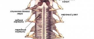
The third (III) ventricle is located in the midline and is a narrow vertical slit. Its lateral walls are formed by the medial surfaces of the thalamus and the subtubercular region, the anterior - by the columns of the fornix and the anterior commissure, the lower - by the formations of the hypothalamus, and the posterior - by the cerebral peduncles and the supracuberous region. The upper wall - the lid of the third ventricle - is the thinnest and consists of the soft membrane of the brain, lined on the side of the ventricular cavity with an epithelial plate (ependyma). The soft shell has a large number of blood vessels here, forming the choroid plexus. In front, the third ventricle communicates with the lateral ventricles (I - II) through the interventricular foramina, and behind it passes into the cerebral aqueduct.
Physiology of the diencephalon (human anatomy)
The thalamus is a sensitive subcortical nucleus. It is called the “collector of sensitivity”, since afferent pathways from all receptors converge to it, excluding olfactory ones. In the lateral nuclei of the thalamus there is a third neuron of the afferent pathways, the processes of which end in the sensitive zones of the cerebral cortex.
The main functions of the thalamus are integration (unification) of all types of sensitivity, comparison of information received through various communication channels, and assessment of its biological significance. The nuclei of the thalamus are divided according to their function into specific (the ascending afferent pathways end on the neurons of these nuclei), nonspecific (nuclei of the reticular formation) and associative. Through the associative nuclei, the thalamus is connected with all the motor subcortical nuclei: the striatum, the globus pallidus, the hypothalamus - and with the nuclei of the midbrain and medulla oblongata.
The study of the functions of the thalamus is carried out by cutting, irritation and destruction. A cat in which the incision is made above the diencephalon is very different from a cat in which the highest part of the central nervous system is the midbrain. She not only gets up and walks, that is, performs complexly coordinated movements, but also shows all the signs of emotional reactions. A light touch causes an angry reaction: the cat whips its tail, bares its teeth, growls, bites, and extends its claws. In humans, the thalamus plays a significant role in emotional behavior, characterized by peculiar facial expressions, gestures and shifts in the functions of internal organs. During emotional reactions, blood pressure rises, pulse and breathing quicken, and pupils dilate. The facial reaction of a person is innate. If you tickle the nose of a fetus for 5-6 months, you can see a typical grimace of displeasure (P.K. Anokhin). In animals, when the thalamus is irritated, motor and pain reactions occur: squealing, grumbling. The effect can be explained by the fact that impulses from the visual thalamus easily transfer to the associated motor subcortical nuclei.
In the clinic, symptoms of damage to the thalamus are severe headache, sleep disturbances, disturbances in sensitivity (increased or decreased), movements, their accuracy, proportionality, and the occurrence of violent involuntary movements.
The hypothalamus is the highest subcortical center of the autonomic nervous system. In this area there are centers that regulate all vegetative functions, ensuring the constancy of the internal environment of the body, as well as regulating fat, protein, carbohydrate and water-salt metabolism. In the activity of the autonomic nervous system, the hypothalamus plays the same important role as the red nuclei of the midbrain play in the regulation of skeletal-motor functions of the somatic nervous system.
The earliest studies of the function of the hypothalamus belong to Claude Bernard. He discovered that an injection into the diencephalon of a rabbit caused an increase in body temperature of almost 3°C. This classic experiment, which made it possible to discover the thermoregulation center in the hypothalamus, was called heat injection. After the destruction of the hypothalamus, the animal becomes poikilothermic, that is, it loses the ability to maintain a constant body temperature.
Later it was found that almost all organs innervated by the autonomic nervous system can be activated by irritation of the subtubercular region. In other words, all the effects that can be obtained by irritating the sympathetic and parasympathetic nerves are observed when irritating the hypothalamus.
Currently, the method of implanting electrodes is widely used to stimulate various brain structures. Using a special, so-called stereotaxic technique, electrodes are inserted into any given area of the brain through a burr hole in the skull. The electrodes are insulated throughout, only their tip is free. By connecting electrodes in a circuit, you can locally irritate certain areas.
When the anterior parts of the hypothalamus are irritated, parasympathetic effects occur: increased intestinal movements, separation of digestive juices, slowing down heart contractions, etc.; when the posterior sections are irritated, sympathetic effects are observed: increased heart rate, constriction of blood vessels, increased body temperature, etc. Consequently, parasympathetic centers are located in the anterior sections of the hypothalamus, and sympathetic centers in the posterior sections.
Since stimulation with the help of implanted electrodes is carried out on the animal without anesthesia, it is possible to judge the behavior of the animal. In Andersen's experiments on a goat with implanted electrodes, a center was discovered, the irritation of which causes unquenchable thirst - the thirst center. When irritated, the goat could drink up to 10 liters of water. By stimulating other areas, it was possible to force a well-fed animal to eat (hunger center).
The experiments of the Spanish scientist Delgado on a bull became widely known. An electrode was implanted into the bull's fear center. When an angry bull rushed at a bullfighter in the arena, the irritation was turned on and the bull retreated with clearly expressed signs of fear.
American researcher D. Olds proposed modifying the method: allowing the animal itself to make contact (self-irritation method). He believed that the animal would avoid unpleasant stimuli and, on the contrary, would strive to repeat pleasant ones. Experiments have shown that there are structures whose irritation causes an uncontrollable desire to repeat. The rats worked themselves to the point of exhaustion by pressing the lever up to 14,000 times. In addition, structures were discovered whose irritation apparently causes an unpleasant sensation, since the rat avoids pressing the lever a second time and runs away from it. The first center is obviously the center of pleasure, the second is the center of displeasure.
Extremely important for understanding the functions of the hypothalamus was the discovery in this part of the brain of receptors that detect changes in blood temperature (thermoreceptors), osmotic pressure (osmoreceptors) and blood composition (glucoreceptors).
Reflexes arise from receptors “turned into the blood” aimed at maintaining the constancy of the internal environment of the body - homeostasis. “Hungry” blood, irritating glucoreceptors, excites the food center: food reactions arise, aimed at searching and eating food.
One of the common manifestations of hypothalamic disease is a violation of water-salt metabolism, manifested in the release of large amounts of low-density urine. The disease is called diabetes insipidus.
The subcutaneous region is closely related to the activity of the pituitary gland. The hormones vasopressin and oxytocin are produced in large neurons of the supravisual and paraventricular nuclei of the hypothalamus. Hormones travel along axons to the posterior lobe of the pituitary gland, where they accumulate and then enter the blood.
A different relationship between the hypothalamus and the anterior pituitary gland. The vessels surrounding the nuclei of the hypothalamus unite into a system of veins, which reach the anterior lobe of the pituitary gland and here again break up into capillaries. With the blood, releasing factors, or releasing factors, enter the pituitary gland, stimulating the formation of hormones in its anterior lobe.
Reticular formation. In the brain stem and diencephalon, between its specific nuclei, there are clusters of neurons with numerous highly branching processes, forming a dense network. This system of neurons is called a network formation, or reticular formation. Special studies have shown that all the so-called specific pathways that carry certain types of sensitivity from receptors to sensitive areas of the cerebral cortex give off branches in the brain stem that end on the cells of the reticular formation. Streams of impulses from the periphery from extero-, intero- and proprioceptors support constant tonic excitation of the structures of the reticular formation.
Nonspecific pathways begin from the neurons of the reticular formation. They go up to the cerebral cortex and subcortical nuclei and down to the neurons of the spinal cord.
By irritating individual structures of the reticular formation, it was possible to reveal its function as a regulator of the functional state of the spinal cord and brain, as well as the most important regulator of muscle tone. The role of the reticular formation in the activity of the central nervous system is compared to the role of a regulator on a TV: without giving an image, it can change the volume of sound and illumination.
Irritation of the reticular formation does not cause a motor effect, but affects existing activity, inhibiting it or enhancing it. If in a cat a protective reflex is induced by short, rhythmic stimulation of the sensory nerve - flexion of the hind leg, and then against this background the reticular formation is irritated, then depending on the zone of irritation, the effect will be different: the spinal reflexes will either sharply intensify, or become weaker and disappear, i.e. e. will slow down. Inhibition occurs when the posterior parts of the brain stem are irritated, and reflexes are strengthened when the anterior parts are irritated. The corresponding zones of the reticular formation are called inhibitory and activating zones.
The reticular formation has an activating effect on the cerebral cortex, maintaining a state of wakefulness and concentrating attention. If you turn on stimulation of the reticular formation in a sleeping cat with electrodes implanted into the diencephalon, the cat will wake up and open its eyes. On the electroencephalogram, slow waves characteristic of sleep will disappear, and fast waves characteristic of the waking state will appear. The reticular formation has an ascending, generalized (encompassing the entire cortex) activating effect on the cerebral cortex. According to I.P. Pavlov, “the subcortex charges the cortex” (Fig. 115). In turn, the cerebral cortex regulates the activity of the network formation.
Rice. 115. Cat's brain (diagram). Facilitating (5) and inhibitory (4) zones of the reticular formation of the brain stem, as well as connections going to it from the cortex (1), subcortical nuclei (2) and cerebellum (3) (according to Magoon)
content .. 101 102 103 104 105 106 ..
Pituitary
The pituitary gland is one of the most important endocrine organs of the body. Its function is to produce tropic hormones, which, acting on target organs (most often the endocrine glands), regulate their activity. The pituitary gland is located in the diencephalon, its structure and functions are anatomically connected to the hypothalamus through the infundibulum, forming the hypothalamic-pituitary system. The pituitary gland itself lies in a bone formation - the sella turcica. Has three parts:
- Adenohypophysis (anterior lobe) - here tropic hormones are synthesized that regulate the activity of the glands: thyroid-stimulating, adrenocorticotropic, gonadotropic, somatotropic, luteotropic (prolactin). A tumor of the pituitary gland can develop from this part (link to one of the articles);
- The middle lobe is where melanocyte-stimulating hormone is synthesized, which affects pigment metabolism.
- Neurohypophysis (posterior lobe) - antidiuretic hormone and oxytocin are stored here, and from here these hormones are released into the blood. It is this part that is connected to the hypothalamus through the infundibulum.
The pituitary gland is called the most important gland of the body; the work of the other endocrine glands depends on its activity. Damage to this organ causes serious diseases: acromegaly, hyperthyroidism, premature puberty.
Morphofunctional organization of the diencephalon.
Intermediate the brain develops during embryogenesis. from the front brain bladder and forms the walls of the third brain. ventricle Topographically and functionally it is divided into epithalamus, thalamus and hypothalamus.
Epithalamus, or suprathalamic region.
, comp.
from the fornix located under the corpus callosum and from the internal gland. secretions of the epiphysis, which form the upper wall of the third ventricle. Thalamus
, or vision.
The tubercle is a voluminous, ovoid body consisting of a cluster of gray matter. The lower and lateral surface of the thalamus is fused with neighboring parts of the brain. The medial surface is visible. The tuberosity forms the lateral wall of the cavity of the third ventricle. The thalamus is a large subcortical formation, through which it enters the cortex. hemispheres pass through a variety of afferent pathways. The bottom of the third ventricle is formed by a group of structures, which are collectively called the hypothalamus or hypothalamus. The hypothalamus
contains a large number of nuclei and is the center of regulation of the visceral functions of the body.
Morphofunctional organization of the thalamus
Nerve. thalamic cells are grouped into large groups. number of nuclei, which are topographically divided into anterior, posterior, middle, medial and lateral groups. According to their function, thalamic nuclei can be differentiated into specific, nonspecific, associative and motor
. In the specific, or projection, nuclei of the thalamus, synaptic activity occurs. switching of sensory information from axons rising. afferent pathways to the next final neurons, the processes of which go in response. sensory projection areas of the cortex. hemispheres. The damage is specific. nuclei leads to irreversible loss of certainty. types of feelings. These facts indicate that it is specific. the nuclei are a transfer station on the path of afferent impulses from the peripheral. receptors for the bark. hemispheres. Among the main projection nuclei of the thalamus, one can distinguish the ventrobasal nucleus, which is specific. the core of the somatosensory system. It is divided into two parts - the ventral posteriolateral nucleus, to which the ascending fibers of the spinothalamic approach. tract and the medial loop, carrying information from the skin receptors of the trunk, proprioceptors of the muscles and articular apparatus, and the ventral posteriomedial nucleus, which are approached accordingly. paths from the nuclei of the trigeminal nerve, providing innervation to the facial part of the head. In specific The nuclei of the thalamus project afferents not only from exteroceptors and motor receptors. apparatus. Electrophysiological studies have shown that in the ventrobasal complex of the thalamus there is a region. projections of the vagus and splanchnic nerves, senses. fibers that carry information from interoceptors. At the same time, the thalamus, as a suprasegmental center of reflex activity, has connections with the hypothalamus, where the hl. vegetative centers. These connections are characteristic of the anterior group of thalamic nuclei and creation. a material prerequisite for the participation of this structure in the system of regulation of the visceral functions of the body.
Associative nuclei.
Unlike specific nuclei, they cannot be attributed to any one sensory system and receive afferent impulses from specific ones. projection nuclei. The three cores of this group have connections with Ch. associative regions cortex: the pillow core is connected with the associative zone of the parietal and temporal. cortex, the posterior lateral nucleus - with the parietal cortex, the medial dorsal nucleus - with the frontal lobe. The fourth nucleus - the anterior one - has connections with the limbic cortex. hemispheres. Apparently, the associative nuclei of participation. in higher integrative processes, but their function has not yet been sufficiently studied.
To motor
relative to nuclei
ventrolateral nucleus, which has input from the cerebellum and basal ganglia and at the same time gives projections to the motor. bark zone bol. hemispheres. This nucleus is involved in the regulation of movements. The last pain a group of thalamic nuclei is formed by nonspecific
nuclei, which are functionally associated with the reticular formation of the trunk. These nuclei include the median and introlaminar group of thalamic nuclei, which receive afferent input from fibers ascending from the reticular formation, and, in addition, have bilateral connections with specific. nuclei of the thalamus. Unlike specific nuclei with local projections in the cortex are phylogenetically more ancient, nonspecific. the nuclei exhibit diffuse projections throughout the entire region. bark. This structural feature determines their name and function, which consists of regulating excitability and electrical activity. activity of cortical neurons. Evidence of influence is non-specific. nuclei of the thalamus to the cortex were first obtained by researchers E. Dempsey and R. Morrison.
When comparing functions specifically. and nonspecific. The nuclei of the thalamus raise the question of the interaction of these two systems, which can influence the same neurons of the cortex. hemispheres. As shown by electrophysiological studies, bottom-up influences are non-specific. nuclei of the thalamus are manifested not in causing a discharge of the cortical neuron, but in changing its excitability. Non-specific. influences from the thalamus, increased. the excitability of cortical neurons facilitates their activity, while the responses of cortical neurons to impulses coming from specific. projection nuclei are intensified. However, it is nonspecific. influences may also have the opposite sign and exhibit an inhibitory effect on the discharges of cortical neurons. There is a point of view that is nonspecific. the nuclei are included in the ascending activating system and are intermediaries between the cortex and the reticular formation of the trunk, which receives information from all senses. T.O., non-specific. the nuclei transmit the activating influences of the reticular formation and participate in maintaining optimal tone of the cortex. However, this point of view is not generally accepted, and some researchers consider the reticular formation nonspecifically. the nuclei of the thalamus as two separate systems that control the excitability of cortical neurons.
Third ventricle
The structure of the diencephalon suggests the presence of a cavity through which the outflow of cerebrospinal fluid (CSF) occurs. The third ventricle is a narrow slit-like formation. It is connected to the first and second ventricles through the foramina of Monroy, and to the fourth through the aqueduct. There is a well-developed choroid plexus here. A tumor in this department may result in the diencephalon not being able to perform its functions correctly. The outflow of fluid will be impaired, and the optic tracts and other organs of this part of the brain may be compressed.
Thus, we can distinguish five main functions that the diencephalon has:
- Regulation of the activity of all major endocrine glands;
- Center for adaptation - regulation of temperature, water-salt balance, time of sleep and wakefulness, and other characteristics;
- Neurohumoral regulation - stimulation or inhibition of the activity of the external and internal secretion glands based on information from the environment and the state of the body;
- Center of sexual desire and pleasure;
- Center for the formation of protective reflexes: coughing, lacrimation, sneezing.
Nuclei and pathways of the midbrain
Midbrain
consists of ventral and dorsal parts. The ventral part represents the cerebral peduncles, between which the anterior perforation is located.
It is divided into a base and a tire. The pyramidal and frontopontine tracts pass through the base of the cerebral peduncle.
The tegmentum contains the spinothalamic and nuclear-thalamic tracts, the medial longitudinal fasciculus, extrapyramidal tracts (tr. tectospinalis, tr. rubrospinalis, tr. reticulospinahs), as well as gray matter (tegmental nuclei).
The dorsal part of the midbrain is formed by the roof plate; the anterior and posterior tubercles of the quadrigeminal are located here.
The midbrain cavity is the midbrain aqueduct
(aqueductus mesencephali) (Aqueduct of Sylvius). It is oriented along the axis of the brain, connecting the third and fourth ventricles. Its length is about 15 mm, the average diameter is 1-2 mm. There is a slight expansion in the middle part of the cerebral aqueduct.
The beginning of the aqueduct is located in the upper corner of the IV ventricle
, it is covered by the superior cerebral velum.
The hole through which the aqueduct opens into the third ventricle is located under the posterior commissure of the cerebrum.
Cranial nerve nuclei and other gray matter formations located in the midbrain
The following groups of gray matter formations are located in the midbrain: 1.
Nuclei of the oculomotor and trochlear nerves, partially the nuclei of the trigeminal nerve (mesencephalic nucleus, oral part of the nucleus of the spinal tract). 2. Mesencephalic gaze center. 3. Nuclei of the anterior and posterior colliculi of the quadrigeminal. 4. Nuclei of the posterior commissure (Darkshevich’s nucleus) and interstitial (Cajal’s nucleus). 5. Structures of the extrapyramidal system: caudate nucleus, lenticular nucleus (includes the putamen and globus pallidus), substantia nigra, subthalamic nucleus, part of the gray matter of the midbrain and diencephalon, as well as numerous connections of these formations with the superior and underlying structures of the brain and spinal cord.
6. Mesencephalic nuclei of the reticular formation of the trunk. 7. Central gray matter along the banks of the Sylvian aqueduct (subst. grisea centralis).
Midbrain pathways
Ascending pathways of the midbrain
: 1.
Spinothalamic tract (tr. spinothalamicus) (spinal loop - lemniscus spinalis). 2. Bulbothalamic tract (tr. bulbothalamicus) (medial loop - lemniscus medialis). The division of sensory pathways into spino- and bulbothalamic is arbitrary, since here they almost completely merge and represent a single tract. 3. Nuclear-thalamic tract (tr. nucleothalamicus) (trigeminal loop - lemniscus trigeminalis).
4. Auditory tract (tr. acusticus) (lateral loop - lemniscus lateralis). 5. Anterior spinocerebellar tract (tr. spinocerebellaris anterior) - Govers' tract. 6. Part of the visual pathway ending in the structures of the midbrain (the other part goes in transit to the lateral geniculate body and further to the cerebral cortex).
Along with the main pathways listed above, a number of other ascending pathways are also described in the bridge - the dorsosopercular (tr.
spinotectal), spinoreticular (tr. spinoreticularis).
Descending pathways of the midbrain
: 1.
Corticospinal pyramidal tract (tr. corticospinalis). 2. Corticonuclear pyramidal tract (tr. corticonuclearis). 3. Red nuclear spinal tract (tr. rubrospinalis). 4. Roof-spinal tract (tr. tectospinal). 5. Reticulospinal tract (tr. reticulospinalis).