The main venous collector, where venous blood from the head and neck collects, is the internal jugular vein, v. jugularis interna. It extends from the base of the skull to the supraclavicular fossa, where it merges with the subclavian vein, v. subclavia, forming the brachiocephalic vein, v. brachiocephalica.
The internal jugular vein collects most of the venous blood from the cranial cavity and from the soft tissues of the head and neck organs.
In addition to the internal jugular vein, venous blood from the soft tissues of the head and neck is also collected by the external jugular vein, v. jugularis externa.
Anatomy of the head and neck
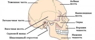
The head is presented as:
- The base of the skull, consisting of the frontal, temporal, zygomatic, maxillary and mandibular lobes. The parietal and occipital bones are also distinguished.
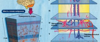
- The muscle layer covering the skeleton is presented in the form of striated tissue. They help fix the head and extend into the cervical region.
- Nerve endings line the head both on top of the muscle layer and inside the cranial vault.
- The facial part is “overloaded” with facial muscles that allow you to express emotions.
The cranial cavity contains the brain, without which normal human existence is impossible.
For its development and functioning, a constant supply of nutrients and oxygen is necessary. This can be provided by the circulatory system.
The neck is the part of the human body that connects the head to the body. It contains many structures that contribute to the development of the human brain and head as a whole.
The structure of the neck includes bone bases - vertebrae, a layer of striated muscles, and nerve endings. Circulatory and lymphatic system. The presented description is deeply superficial, because both the head and neck have a complex anatomical structure.
Functions of the veins of the head and neck
The main functions of the large blood circulation as a whole:
- removal of hormones;
- maintaining the tone of organs and systems;
- filling and deposition of blood;
- removal of breakdown products during metabolism;
- maintaining the reflexogenic zone;
- regulation of the circulatory process when blood pressure decreases due to heavy blood loss;
- transportation of hemomicrocirculatory bed to the heart muscle.
The blood supply to the cervical and head regions is carried out by arterial and venous vessels.
The presence of valves in the lumen of the vessel prevents blood from flowing back.
Perform specific functions including:
- supplying nutrients to the brain and other tissues of the head;
- delivery of blood from the heart and back;
- collection of carbon dioxide.
The composition of venous blood is represented by a gas mixture - carbon dioxide molecules. As well as waste products of formed elements - glucose, albumin.
Structure and features of work
The distribution of superficial and deep vessels is represented by a large number. They provide high reliability of uninterrupted power supply to the tissues of the skull. The superficial ones include:
- upper outlet;
- inferior outlet;
- superficial average;
- superior anastomotic;
- lower anastomotic.
The deep cerebral veins are represented by a large list. They are divided into upper and lower.
The main task of deep venous vessels is to collect blood. Flowing from the basal ganglia, vascular plexuses and diencephalon.
The upper group includes the following veins:
- Lateral ventricle;
- internal brain;
- superior thalamostriatal.
Representatives of the lower group:
- Paired basal, formed from the vessels of the medulla oblongata, pons;
- large brain
Other types:
- Cerebellar;
- sinuses of the dura mater;
- superior inferior and sagittal sinus;
- straight, transverse, occipital sinus;
- sigmoid and sinus drainage.
The veins of the neck are divided into vessels of the anterior and posterior sections. The vascular network of the upper body has some differences with the base of the body. Since the arterial branches are not duplicated with the venous branches and are located completely differently.
Internal jugular vein
Refers to the anterior cervical region, like the outer one.
Anatomically located in the jugular foramen and occupies most of the space. It is the largest main vessel of the cervical spine.
IJVs are presented in the form of slit-like channels, placed in a connective tissue membrane of increased density.
The lumen of the vessel is constantly open, due to which the outflow of blood occurs continuously, preventing stagnation.
At the levels of the larynx, the PU of the internal section comes into contact with the carotid artery on both sides. The outflow of blood is carried out through the sinus system. From the left and right sides, the collected blood enters the superior vena cava.
The upper section is equipped with valves. Here there is a merger with the subclavian vascular system. The IJV is divided into intracranial and external cranial branches.
Ducts of the IJV:
- snail plumbing;
- pharyngeal;
- meningeal;
- lingual;
- superior thyroid;
- middle thyroid;
- sternocleidomastoid.
Diploic veins of the brain
Located in the diploic substance of the cranial bones, they are represented by a developed canal system.
There are no valves in the lumens of the vessels, since the outflow of blood occurs from the bones of the skull. Inside the cranium, they communicate with the meningeal and sinuses of the meninges. The outside is covered with emissary veins.
The group includes the following representatives of the venous system:
- Frontal diplodic;
- Anterior temporal diploic;
- Posterior temporal;
- Occipital.
Diploic veins of the brain are their own cerebral veins. They lie deep in the canals of the bone, originating from the spongy substance.
Emissary veins
The main function is to connect the venous vessels of the skin with deeply located vessels.
To perform its task, the emissary system passes through a series of cranial foramina. According to their localization, there is a classification:
- Occipital;
- mastoid;
- parietal;
- condylar.
The name corresponds to the location of the bones. The emissary veins are designated in the diagram as vessels of the systemic circulation of the head vault.
Superior and inferior ophthalmic veins
Due to the branched network, they connect with the facial, frontal and paranasal veins. As in previous cases, an inextricable connection is formed with the sinuses of the dura mater.
The vessels do not have a valve apparatus and therefore the blood flow can vary from facial to cavernous sinusitis. Due to this structure of the orbit, the upper and lower eyelids are susceptible to inflammatory processes.
Preventive actions
The best prevention of swollen vessels on the head is proper nutrition, adherence to a work and rest schedule, and giving up bad habits. In addition, you must:
- If possible, avoid stressful situations and emotional stress;
- undergo timely vascular diagnostics;
- monitor blood pressure levels;
- avoid heavy physical labor;
- take a contrast shower daily;
- tune in to a positive mood.
If the veins on the forehead are swollen and there is a deterioration in the general condition, then you should immediately seek help from a doctor. Only a specialist can make an accurate diagnosis and prescribe appropriate treatment. Therapy in the early stages of the disease will prevent complications and stop the development of pathological processes.
Vein diseases of the head and neck
The main disease is a violation or difficulty in the outflow of venous blood. Pathology develops for a number of reasons:
- Tumors compressing blood vessels;
- skull injuries of various types;
- hypertension in combination with arrhythmia;
- violations of the venous supply system;
- alcohol intoxication.
Venous stagnation brings many problems and manifests itself in the form of the following symptoms:
- noise in ears;
- headache, worse after exercise;
- muscle weakness;
- poor memory;
- swelling and cyanosis of the skin;
- dizziness to the point of fainting.
Osteochondrosis is the main cause of blood stagnation and difficulty in its outflow in the lumen of the venous vessels.
The disease must be treated on time. Since congestion leads to ischemia of brain tissue or the entire organ.
Clinical manifestations of cerebral venous insufficiency
Symptoms and methods of eliminating cerebral venous insufficiency
At the initial stage of development of the disease, there are no obvious signs. Characteristic symptoms appear with a significant deterioration in the condition of blood vessels:
- discomfort when tilting the head;
- headache or dizziness with a sudden change in body position or changes in weather conditions;
- worsening sleep, fainting;
- blurred vision, noise in the head;
- redness of the whites of the eyes, swelling of the eyelids;
- cyanosis of the facial skin;
- numbness of the limbs;
- epilepsy attacks.
At an advanced stage, mental disorders develop - delusions, hallucinations. For a patient, even a mild experience can develop into neurosis, hysteria, or depression.
Features and structure of the superficial vein system
The structure of the system of superficial vessels is presented in the form of several groups:
- Drainage of blood from the cerebral cortex. More precisely, the upper and lower superficial veins originate from the white matter of the hemisphere;
- The superficial mesencephalon collects biological fluid from the telencephalon.
As a result of anastomosis, a venous network is formed on the surface of the skull.
Collateral blood flow is possible in any direction. The superior anastomotic vein plays a special role. It connects the superior sagittal, cavernous and parietal sinuses with the temporal sinuses.
The lower anastomotic connects the transverse venous sinus with the cavernous or sphenoparietal sinus. And also temporal and parietal with occipital.
Treatment
Swelling of the veins in the temples is not a separate disease. This may be just one of the symptoms of various pathologies. Therefore, treatment will depend on the cause of this manifestation.
For temporal arteritis, corticosteroids (Prednisolone) and cytostatics (Azathioprine, Methotrexate) are prescribed. This treatment is aimed at stopping the autoimmune process. At the same time, it is recommended to take blood thinners (Aspirin).
Treatment of vein thrombosis is carried out using Heparin injections. This remedy prevents the formation of clots in blood vessels. Venoprotectors are also prescribed: Hepatrombin, Ascorutin and vasodilating drugs: Dibazol, Agapurin.
If pain and throbbing in the temples is associated with hypertension, then antihypertensive drugs are prescribed: Enap, Enalapril. For atherosclerosis, antilipid drugs are prescribed: Lovastatin, Rosuvastatin. It is recommended to follow a diet with limited fat.
When veins are swollen due to spasms and neurological pathologies, the prescription of antispasmodics and anti-inflammatory drugs is indicated: Spazgana, Ketanova, Pentalgina.
If there is no effect from conservative treatment, surgical intervention must be resorted to. Vascular surgery is performed - angioprosthetics or bypass surgery. This treatment is indicated in the presence of complications or severe thrombosis. Surgery is also necessary when a vessel is compressed by a tumor. In this case, the tumor must be removed.
Other veins located in the head and neck
A huge number of vessels are present in the muscle and bone layers of the brain. The name of which refers to the organs adjacent to them. In order to know all the names and functions, you must carefully study anatomy textbooks.
Large classification of veins and their functions
Eyes and sockets:
- Top, bottom, central;
- vorticose;
- episcleral.
Functions: outflow of blood from the contents of the orbits into the inferior ophthalmic vein.
Face:
- The postmandibular and its tributaries - the parotid, anterior auricular, temporomandibular;
- Palatine external;
- Submental;
- Deep facial;
- Superior inferior labial;
- Lower and upper eyelids;
- External nasal.
Veins play a huge role in the human body. Each vein has its own name and performs a specific function.
If a person has diseases that are associated with venous problems. It is necessary to begin their treatment immediately. Otherwise, irreversible consequences are possible.
Temporal arteritis
Temporal arteritis occurs more often in older people. Its cause may be autoimmune processes or past infectious diseases. Inflammation occurs in the walls of blood vessels, accompanied by the formation of pathological large cells. Therefore, doctors call this disease giant cell temporal arteritis.
The patient exhibits the following signs of illness:
- swelling of the veins in the temples;
- severe pain in the temple, radiating to the neck, shoulder, forehead and cheeks;
- increased pain when chewing;
- drooping eyelids;
- double vision;
- decreased visual acuity;
- swelling and redness of the temporal region.
The disease must be treated immediately at the diagnostic stage. Without treatment, this pathology can lead to vision loss and stroke.