Diseases of the central nervous system of the organic type occur in the practice of doctors in approximately 20% of cases. They are represented by strokes, neuroinfections, tumors, benign and malignant (less often). Manifestations of pathological processes are variable in nature, always threatening.
Symptomatic epilepsy is a variant of a neurological disease. Accompanied by a sharp increase and chaotization of electrical impulses generated by the brain. Typical signs are always the same, depending on the location of the outbreak.
Unlike the cryptogenic form, the secondary one is always caused by the presence of structural, anatomical changes in the cerebral structures. In the first case, the cause is either not clear, or it is not possible to identify it at all (idiopathic variety).
Another name for the pathology is secondary symptomatic epilepsy, which indicates its origin and conditionality with other diseases. Diagnosis in this case is not very difficult. The prevalence is up to 70% of all identified cases. With a total number of patients in the world of about 50 million people.
The disease has its own diagnosis code according to ICD-10 - G40.2.
Causes
Causes of the disease:
- brain tumors, predominantly temporal or frontal localization;
- cancerous lesions of the vessels of the neck and/or brain;
- traumatic brain injury - possible delayed onset of attacks (after several months or even years);
- Bourneville disease (tuberous sclerosis), characterized by the formation of benign tumors in various organs;
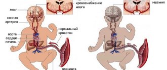
- intrauterine hypoxia;
- fetal asphyxia;
- intrauterine infection;
- injuries received by the child during childbirth;
- neuroinfections (meningitis, encephalitis, arachnoiditis);
- abscesses in the brain;
- brain damage due to rheumatism, malaria;
- childhood infections (measles, scarlet fever, diphtheria);
- poisoning with toxic substances;
- intoxication with endotoxins in decompensated failure (renal, liver), diabetes mellitus;
- organic aciduria (disorder of amino acid metabolism);
- violation of organogenesis (the last phase of embryonic development);
- violation of histogenesis (a complex of processes leading to the formation of tissue in the embryo).
An unfavorable background for the manifestation of the disease is atherosclerosis, stroke, cerebral palsy, hepatocerebral dystrophy (pathological accumulation of copper in the liver), torsion dystonia (uncontrolled contractions of different muscle groups), chorea (erratic unbalanced movements similar to dancing), hippocampal sclerosis.
General information about focal epilepsy
The definition of “focal epilepsy” combines all forms of epileptic paroxysms that arise due to the presence of a local focus of increased epi-activity in cerebral structures.
Epileptic activity begins focally, but can spread from the focus of excitation to the surrounding brain tissue, which causes secondary generalization of the seizure. It is important to differentiate paroxysms of FE and attacks of generalized epilepsy with a primary diffuse nature of excitation. In addition, there is a multifocal form of epilepsy. In this form of epilepsy, there are several local epileptogenic zones in the brain. Approximately 82% of all epileptic syndromes are focal epilepsy, and in 75% of cases it begins in childhood. Most often it occurs against the background of traumatic, ischemic or infectious damage, or disorders of brain development. Secondary focal epilepsy of this nature is diagnosed in 71% of all patients suffering from epilepsy.
Classification
According to the ICD-10 (International Classification of Diseases) code, symptomatic epilepsy is classified as G40.
Types of epilepsy according to the prevalence of the process:
- symptomatic generalized epilepsy (the lesion is distributed throughout the brain);
- symptomatic focal epilepsy (the focus is localized in one lobe).
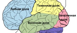
The affected area differs:
- symptomatic temporal lobe epilepsy (44% of all cases, in children - 25.2%) - characterized by impaired consciousness, automatisms, hallucinations;
- symptomatic frontal epilepsy (24%, in children - 27.1%) - characterized by rapid onset, short duration (up to one minute), high frequency, repetitive movements;
- localized in the occipital lobe (10%, in children - 7.5%) - characterized by visual impairment, hallucinations, automatic blinking, head twitching;
- localized in the parietal lobe (1%, in children - 14.9%) - characterized by pain, muscle spasms, incorrect perception of temperature, in adults - an acute desire for sexual intimacy.
Idiopathic or cryptogenic epilepsy occurs in 60–70% of cases of epileptic seizures. In this form, the source of spread of the spike wave (discharge of excitation) is known, but the causes of the development of the disease are unknown.
Preventive measures
Specific measures to prevent symptomatic epilepsy have not been developed. To reduce the risk of such a problem, it is necessary to promptly treat infectious and rheumatological diseases. When staying in nature, you need to use special clothing and protection against ticks, whose bites can cause the development of tick-borne encephalitis.
To prevent the development of pathology, it is necessary to get rid of alcohol and drug addiction. To reduce the risk of developing epilepsy in a child, a woman needs to plan her pregnancy to reduce the risk of developing hypoxia in the fetus and birth injuries.
Symptoms
The clinical picture depends entirely on the form of the disease and the location of the source of excitation. The main signs of a generalized seizure are loss of consciousness and severe autonomic disorders; convulsions may be absent.
Depending on the motor phenomena, epileptic seizures are distinguished:
- Convulsive deployed. They are characterized by a large convulsive attack with loss of consciousness, tonic-clonic convulsions, which occur in two phases. The first phase, tonic, lasts up to 20 seconds, convulsive tension of the entire skeletal muscles is observed. The second phase, clonic, lasts up to 40 seconds, alternating muscle spasms with muscle relaxations is observed. During the attack and after the patient is in an epileptic coma. When emerging from a coma, disorientation (confused consciousness) is initially observed.
- Convulsive, undeveloped. Only tonic or only clonic convulsions are characteristic. After the attack, the patient is agitated. There is no epileptic coma.
- Massive myoclonus. The attack develops at lightning speed. Characteristic are myoclonic serial twitches, following each other.
- Nonconvulsive typical (simple) absence seizure. Characterized by loss of consciousness, lack of motor activity, moderate dilation of the pupils, hyperemia or pallor of the skin. Absence lasts for several seconds. People around and even the patient himself usually do not notice the seizure.
- Nonconvulsive complex absence. Automatisms (fingering with hands, rolling of eyes, twitching of eyelids) are added to the manifestations of simple absence seizure. With a sharp decrease in muscle tone, the patient may fall as if knocked down.
Symptomatic partial epilepsy is characterized by different symptoms, depending on in which part of the brain the epileptic focus arose.
Characteristic manifestations of damage:
- premotor cortex (in front of the precentral gyrus): the patient turns his head and eyes in the direction opposite to the source of excitation;
- postcentral gyrus: Jacksonian seizures (motor, sensory, sensorimotor disorders);
- occipital lobe cortex: photopsia (visual hallucinations) in the field of vision opposite to the focus of excitation;
- superior temporal gyrus: auditory hallucinations;
- mediobasal temporal cortex: taste and olfactory hallucinations.
Symptomatic epilepsy can be mild or severe. In a mild form, consciousness is not lost; there is a loss of control over different parts of the body and unusual sensations. In severe cases, the patient loses touch with reality, the person is unable to control movements, and muscle contractions are observed.
Partial seizures are simple, involving a small number of neurons, and occur mostly without loss of consciousness. Complex partial seizures occur with impaired consciousness, characterized by disorientation, unusual perception of the body and the surrounding space, various hallucinations, and the “déjà vu” effect.
The patient cannot describe unusual perceptions, does not recognize objects, or perceives them as unrealistic. Sometimes the attack is perceived as a dream. Another characteristic manifestation is automatic, stereotypical inappropriate movements.
Other psychic phenomena:
- unusual sensations in various parts of the body;
- nausea;
- intrusive memory;
- attack of fear;
- mental leaps.
Secondarily generalized partial seizures end in convulsions. An attack can be preceded by an “aura” - a symptom of epilepsy, characterized by any patient sensation or experience (depending on the location of the focus) that regularly appears immediately before an attack. After the attack, the patient may remember the aura.
In some cases, symptomatic focal epilepsy in children manifests itself in the form of West syndrome, which is characterized by frequent seizures recorded by an encephalogram. Male infants under eight months of age are more affected. Psychomotor development is impaired, anticonvulsant therapy is ineffective.
In 10.3% of children, simultaneous excitation of several epifoci is noted. In almost 15%, the focus during an attack is not diagnostically identified due to the absence of EEG abnormalities or the presence of conflicting data. Of this group, 71% had a clinical picture consistent with temporal lobe epilepsy.
Parietal epilepsy, seizures
In two thirds of cases, patients suffer from somatosensory attacks: paresthesia, dysesthesia, painful sensations (burning, numbness or tingling) that spread to the face and hands.
Somatic illusions accompany parietal epilepsy slightly less frequently than somatosensory seizures.
A person feels an “incorrectness” in posture, movements or position of the limbs, or less often, he does not feel the limbs or the whole body at all. Similar illusions occur if the non-dominant hemisphere is affected.
Cases of sensations in the genitals and orgasm are described. If the dominant hemisphere is affected, linguistic difficulties occur - alexia with dyscalculia and agraphia. In case of damage to the non-dominant hemisphere of the brain, orientation in space is disrupted.
Simple focal sensory seizures can spread to extraparietal areas and cause unilateral focal cloning (in more than half of patients), eye and head version (40%), tonic settings of any limb (about 30%) and automatisms (about 20%) .
Most patients experience tonic-clonic seizures of a secondary generalized nature.
Diagnostics
Epilepsy is treated and diagnosed by an epileptologist, psychiatrist or neuropsychiatrist. During the examination, consultation with other highly specialized specialists (ophthalmologist, otoneurologist, neurosurgeon, geneticist) is often necessary. The doctor takes a detailed history, including family history, and examines the patient.
The following examination options are prescribed:
- EEG (electroencephalogram). The main method for diagnosing different types of epilepsy. The most indicative EEG is in dynamics, with a load, during sleep, with provocation. For different forms of epilepsy, the encephalogram records different data. During a generalized seizure, bilateral synchronized discharges are recorded. Not for all types of disease the EEG shows deviations from the norm. In this case, an EEG with sleep deprivation is used - biorhythms are recorded on an encephalogram after the patient has been awake for a long time. This diagnosis makes it possible to detect the convulsive readiness of neurons, which is not visible on a conventional EEG.
- Visual diagnostics - MRI, CT, PET/CT (positron emission tomography with computed tomography). Allows you to identify organic brain lesions, the consequences of neuroinfections, sclerotic changes, anomalies in the structure of brain structures.
- Angiography. Assessment of the state of cerebral vessels and blood supply to the brain. If angiography does not provide a complete picture, an MRI of the cerebral vasculature is performed.
- Neuropsychological diagnostics. A set of various special tests and techniques (Russian psychiatrists use the Luriev battery), during which higher mental functions are assessed, allowing one to judge disorders in certain lobes of the brain.
- Study of cerebrospinal fluid. Indicated for suspected neuroinfection. The type of pathogen that disrupts the function of the central nervous system and brain is identified in the liquid.
- Laboratory tests: blood, urine, blood biochemistry, karyotyping (genetic screening), hormones (if necessary).
If the diagnosis consistently excludes diseases that can cause epileptic seizures, cryptogenic (idiopathic) epilepsy is diagnosed. Differential diagnosis is carried out between various forms of the disease, based mainly on the characteristic clinical picture and EEG data.
Consequences of gliosis
The main reason for the appearance of gliosis foci is various damage to the central nervous system, which is associated with all kinds of changes and pathological processes. The consequences of cerebral gliosis often include brain encephalitis, pressure surges, multiple sclerosis, circulatory disorders in various tissues and organs, and hypertensive crises. Any disturbance in the functioning of the central nervous system always leads to problems in the functioning of all organs and systems of the body.
Gliosis of the brain
- This is a fairly serious pathology that can be a consequence of various diseases. In order for the treatment to be as effective as possible, it is recommended that all efforts be directed towards eliminating the cause of the development of gliosis - this is what will stop the process that causes irreparable damage to your body.
The condition that we will consider in this article is characterized by the replacement of damaged neurons with special cellular structures, which are called gliosis. It is a scar-like body with a fine structure that prevents damage to adjacent brain tissue and protects it. Cells that are deformed or die as a result of any damage to the central nervous system are replaced by other cells called glia. They perform very important functions in the functioning of the brain: delimiting, supporting, protective, trophic, secretory and consist of intercellular substance and different types of cellular elements. Thus, gliosis of the brain is somewhat similar to scars or scars on the tissue of the central nervous system. However, such cells are not able to completely replace lost neurons, so neurological deficits are observed.
This process has been studied by specialists for a long time. Thus, a number of studies were carried out when a special extract from the blood of an elderly person was added to glial cells. The results showed that in this case, glia began to multiply very quickly. In old age, neurons constantly die, and glial cells are formed instead. As a result of this process, the brain acquires a sponge-like structure. And because of this, age-related changes occur: memory weakens, reaction speed deteriorates, and coordination of movements is impaired. The experiment showed that neurons die due to increased proliferation of glia. The death of neurons is caused by constant consumption of large quantities of fatty foods. Many researchers have come to the conclusion that gliosis is an attempt by the hypothalamus to recover.
Gliosis can be congenital, but it is an extremely rare form of the disease. Mutations occur in certain genes that determine the replacement of neurons by glial cells in the fifth month of a newborn’s life. Children with this diagnosis do not live long and rarely live past the age of three.
The central nervous system is formed from the following types of cells:
- neurons that form and transmit impulses;
- ependyma lining the spinal cord and ventricles of the brain;
- neuroglia, acting as auxiliary tissue, the volume of which reaches 50% of the entire central nervous system.
With the proliferation of glial cells, glial foci of the brain are formed. In this case, the amount of gliosis is calculated as the ratio of glial cells to other CNS cells per unit. volume. That is, this indicator characterizes a value proportional to the amount of healing in the body.
Treatment
For single epileptic attacks in children, during a normal pregnancy, or when an attack is provoked by sleep deprivation, specific treatment is not required.
If there have already been episodes of epileptic seizures in the anamnesis, even many years ago, there is any risk factor that can cause paroxysm, there is confidence in the manifestation of idiopathic epilepsy, treatment is prescribed after the first epileptic seizure.
Symptomatic epilepsy is treated with anticonvulsant drugs. Initially, only one medication is selected (monotherapy), which is taken for at least three months, after which the therapeutic effect is assessed. If positive dynamics are recorded, the patient takes the drug regularly for two to three years. Changing the medication is undesirable: this may cause immunity to the drugs of this group and worsen the patient’s condition.
The drug is prescribed with a gradual increase in dosage to the therapeutic one. It is canceled by gradually reducing the dose, otherwise abrupt withdrawal will provoke an epileptic attack. If one drug is ineffective, another drug from the same group is prescribed, but the first one is gradually withdrawn only after the dose of the second drug has been brought to the therapeutic level.
A combination of anticonvulsants is prescribed for life-threatening conditions. Basically, two drugs are used, their interactions and side effects are taken into account. It is believed that a combination of three or more drugs is ineffective because it is almost impossible to assess their cumulative therapeutic effect, and the cumulative side effects can significantly worsen the quality of life.
Children from an early age are shown neuropsychological correction - a set of techniques aimed at replacing impaired brain functions with compensatory ones so that the child is able to control behavior and learn independently.
If the cause of the disease is hemorrhage, neoplasm, aneurysm, or head injury, surgical intervention is resorted to with further prescription of anticonvulsant therapy.
Indications and contraindications for surgical treatment
There are a number of conditions under which surgery is necessary. Surgery is often prescribed when drug therapy is ineffective.
In addition, such a radical approach to the treatment of epilepsy is prescribed when the patient experiences severe side effects from taking medications, or is intolerant to certain components of antiepileptic drugs.
Surgical therapy for epilepsy is required in cases where the pathology occurs in a complicated form and leads to a deterioration in the patient’s quality of life. Operations are also performed in cases where the focus of increased epileptic activity has a clear localization and can be removed without the risk of severe neurological disorders.
Among other things, indications for surgical treatment are cases where epilepsy has developed against the background of hemorrhages, cysts, aneurysms, tumors and abscesses of the brain.
A contraindication for surgical treatment is progressive degenerative or infectious-inflammatory damage to brain structures.
Surgical interventions are not prescribed for patients who have severe mental disorders.
A high risk of stroke or progressive cerebrovascular accident is also a contraindication.
Forecasts
It is difficult to predict the course of epilepsy - it all depends on the etiology, the extent of brain damage, the form of the disease, and the severity of clinical manifestations. The most pessimistic prognosis is for children with seizures due to birth trauma and neuroinfection. Relatively favorable prognosis for TBI.
In the generalized form with frequent attacks, the prognosis is less favorable. Possible deterioration in social adaptation, and in children - a lag in mental development. Remission can be achieved in 35–65% of patients. With localized focal partial epilepsy, the course of the disease and prognosis are more favorable. Much depends on a well-chosen medication, sensitivity to medications, and timely initiation of therapy.
Foci of gliosis
This term refers to pathological proliferations of glial cells - they are the ones that replace destroyed neurons. It should be noted that the appearance of foci of gliosis can be a consequence of various diseases and pathological processes. These include:
- Tuberous sclerosis.
- Multiple sclerosis.
- Inflammations – encephalitis of various origins.
- Birth injuries.
- Hypoxia.
- Long-term hypertension.
- Chronic form of hypertensive encephalopathy.