Structure and functions of the brain
| Name department | Department structures and structure | Functions |
| Medulla | ||
| Bridge | ||
| Cerebellum | ||
| Midbrain | ||
| Diencephalon | Thalamus | |
| Hypothalamus | ||
| Telencephalon: big hemispheres |
Task 2
. Using a dictionary on the topic “Local brain systems and their functional organization,” fill out the table:
Table 2.
Local brain systems and their functional organization
| Lobes of the cerebral cortex | Functional areas of the cortex (sensory, motor, associative) | Characteristic disorders in case of defeat given lobe of the brain |
| Occipital lobe | ||
| Temporal lobe | ||
| Parietal lobe | ||
| Frontal lobe |
Interesting facts about the brain
The brain as a substrate of mental processes is a single supersystem, a single whole, consisting, however, of differentiated sections (areas or zones) that perform different roles in the implementation of mental functions.
Impossibly interesting video from Sergei Vyacheslavovich Savelyev himself
The Mystery of the Two Hemispheres of the Human Brain
Three functional blocks of the brain according to A.R. Luria
We can distinguish 3 main functional blocks, or three main brain apparatuses, the participation of which is necessary for the implementation of speech activity. I block of regulation of tone and wakefulness (level of involuntary self-regulation and self-organization)
Anatomy:
Systems that provide and regulate cortical tone are located in the stem and subcortical regions of the brain: - reticular formation of the brain stem;
- nonspecific structures of the midbrain, its diencephalic sections; - limbic system; - mediobasal cortex of the frontal and temporal lobes of the brain; - striopallidal system. Functions:
In order to ensure the full flow of speech processes, a person must be in a state of wakefulness.
Only under optimal waking conditions can a person receive and process information, recall the necessary selective systems of connections in memory, program his activities and control the course of his mental processes, correcting errors and maintaining the direction of his activities. The systems of the first block of the brain are in a dual relationship with the cortex, toning it and at the same time experiencing its regulatory influence in accordance with the tasks assigned to the organisms.
These systems are built according to the type of nonspecific nervous network, which carries out its function through a gradual, gradual change in states and is not directly related to the reception and processing of information coming from outside, or to the development of intentions, plans and behavior programs.
A significant part of a person’s activity is determined by intentions and plans, prospects and programs that are formed in the process of his conscious life, are social in nature and are carried out with the close participation of first external and then his internal speech. Every idea formulated in a speech pursues a certain goal and evokes a whole program of actions aimed at achieving this goal. Carrying out a plan or achieving a goal requires a certain amount of energy and can only be achieved with a certain level of activity. In 1949, Magoon and Moruzzi discovered that in the brain stem there is a special nerve formation that is adapted to act as a mechanism that regulates the state of the cerebral cortex, i.e. is able to change her tone and ensure her wakefulness. This formation is built like a nervous network, in which the bodies of nerve cells are interspersed, connected to each other by short processes. Through the network of this formation, called the reticular formation
, excitation does not spread in separate, isolated impulses, but gradually, gradually changing its level and, thus, modulating the state of the entire nervous apparatus.
Currently, the point of view about the important and specific role of not only cortical, but also subcortical structures in mental activity with the leading participation of the cerebral cortex has become generally accepted. II block of reception, processing and storage of information (operational) Anatomy:
- visual area (occipital - indicated in blue in the figure below); - auditory region (temporal - indicated in yellow in the figure below); - general sensitive area (parietal - indicated in green in the figure below).
Functions:
According to their functional characteristics, the systems of this block are adapted to receive exteroceptive stimuli coming to the brain from peripheral receptors, to crush them into a huge number of components (i.e., to analyze them into the smallest constituent details) and to combine them into the necessary dynamic functional structures (i.e., to synthesize them into entire functional systems).
This functional block of the brain has high modality specificity. The systems of this block have a hierarchical structure, breaking up into primary (projection) zones, which receive information and split it into the smallest components, secondary (projection-associative) zones, which provide coding (synthesis) of these components and transform the somatotopic projection into a functional organization , and tertiary zones (overlap zones), ensuring the joint work of various analyzers and the development of supramodal (symbolic) schemes that underlie complex forms of cognitive activity. The parietal region ensures the functioning of the skin-kinesthetic analyzer, which is associated with various types of skin sensitivity, touch, muscle-joint sense, and acts as the basis for the formation of a diagram of one’s own body, articulation, and subtle object movements. Also responsible for spatial orientation. The old section (posterior central gyrus) is the center of sensory projections of the body. The new department includes centers in charge of complex types of deep sensitivity (muscular-articular, two-dimensional-spatial sense, sense of weight and range of motion, sense of recognizing objects by touch. The newer department contains centers of praxis. Praxis - which have become automated in the process of repetition and exercises are purposeful movements that are developed during training and constant practice during individual life (walking, eating, dressing, mechanical elements of writing, various types of work activity).From birth to 20 years, neurons in this area undergo significant changes: their size increases, especially in the first seven years, the shape and nature of branching change. The first year of life is considered as the optimal age for the formation of the sensory base of subsequent development. During this period, the development of motor and kinesthetic zones occurs, and their connection with visual-tactile processes is formed. The occipital region provides the work of the central link of the visual analyzer. Responsible for processing visual information and organizing complex processes of visual perception. The development of the nervous structures of the peripheral and central parts of the visual analyzer begins in prenatal development. The width of the cortex in the occipital region varies from birth to 20 years, but its most dramatic growth occurs during the first year of life. The size corresponding to the adult state, depending on the type of neurons, is achieved by 8-12 and 13-16 years. The temporal region is associated with the work of the auditory analyzer. Functions: perception of auditory, gustatory, olfactory sensations; analysis and synthesis of speech sounds; memory mechanisms. After birth, the most important stage is the age of 2 years, when the child’s temporal region begins to approach the size of the temporal region of an adult. After 2 years, there is a slight slowdown in the growth and development of cortical cells and the width of the cortex. By the age of 7, the size of the surface of the child’s temporal cortex almost corresponds to the size of the adult’s cortex. In the posterior parts of the cerebral hemispheres, at the junction of the temporal, parietal and occipital regions, there is the TPO zone, which is a zone of “overlap”, interaction of different analytical systems, and provides complex, supramodal integrative functions. Here, complete differentiation of the cortex occurs most late and the most significant morphological rearrangements occur, associated with the asynchronous development of layers, sublayers and cytoarchitectonics in different fields. In the first two years of life, the width of the fields increases 2 times and by 7 years the width of the fields increases 3 times. From 8 to 12 years, the growth of the cortex in width in the left hemisphere is more intense than in the right. III block of programming, regulation and control of complex forms of activity (level of voluntary self-regulation and self-organization) Anatomy:
Prefrontal parts of the brain (indicated in orange in the figures above): the anterior parts of the cerebral hemispheres, in front of the anterior central gyrus.
Occupies 25-28% of the cortex, weight = 450 g. Functions:
Organization of voluntary movements;
organization of motor mechanisms of speech; regulation of complex behaviors; regulation of memory processes. A person forms plans and programs for his actions, monitors their implementation and regulates his behavior, bringing it in accordance with these plans and programs; finally, he controls his conscious activity by comparing the effect of his actions with his original intentions. The “exit gate” of this block is the motor cortex area (4th Brodmann area), layer V of which contains giant pyramidal cells of Betz, the fibers from which go to the motor nuclei of the spinal cord, and from there to the muscles, forming part of the large pyramidal tract. This zone of the cortex is topographically structured so that its upper sections are the source of fibers going to the lower extremities of the opposite side, the middle sections are the source of fibers going to the upper extremities of the opposite side, and the lower sections are the source of fibers going to the muscles of the face, lips and tongue. To a large extent, this zone contains organs that have the greatest functional significance and require the most fine regulation. The most significant part is the prefrontal parts of the brain, which, due to the absence of pyramidal cells in their composition and the presence of a large number of small cells (granules), are sometimes called the granular frontal cortex. It is these sections of the brain that play a decisive role in the formation of intentions and programs, in the regulation and control of the most complex forms of human behavior. The prefrontal region of the brain has a rich system of connections both with the underlying parts of the brain and the corresponding parts of the reticular formation, and with almost all other convexital parts of the cortex. The frontal lobes of the brain have powerful bundles of ascending and descending connections with the reticular formation.
These areas of the neocortex receive impulses from the systems of the first functional block, “charging” from it, at the same time they have an intense modulating effect on the formation of the reticular formation, giving its activating impulses a differentiated character and bringing them into line with those dynamic patterns of behavior that are formed directly in the frontal cortex of the brain. A distinctive feature of the processes of regulation of conscious activity in a person is that this regulation occurs in him with the close participation of speech, since many higher mental processes are formed and proceed on the basis of speech activity, which in the early stages of development is developed in nature, and then becomes increasingly reduced . Significant stages of microstructural changes in the ensemble organization of the prefrontal regions of the frontal region occur at 1 year, 3 years, 5-6 years, 9-10 years, 12-14 years, 18-20 years. A number of authors, based on a comparison of data on increases in brain weight, skull size, and changes in nervous activity, have identified individual periods of accelerated development of the frontal lobes of the brain. At the age of 3-4 months, the first such segment is observed, at this time the child begins to reach the objects around him. The next acceleration occurs at about 8 months, when the child begins to crawl and search for hidden objects, then at 12 months, when there is a significant improvement in finding objects. The interval between 1.5 and 2 years correlates with the rapid development of speech. The period between 3 and 6 years is accompanied by the consistent involvement of language as a means of planning actions. Subsequent periods of activity in the frontal lobes of the brain at 9, 12, 15 and 18-20 years are associated with different phases of improvement in thinking.
The main pathways of the brainstem and spinal cord
In the white matter of the brain stem and spinal cord there are conductors of the ascending and descending directions. Descending pathways carry motor impulses from the cerebral cortex (corticospinal, pyramidal tract) to the reflex apparatus of the spinal cord, as well as impulses that contribute to the implementation of a motor act (extrapyramidal tracts) from various parts of the subcortical formations and the brain stem. Descending motor conductors end on peripheral motor neurons of the spinal cord segment by segment. The overlying parts of the central nervous system have a significant influence on the reflex activity of the spinal cord. They inhibit the reflex mechanisms of the spinal cord's own apparatus. Thus, with pathological switching off of the pyramidal tracts, the spinal cord’s own reflex mechanisms are disinhibited. At the same time, spinal cord reflexes and muscle tone are enhanced. In addition, protective reflexes are identified that are observed in the burrow only in newborns and children in the first months of life. The ascending pathways transmit sensitive impulses from the spinal cord from the periphery (from the skin, mucous membranes, muscles, joints...) to the overlying parts of the brain. Eventually these impulses reach the cerebral cortex. From the periphery, impulses enter the cerebral cortex in two ways: through the so-called specific conduction systems (through the ascending conductor and the visual thalamus) and through a nonspecific system - through the reticular formation of the brain stem. All sensory conductors give off collaterals to the reticular formation.
Interhemispheric connections
All mental functions are updated thanks to the functional interaction of the right and left hemispheres of the brain, during which each makes its own specific contribution, “individual talent” to the course of any stage, level, form, modal and semantic organization, etc. The formation of interhemispheric interaction in human ontogenesis contains a number of steps, the evolutionary content of which consists in the gradual inclusion - with subsequent assimilation - of commissural structures of different levels and phylogenetic maturity in ensuring holistic mental activity. At the first stage (from the prenatal period to 2-3 years, transcortical connections of the stem level, brain adhesions of the hypothalamic-diencephalic region and basal ganglia are fundamental. Here, within the framework of the 1st functional block of the brain, the basis is laid for the interhemispheric provision of neurophysiological, neurohumoral and neurochemical asymmetries underlying the somatic, affective and cognitive status of the child. The second stage - the age period from 3 to 7-8 years - is characterized by the activation of interhippocampal commissural systems coming to the fore. Thanks to progressive afferent and efferent ipsi- and contralateral projections, as well as intimate connections with the commissural formations of the fornix and septum pellucidum; being the most important formation of the limbic system, the interhippocampal complex begins to play a leading role in the organization of interhemispheric support for polysensory, intermodal cognitive and/or emotional-motivational integration. The final stage in the formation of interhemispheric interactions of the child is the stage of priority importance of the complex of transcallosal connections , lasting from 7 to 12-15 years. Neurophysiologically, this is reinforced by the formation of “Walter’s will” - the central mechanism of voluntary attention. Previously, the corpus callosum, the main cerebral commissure for humans, was included in the actualization of interhemispheric exchange mainly between the homotopic areas of the posterior parts of the right and left hemispheres, increasing its controlling function in relation to the underlying commissural levels. Now the picture is qualitatively changing.
The main vectors of corticalization of mental functions
Fig.: Formation of the brain organization of mental processes in ontogenesis
The formation of the cerebral organization of mental processes in ontogenesis occurs from the stem and subcortical formations to the cerebral cortex (from bottom to top), from the right hemisphere of the brain to the left (from right to left), from the posterior parts of the brain to the anterior (back to front). The apotheosis of cerebral functional ontogenesis is the descending controlling and regulatory influences from the anterior (frontal) parts of the left hemisphere to the subcortical. But, unfortunately, all these processes will become simply impossible or distorted if there is no neurobiological preparedness of the brain systems and subsystems that provide them. In other words, the development of certain aspects of the child’s psyche clearly depends on whether the corresponding brain substrate is sufficiently mature and complete. Consequently, for each stage of a child’s mental development, the potential readiness of a complex of certain brain formations to support it is first of all necessary. But, on the other hand, there must be a demand from the outside (from the outside world, from society) for a constant increase in the maturity and strength of one or another psychological factor. If this is absent, distortion and inhibition of psychogenesis in different variants are observed, entailing secondary functional deformations at the level of the brain. Moreover, it has been proven that in the early stages of ontogenesis, social deprivation leads to brain degeneration at the neural level.
Before you are born, your brain creates a neural map of every part of your body.
If you could harness the power in your brain, it would be enough to light a 10-watt light bulb. When we touch something, we send impulses to the brain traveling at a speed of 200 kilometers per hour. It is estimated that the human brain generates approximately 70,000 thoughts during the day. The brain weighs only 2% of your body weight, but 20% of the blood passing through the heart carries nutrients to the brain. Robert Frost: The brain is an amazing organ. It starts working as soon as you wake up in the morning and doesn't stop until you come to work. At age 7, your brain reaches almost 100% size. At first, every person's brain is formed as a woman's. It is known that the typical male brain differs in biochemical composition from the typical female brain. Bombarding the fetal brain with testosterone masculinizes the body. If there is not enough testosterone in the early stages, masculinization does not occur. The central nervous system discards 99% of the information your senses register so as not to bother your brain with unimportant details. Brain waves have something to do with your attractiveness. If these waves somehow coincide with the waves of a possible partner, then contact is established instantly. At least, this is what some specialists working with the encephalograph think. They are trying to understand whether it is possible to find a suitable match based on analyzes of encephalograms. The brain uses 10 times more oxygen than all other parts of the body combined. There is a strong belief that the human brain uses approximately 15% of its capacity. Every day you use 7 million brain cells to function. To simply recognize a geometric shape—circle, square, or triangle—your brain uses about 25 million nerve cells. It is enough to understand only 70% of what you hear - the brain will fill in what is missing. Over the course of a person's life, brain cells gradually disappear. However, the brain is good at adapting. This plasticity helps it cope with cell loss. The brain can compensate for aging or even injury by changing its own structure. Typically, in such cases, the number and efficiency of connections between neurons change. Your brain constantly separates true information from false information. Memory tests show that people easily remember what they believe to be true and are more likely to forget what they believe to be false. It is possible that the human brain contains more than 1014 synapses. There are simply not enough genes in our bodies to match this complexity. Researchers have determined how nongenetic factors (including neural activity, contact with other cells, radiation, and chemical exposures) influence brain function, especially as it grows and develops. Using computer terminology, we can say that our brain is constantly reprogramming itself in response to external and internal influences. A quarter of the human brain is associated with vision function. Sources: Semenovich A.V. Introduction to childhood neuropsychology. Tutorial. - M., 2005. Luria A.R. Fundamentals of neuropsychology. Textbook aid for students higher textbook establishments. - M., 2003. Khomskaya E.D. Neuropsychology: Textbook for universities. 4th ed. - St. Petersburg, 2010. Badalyan L.O. Neuropathology: a textbook for students. higher textbook institutions - M., 2007. Mikadze Yu.V. Neuropsychology of childhood. Tutorial. - St. Petersburg, 2008. Rotenberg R. Grow healthy: Children's encyclopedia of health. - M., 1992. Juan St. Oddities of our brain. - M., 2009.
The brain, its structure and functions Structure of the brain
Spinal cord
, located in the spinal column, regulates the simplest automated muscle-motor reactions; it passes into the medulla oblongata of the brain.
Brain
– the anterior section of the central nervous system of vertebrates, located in the cranial cavity; the main regulator of all vital functions of the body and the material substrate of its higher nervous activity. The human brain is most highly developed due to an increase in mass and a more complex structure of the cerebral cortex.
Stages of brain formation
The GM begins to form during the period of embryonic development, which occurs on the 27th day of pregnancy. Brief description of the process: the anterior, middle and posterior tubes of cerebral vesicles are first formed, and then the frontal and occipital regions are created.
At the 5th week of pregnancy, secondary vesicles form, which form the human brain. The frontal part is divided into the terminal and intermediate, and the posterior part is the cerebellum. Chambers are responsible for the formation of cerebrospinal fluid.
If we compare GM parameters between the sexes, the female brain may be 100 g smaller than the male brain. To understand its location in the head, a biology textbook provides a diagram of a section of the GM. The brain itself is located in the skull and is protected from damage by cerebrospinal fluid.
The structure of the brain is considered complex. The organ consists of the following elements:
- cortex, which makes up 2 hemispheres with different functions;
- gray and white matter.
The functions of the right hemisphere include: expression of emotions, perception of colors, photos and music, sensitivity. When faced with a problem, a person’s right hemisphere begins to work. The left side of the brain is considered scientific and is responsible for analytical, logical thinking, and intelligence.
Functions of GM substances
| Substance name | Functions |
| Gray | Participates on the surface of the organ in the production of the cortex. |
| White | It consists of axons and myelon sheaths. It is located under the gray matter. Its ligaments, which pass through the nervous system, are generalized into nerve tracts (transmit signals to other structures of the central nervous system). |
The brain is covered with 3 membranes: hard, arachnoid and soft. They contain connective tissues. The membranes protect the central nervous system and blood vessels (CV). At the same time, they collect cerebrospinal fluid. Its volume is 160 ml. Its composition is similar to plasma and contains:
- chloride and sodium ions;
- proteins.
The brain itself consists of nerve and glial cells. The former occur in different shapes and sizes, but they consist of an axon, dendrites and a body. The transmission of information from neurons occurs through chemical and electrical signals. During the process of excitation, nerve cells transfer energy to each other through the synapse (the space between neurons).
Information collection is carried out by dendrites. Signals enter the body, which determines the degree of their importance and the need for subsequent transmission. If the information is important, it travels to the axon and then to the synapse. It is crossed by biological transmitters that stimulate nerve cells to transmit information.
For the process to function normally, neurons need protection and nutrition. Glial cells are endowed with similar functions. Their number is several times greater than the number of neurons.
Functions of the cerebrum
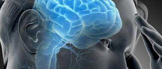
Thanks to the work of the brain, a person can think, feel, hear, see, touch, and move. The large (final) brain controls all vital processes occurring in the human body, and is also the “receptacle” of all our intellectual abilities. What distinguishes humans from the animal world is, first of all, developed speech and the ability for abstract thinking, i.e. the ability to think in moral or logical categories. Only in human consciousness can various ideas arise, for example, political, philosophical, theological, artistic, technical, creative.
In addition, the brain regulates and coordinates the work of all human muscles (both those that a person can control through willpower and those that do not depend on a person’s will, for example, the heart muscle). The muscles receive a series of impulses from the central nervous system, to which the muscles respond by contracting with a certain strength and duration. Impulses enter the brain from various sense organs, causing the necessary reactions, for example, turning the head in the direction from which the noise is heard.
The left cerebral hemisphere controls the right half of the body, and the right hemisphere controls the left. The two hemispheres complement each other.
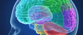
The brain resembles a walnut; it has three large sections - the brainstem, the subcortical section and the cerebral cortex. The total surface of the cortex increases due to numerous grooves that divide the entire surface of the hemisphere into convex convolutions and lobes. Three main sulci - central, lateral and parieto-occipital - divide each hemisphere into four lobes: frontal, parietal, occipital and temporal. Individual areas of the cerebral cortex have different functional significance. The cerebral cortex receives impulses from receptor formations. Each peripheral receptor apparatus in the cortex corresponds to an area called the cortical nucleus of the analyzer. An analyzer is an anatomical and physiological formation that provides perception and analysis of information about phenomena occurring in the environment and (or) inside the human body, and generates sensations specific to a particular analyzer (for example, pain, visual, auditory analyzer). The areas of the cortex where the cortical nuclei of the analyzers are located are called the sensory areas of the cerebral cortex. The motor zone of the cerebral cortex interacts with sensory zones; when it is irritated, movement occurs. This can be shown with a simple example: when a candle flame approaches, the pain and heat receptors of the fingers begin to send signals, then the neurons of the corresponding analyzer identify these signals as pain caused by a burn, and the muscles are “given the order” to withdraw the hand.
Association zones
Association zones are functional areas of the cerebral cortex. They connect incoming sensory information with previously received and stored in memory, and also compare information received from different receptors. Sensory signals are comprehended, interpreted and, if necessary, transmitted to the associated motor area. Thus, associative zones are involved in the processes of thinking, remembering and learning.
Telencephalon lobes
The telencephalon is divided into the frontal, occipital, temporal and parietal lobes. The frontal lobe contains areas of intelligence, concentration, and motor areas; in the temporal - auditory zones, in the parietal - zones of taste, touch, spatial orientation, and in the occipital - visual zones.
Speech zone
Extensive damage to the left temporal lobe, for example as a result of serious head injuries and various diseases, as well as after a stroke, is usually accompanied by sensory and motor speech disorders.
The telencephalon is the youngest and most developed part of the brain, which determines a person’s ability to think, feel, speak, analyze, and also controls all processes occurring in the body. The functions of other parts of the brain primarily include control and transmission of impulses, many vital functions - they regulate the exchange of hormones, metabolism, reflexes, etc.
Oxygen is required for the normal functioning of the brain. For example, if cerebral circulation is disrupted during cardiac arrest or injury to the carotid artery, then after a few seconds the person loses consciousness, and after 2 minutes, brain cells begin to die.
Functions of the diencephalon
The thalamus and hypothalamus are parts of the diencephalon. Impulses from all receptors in the body enter the nuclei of the thalamus. The received information is processed in the thalamus and sent to the cerebral hemispheres. The thalamus connects to the cerebellum and the so-called limbic system. The hypothalamus regulates the autonomic functions of the body. The influence of the hypothalamus is carried out through the nervous system and endocrine glands. The hypothalamus is also involved in the regulation of the functions of many endocrine glands and metabolism, as well as in the regulation of body temperature and the activity of the cardiovascular and digestive systems.
Limbic system
The limbic system plays an important role in shaping human emotional behavior. The limbic system includes nerve formations located on the medial side of the telencephalon. This area has not yet been fully explored. It is assumed that the limbic system and the subthalamus controlled by it are responsible for many of our feelings and desires, for example, under their influence thirst and hunger, fear, aggressiveness, and sexual desire arise.
Brain stem functions
The brain stem is a phylogenetically ancient part of the brain, consisting of the midbrain, hindbrain and medulla oblongata. The midbrain contains the primary visual and auditory centers. With their participation, orienting reflexes to light and sound are carried out. The medulla oblongata contains centers for the regulation of respiration, cardiovascular activity, functions of the digestive organs, and metabolism. The medulla oblongata takes part in the implementation of such reflex acts as chewing, sucking, sneezing, swallowing, vomiting.
Functions of the cerebellum
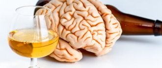
The cerebellum controls body movements. The cerebellum receives impulses from all receptors that are stimulated during body movements. Cerebellar function may be impaired by drinking alcohol or other substances that cause dizziness. Therefore, under the influence of intoxication, people are not able to normally coordinate their movements. In recent years, more and more evidence has emerged that the cerebellum is also important in human cognitive activity.
Cranial nerves
In addition to the spinal cord, twelve cranial nerves are also very important: pairs I and II - the olfactory and optic nerves; III, IV VI pairs - oculomotor nerves; V pair - trigeminal nerve - innervates the masticatory muscles; VII - facial nerve - innervates facial muscles, also contains secretory fibers to the lacrimal and salivary glands; VIII pair - vestibulocochlear nerve - connects the organs of hearing, balance and gravity; Pair IX - glossopharyngeal nerve - innervates the pharynx, its muscles, parotid gland, taste buds of the tongue; X pair - the vagus nerve - is divided into a number of branches that innervate the lungs, heart, intestines, and regulate their functions; Pair XI - accessory nerve - innervates the muscles of the shoulder girdle. As a result of the fusion of the spinal nerves, the XII pair is formed - the hypoglossal nerve - which innervates the muscles of the tongue and the hypoglossal apparatus.