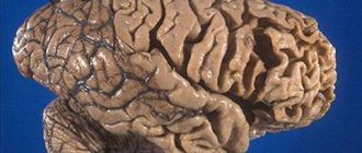
The brain stem is one of the important parts of the human central nervous system, since its sections contain nerve centers responsible for the regulation of all vital processes in the body, as well as the nuclei of the oculomotor and other cranial nerves, the function of which is to control the muscles of the face. For this reason, even minor damage to the brain stem by various neoplasms threatens the patient with disorders in the functioning of internal organs.
Types of lesions
Most often, brain stem tumors are diagnosed in children in the first years of life and in adolescence, with the peak incidence usually occurring at 4-6 years of a child’s life. Most of these neoplasms histologically represent gliomas of various formations:
- Astrocytomas. Occurs in more than 50% of clinical cases. It develops as a result of tumor damage to glial tissue cells - astrocytes, which perform a protective and auxiliary function in relation to neurons. Depending on the structural structure, all astrocytomas are divided into 2 categories: malignant and benign tumors. The former include pilocytic and fibrillar neoplasms, and the latter include anaplastic and multiform neoplasms.
- Ependymomas. They are rare, but they cause hydrocephalus and increased intracranial pressure even in the early stages of development.
- Oligodendrogliomas. They can reach large sizes, while the neoplasm has clear boundaries and does not grow together with the surrounding tissues.
- Glioblastoma. It is an aggressive type of glial malignant tumor.
According to the nature of their location in the tissues of the trunk, glial tumors are of 2 types:
- Expansive neoplasms. They have a clear location and do not merge with surrounding structures. Thanks to this feature, they can be easily removed surgically, after which in half of the cases the patient recovers completely.
- Infiltrative neoplasms. These include diffuse glioma of the brain stem, which, on the contrary, does not have a clear location, and its cells grow into the surrounding tissues and displace them, which is why the affected part ceases to function properly. Due to the fact that such a tumor cannot be removed without affecting the functional parts, the prognosis of the disease in most cases is unfavorable.
Brainstem stroke
Conservative therapy
Acute disorders of cerebral circulation of the brainstem zone require urgent hospitalization in a specialized vascular center or neurological hospital - optimally in the first 3 hours from the moment of onset.
The basis of therapy is conservative measures, which begin at the prehospital stage with the provision of emergency care, oxygenation, symptomatic correction (hypotensive, anticonvulsant). Early treatment helps improve the prognosis even with severe lesions. Restoration of vital functions, including mechanical ventilation, is carried out in the intensive care unit, where, after determining the characteristics of the brainstem stroke, specific treatment begins. It is aimed at restoring blood flow through the vessels of the vertebrobasilar bed, preventing complications and eliminating them, minimizing the risk of relapse, and normalizing basic physiological and biochemical constants. The key areas of drug therapy are:
- Intravenous thrombolysis.
The administration of thrombolytic agents (recombinant plasminogen activator) is indicated in the most acute period of the ischemic process. Despite the high efficiency of recanalization of arterial stenoses, such treatment has many contraindications and increases the risk of hemorrhagic complications. - Anticoagulants and antiplatelet agents.
The progressive course of brainstem stroke requires the use of anticoagulants - first low-molecular-weight heparins, then oral agents. They are also prescribed for preventive purposes after stabilization of cerebral hemorrhages. In all cases of acute brain stem ischemia, antiplatelet agents (acetylsalicylic acid) are required. - Neuroprotectors.
To improve cerebral perfusion, increase the regenerative potential of the affected nervous tissue, and accelerate the recovery of impaired functions, neuroprotectors are used. These include vasoactive (vinpocetine, pentoxifylline), neurotrophic agents (a complex of cerebral peptides or animal blood hemoderivat), antioxidants (thioctic acid, ascorbic acid, inosine).
Neurorehabilitation
Early comprehensive rehabilitation is of utmost importance for eliminating the consequences of brainstem stroke and restoring the functional capabilities of patients. The medical direction is based on ongoing neuroprotective therapy, spasticity and contractures are treated with muscle relaxants, and concomitant emotional and mental disorders are corrected with antidepressants.
Neurorehabilitation involves the use of kinesiotherapy, exercise therapy, and massage. Modern hardware technologies are being introduced into everyday practice in the form of robotic simulators operating using a biofeedback mechanism. Cyclic exercises allow you to activate the patient, restore movement, coordination and walking function. The possibilities for comprehensive rehabilitation are expanded by physical procedures, psychotherapy, social and labor adaptation.
Surgery
Radical correction of brainstem ischemic stroke involves the use of intravascular recanalization methods. Taking into account the clinical situation, selective intra-arterial thrombolysis, mechanical or aspiration thrombectomy, balloon angioplasty with stenting of extracranial vessels can be used. After intracranial hemorrhage with dislocation of brain structures, the hematoma is removed and decompressed through hemicraniectomy. Ventricular drainage surgery helps eliminate acute hydrocephalus.
Experimental treatment
Given the low level of evidence for existing drugs for neuroprotection, a search for new drugs is being conducted. The possibilities of antagonists of glutamate and NMDA receptors (eliprodil, selfotel), cytoprotective agents (lubeluzole), antioxidants (tirilazide) are being studied. Methods of stereotactic and endoscopic removal of hematomas require further study; ventricular thrombolysis and local hemostasis with recombinant factor VIIa are at the testing stage.
Specifics of the disease
Due to the fact that the sections of the trunk contain nuclei of the regulatory formation, which are responsible for performing a large number of functional tasks of the human central nervous system, their damage by neoplasms is manifested primarily by the appearance of deviations in the functioning of certain internal organs, which significantly complicates diagnosis.
Initially, damage to the brain stem is manifested by the appearance of characteristic neurological abnormalities, for example, it may be a hearing disorder or uncontrollable twitching of the facial muscles. Further, as the tumor grows, the symptoms will intensify, headache and increased intracranial pressure will appear, which indicates the development of edema of healthy tissues.
As the situation worsens, the patient, to one degree or another, develops a syndrome of damage to the medulla oblongata, which is expressed in disruption of the functioning of internal organs and the development of central hemiplegia on the side of the body opposite to the tumor. For these reasons, when the first signs of damage to the trunk appear, the patient should seek medical help as soon as possible.
As practice shows, in a quarter of patients, the brain stem tumor is a benign neoplasm that can be removed surgically. At the same time, the remaining diffusely developing neoplasms are usually treated with radiation therapy.
Brainstem lesion syndromes. Methodology for studying the coordination sphere.
Brainstem syndromes include symptoms of damage to the midbrain, pons, medulla oblongata,
Midbrain lesion syndromes . Symptoms associated with oculomotor nerve damage
(external, internal, total ophthalmoplegia), trochlear nerve (convergent strabismus, double vision when looking down).
Quadrigeminal syndrome
: increased righting reflexes, paresis of upward or downward gaze, vertical nystagmus, incoordination of movements of the eyeballs, ophthalmoplegia, horizontal nystagmus, Nothnagel syndrome (impaired balance, hearing, paralysis of the oculomotor muscles, choreic hyperkinesis), paresis and paralysis of the limbs, cerebellar disorders, decerebrate rigidity associated with damage to the mesencephalic centers regulating muscle tone below the red nucleus.
Red core syndrome
: intentional hemitremor, hemihyperkinesis, Claude's syndrome (inferior red nucleus syndrome) - damage to the gosomotor nerve on the side of the lesion, on the opposite side there is intentional hemitremor, hemiataxia. Foix syndrome (superior red nucleus syndrome)—intentional hemitremor, hemihyperkinesis.
Substantia nigra syndrome
characterized by plastic muscle hypertension and the development of akinetic-rigid syndrome.
Tegmental syndrome
: homolateral - ataxia, Horner's syndrome, tremor, myoclonus, contralateral - hemihypesthesia, violation of the quadrigeminal reflexes - rapid orienting movements in response to unexpected visual and auditory stimuli (start - reflexes).
Syndromes of damage to the pons include symptoms associated with damage to the nuclei of the V, VI, VII and VIII nerves, the medial lemniscus, the pyramidal tract, the posterior longitudinal fasciculus, paralysis of the muscles innervated by the facial and abducens nerves, paresis or paralysis of the gaze (pontine center of gaze, posterior longitudinal fasciculus), sensory disturbances on the face, hearing loss, vestibular disorders, vegetative-trophic disorders—hyperthermia, sphincter disorders, sweating disorders, convulsions, hormetonia.
When the lesion is localized in the area of the cerebellopontine angle
symptoms are observed from the VII, VIII, less often VI and V nerves, cerebellar disorders; on the opposite side - spastic hemiplegia.
The medulla oblongata syndrome includes symptoms of damage to the nuclei of the IX, X, XI and XII nerves, the inferior olive, the spinothalamic tract, the nuclei of Gaulle, Burdach, the pyramidal and descending extrapyramidal tracts, the descending sympathetic fibers to the ciliospinal center, the Flexig and Govers tracts.
Hemiparesis, tetraparesis, or paralysis of the limbs may be observed; if the lesion is localized in the area of the pyramidal decussation, alternating hemiplegia (paralysis of the arm on the affected side, paralysis of the leg on the opposite side); sensitivity disorder: hemianesthesia, alternating hemianesthesia; when the lesion is localized in the lateral parts of the spinal cord - dissociated loss of superficial sensitivity on the opposite half of the body, when the lesion is localized in the medial parts of the medulla oblongata - dissociated disorders of deep sensitivity on one or both sides. Impairments in coordination, movements on the side of the lesion, and Bernard-Horner syndrome are also detected. Damage to the caudal part of the medulla oblongata is accompanied by respiratory failure (respiratory paralysis, disturbance of rhythm and frequency of breathing), and cardiovascular activity. Bulbar and pseudobulbar syndrome.
Methodology for studying the coordination system:
Tests are used to determine static ataxia , dynamic ataxia , dysmetria , asynergia muscle tone studies .
To determine static ataxia, the Romberg test is used : legs together, arms at your sides, head straight, eyes closed - stability is assessed. Extend your arms in front of you at shoulder level, close your eyes. The pose becomes more complicated - the heel of one leg is brought to the toe of the other. Stability in the Romberg position .
Gait assessment tests are used:
Normal gait.
Walk in one line, bringing your heels to your toes.
Possibility of flanking gait.
Tests to determine dynamic ataxia : hands in front of you, close your eyes, reach for the tip of your nose with your index finger. A hit, a missed hit, and the presence of inversion tremor are assessed. Similarly, pointing test : touch the tip of the hammer with one and the other hand.
Test for asynergy . Adiadochokinesis is assessed: arms in front of you, a rotational movement is made with the hands, such as rotating and screwing in light bulbs. The speed and symmetry of movements is assessed.
Dysmetria test arms in front of you, palms up and down, then a little faster. The next test for dysmetria is the Schilder : hands in front of you, the right hand rises up, and then lowers to the level of the left hand. Then with the other hand.
When the cerebellar system is damaged in a person, nystagmus . When the cerebellum is damaged, horizontal nystagmus .
The state of speech is assessed (say the 333rd Cavalry Division). If the cerebellum is damaged, speech will not be smooth, intermittent, or scanned.
In a lying position, tests for dynamic ataxia in the legs are examined: you need to raise your right leg and place your right heel on the knee of your left leg, then carry it down the leg. The presence of misses and slips is assessed. Same thing with the other leg.
Test for asynergy - Babinsky's asynergic phenomenon . Lying down, the patient crosses his arms and sits down: when the cerebellum is affected, the legs are raised, not the upper body.
Test for absence of reverse shock. When the cerebellum is damaged, the patient hits himself in the chest with his fist (see video).
Muscle tone is examined by palpation of muscles and passive movements in small and then large joints. When the cerebellum is damaged, muscle hypotonia is observed.
3.Task:
1. Leading syndrome - generalized petit mal seizures such as absences
2. Topical diagnosis - the pathological focus is localized in the medio-basal regions of the frontal
3. Clinical diagnosis: Epileptic disease.
4. Treatment: anticonvulsant therapy (Depakine, Topomax, lamotrigine).
5. Prof: rational work and rest regime, exclusion of alcohol, normal sleep, regular use of anticonvulsants.
Ticket 22
1. Severe traumatic brain injury. Clinic, diagnosis, treatment, clinical manifestations of the consequences of TBI.
Severe TBI is accompanied by prolonged loss of consciousness, coma of varying degrees, the presence of brainstem disorders => gross impairment of vital functions.
severe - severe brain contusion, diffuse axonal damage and acute compression of the brain.
In case of severe contusion, CT scan reveals areas of heterogeneous increase in density (alternating areas of increased and decreased density). Perifocal cerebral edema is severe. A hypodense track is formed in the area of the nearest section of the lateral ventricle. Through it, fluid with breakdown products of blood and brain tissue is discharged.
Diffuse axonal brain damage is typically characterized by a prolonged coma after a traumatic brain injury, as well as pronounced brain stem symptoms. Coma is accompanied by symmetrical or asymmetrical decerebration or decortication, both spontaneous and easily provoked by irritations (for example, painful ones). Changes in muscle tone are very variable (hormetonia or diffuse hypotension). A typical manifestation is pyramidal-extrapyramidal paresis of the limbs, including asymmetric tetraparesis. In addition to gross disturbances in the rhythm and frequency of breathing, autonomic disorders also appear: increased body temperature and blood pressure, hyperhidrosis, etc. A characteristic feature of the clinical course of diffuse axonal brain damage is the transformation of the patient’s condition from a prolonged coma to a transient vegetative state. The onset of this state is indicated by spontaneous opening of the eyes (with no signs of tracking or fixation of gaze).
The CT picture of diffuse axonal brain damage is characterized by an increase in brain volume, as a result of which the lateral and third ventricles, subarachnoid convexital spaces, and also the cisterns of the base of the brain are under compression. The presence of small focal hemorrhages in the white matter of the cerebral hemispheres, corpus callosum, subcortical and brain stem structures is often detected.
Brain compression
Brain compression develops in more than 55% of cases of traumatic brain injury. The most common cause of brain compression is an intracranial hematoma (intracerebral, epi- or subdural). Rapidly increasing focal, brainstem and cerebral symptoms pose a danger to the life of the victim. Availability and duration of the so-called the “light gap” - expanded or erased - depends on the severity of the victim’s condition.
A CT scan reveals a biconvex, less often a flat-convex, limited zone of increased density, which is adjacent to the cranial vault and is localized within one or two lobes. However, if there are several sources of bleeding, the area of increased density can be significant in size and have a crescent shape.
When a patient with a traumatic brain injury is admitted to the intensive care unit, the following measures must be taken:
Examination of the victim’s body, during which abrasions, bruises, joint deformities, changes in the shape of the abdomen and chest, bleeding and/or liquor leakage from the ears and nose, bleeding from the rectum and/or urethra, and a specific odor from the mouth are detected or excluded.
Comprehensive x-ray examination: skull in 2 projections, cervical, thoracic and lumbar spine, chest, pelvic bones, upper and lower extremities.
Ultrasound of the chest, ultrasound of the abdominal cavity and retroperitoneal space.
Laboratory tests: general clinical analysis of blood and urine, biochemical blood test (creatinine, urea, bilirubin, etc.), blood sugar, electrolytes. These laboratory tests must be carried out in the future, daily.
ECG (three standard and six chest leads).
Testing urine and blood for alcohol content. If necessary, consult a toxicologist.
Consultations with a neurosurgeon, surgeon, traumatologist.
Treatment: mechanical ventilation, normal ICP, improvement of cerebral blood flow, elimination of agitation and convulsions, control of diuresis, hemodynamic norms, inflammation of the blood circulation, vasopressors (dopamine, dobutamine, adrenaline), blood transfusions with blood loss of at least 20-30%, maintaining blood pressure above 90, elimination of pain impulses, double excitation (relanium, anesthesia). For a victim with impaired consciousness of 8 points or less on the Glasgow scale, tracheal intubation is indicated, due to which normal oxygenation is maintained. Depression of consciousness to the level of stupor or coma is an indication for auxiliary or controlled mechanical ventilation (at least 50% oxygen). With its help, optimal cerebral oxygenation is maintained. Patients with severe traumatic brain injury (hematomas, cerebral edema, etc. detected on CT) require monitoring of intracranial pressure, which must be maintained below 20 mmHg. For this purpose, mannitol, hyperventilation, and sometimes barbiturates are prescribed. To prevent septic complications, escalation or de-escalation antibacterial therapy is used. For the treatment of post-traumatic meningitis, modern antimicrobial drugs approved for endolumbar administration (vancomycin) are used.
Symptoms of damage to the thoracic spinal cord, Brown-Séquard syndrome. Methods for studying sensitivity, characteristics of conductive and segmental types of sensitivity disorders.
Brown-Séquard syndrome is a symptom complex observed when half the diameter of the spinal cord is affected: on the affected side, central paralysis (or paresis) and loss of muscle-articular and vibration sensitivity are noted, on the opposite side - loss of pain and temperature sensitivity.
Etiology • Trauma and penetrating injuries of the spinal cord • Circulatory disorders of the spinal cord • Infectious and parainfectious myelopathies • Tumors of the spinal cord • Irradiation of the spinal cord • Multiple sclerosis (sclerosis).
Pathogenesis • Radicular and segmental disorders on the side of the injury • Conductive disorders at the level of the injury below.
Clinical picture. In the acute period - phenomena of spinal shock (below the level of the lesion, complete flaccid paralysis and loss of all types of sensitivity are noted). Subsequently, the following develop: • Spastic paralysis (or paresis) and a disorder of deep sensitivity below the level of the lesion on the side of the same name • On the opposite (healthy) side there is a loss of pain and temperature sensitivity to the level of conduction-type damage • Development of flaccid paresis and segmental loss of sensitivity at the level damage • Ataxia, paresthesia, and radicular pain may occur.
Treatment is surgical (decompression).
Symptoms of damage: Segmental (dissociated) type of sensitivity disorder - observed with damage to the sensitive apparatus of the spinal cord (posterior horn, white commissure, dorsal root, spinal ganglion) and the sensitive nuclei of the cranial nerves of the brain stem, only pain and temperature sensitivity is affected, deep sensitivity is preserved.
Conductive type of sensitivity disorder - observed when sensitive pathways are damaged, sensitivity disorders are found downward from the level of the lesion; in this case, deep sensitivity is upset on the side of the same name, and superficial sensitivity on the opposite side.
3. Task:
1. The leading syndrome is motor Jacksonian seizures
2. Topical diagnosis - the lower parts of the anterior central gyrus on the left are affected.
3. Clinical diagnosis: brain tumor in the left frontal lobe.
4. Treatment: surgical.
5. Rehabilitation exercise therapy, massage, kinesiotherapy for the right hand, anticonvulsant therapy (carbamazepine) under EEG control.
Ticket 23
1. Myopathies: etiology, classification, clinical picture, diagnosis, treatment, prognosis.
Degenerative diseases with predominant damage to the neuromuscular system. They are based on progressive degenerative changes in muscles in the absence of primary pathology of the peripheral motor neuron.
Muscular dystrophy is a disease characterized by increasing muscle weakness and atrophy. Primary damage to muscles, but two neurons are spared.
Duchenne muscular dystrophy is an X-linked recessive disease that affects boys. The onset of the disease is in childhood, often in adolescence. Gradual symmetrical increase in weakness of the muscles of the proximal limbs, pelvic and shoulder girdle. Characteristic symptoms: duck gait, winged shoulder blades, emphasized lumbar lordosis. The knee reflexes disappear early, and then the shoulder girdle reflexes. Development of pseudohypertrophy of the calf, deltoid muscles, contractures . The heart muscle suffers, and intelligence decreases.
Erb-Roth muscular dystrophy is transmitted in an autosomal recessive manner. The onset of the disease is 14-16 years old. The disease begins with damage to the muscles of the pelvic girdle and proximal parts of the legs or shoulder girdle. The muscles of the back and abdomen suffer. Characterized by a duck's gait, difficulty getting up from a lying or sitting position, and accentuated lumbar lordosis. Development of contractures and pseudohypertrophies. Intelligence preserved.
Spinal amyotrophies – progressive degeneration of motor neurons s.m. anterior horns due to defects in programmed cell death. => development of flaccid paralysis and denervation muscle atrophy.
Treatment is aimed at maintaining muscle strength, reducing the rate of atrophy, and preventing the development of contractures. Medicinally compensate for energy deficiency, improve tissue metabolism, blood circulation. Vit gr VA E, a/k, orotate K, ATP, riboxin, trental, nootropil.
Main symptoms
A glial tumor of the brain stem leads to organic damage to brain tissue, displacement of sections, and also causes circulatory problems in the organ, which causes symptoms characteristic of this disease:
- Headache. The appearance of this sign of damage to the brain substance is observed in 90% of clinical cases and is caused by an increase in the pressure of the neoplasm on the cranial nerves and blood vessels. Dizziness, darkening of the eyes, tinnitus and other manifestations of oxygen deprivation may also be present.
- Nausea, vomiting. Usually occurs during a headache attack and does not depend on food intake.
- Psychological deviations. It is observed in 65% of patients and manifests itself in changes in consciousness, irritability and apathy towards others.
- The patient experiences deterioration in clarity of vision, disordered pupillary reactions, loss of the inner halves of the visual fields, and paralysis of the facial muscles.
- The development of bulbar syndrome, which is characterized by the appearance of disturbances in the functioning of the functional centers of the medulla oblongata, that is, the patient has dysgraphia, aphonia, anarthria with impaired swallowing and articulation. Further, other manifestations of brainstem lesions arise: impaired cardiac activity, decreased blood pressure and the appearance of peripheral paralysis of the tongue muscles.
Also a distinctive feature of brain stem damage is an increase in the amount of cerebrospinal fluid in the subarachnoid space and, accordingly, an increase in the volume of the head.