The brain stem is one of the important parts of the human central nervous system, since its sections contain nerve centers responsible for the regulation of all vital processes in the body, as well as the nuclei of the oculomotor and other cranial nerves, the function of which is to control the muscles of the face. For this reason, even minor damage to the brain stem by various neoplasms threatens the patient with disorders in the functioning of internal organs.
Brainstem stroke - what is it?
Chemical reactions constantly occur in the brain; even with a slight decrease in blood flow, the organ begins to experience oxygen deficiency. In a healthy person, 50 ml of blood flows through 100 grams of brain matter; in a cerebral accident, 10 ml. This leads to the loss of some functions.
Interesting fact! A nerve cell can exist without oxygen for no more than 7 minutes, then it dies.
The brainstem is the main part of the brain located at the deep base of the skull. This zone contains the midbrain and medulla oblongata, nerve ducts, cranial nerve nuclei, and pons.
The medulla oblongata regulates the process of breathing, blinking, sneezing, coughing, and swallowing. Damage to this area causes cardiac arrest and blockage of breathing.
The pons is responsible for the perception of auditory information; at this point the bridge connects to the cerebellum and spinal cord. If the functioning of the bridge is impaired, deafness and facial paralysis develop. The midbrain regulates unconscious motor acts and movements of body parts. Dysfunction of this area leads to problems with movement.
The trunk is localized in a hard-to-reach place under the cerebral hemispheres.
Attention. If the area swells, it quickly moves and is compressed, which is fatal for the victim. This part of the brain is adjacent to the spinal cord and is associated with the activities of the central nervous system and internal organs.
The brain stem controls:
- heart muscle;
- respiratory tract organs;
- auditory and visual organs;
- body temperature;
- muscle tone;
- motor ability;
- vegetative functions;
- chewing and swallowing functions;
- organs of the reproductive system.
The brainstem region receives information from the sensory organs, then the data passes to the cerebral cortex, from which signals proceed to the spinal cord.
Brainstem stroke develops due to the inability of blood vessels to deliver oxygen and nutrients to the brainstem. Tissues begin to die, their functionality is lost.
Treatment
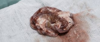
It is performed conservatively or surgically, depending on its type, volume, as well as the individual characteristics of the patient. In the acute form, removal of the subdural hematoma is often indicated. Detection of displacement and compression of brain structures is a stimulus for surgery as soon as possible from the moment of injury (or rupture of a vessel).
a) With MRI without contrast, the image reveals areas of fluid accumulation indicated by white arrows - subacute subdural hematomas. b) MRI visualizes foci of increased signal intensity (indicated by white arrows), as well as foci of reduced MRI signal intensity (indicated by black arrows), these signs are characteristic of acute subdural hematomas.
The absolute indication for surgical treatment of a subdural hematoma is the thickness of the accumulated blood more than one centimeter, determined by imaging studies (MSCT, MRI). The postoperative period should be accompanied by the maintenance of vital functions and control of intracranial pressure.
The operation is also indicated for subacute subdural hemorrhage, if there is an increase in focal symptoms and the appearance of signs of intracranial hypertension.
Classification and causes of attack
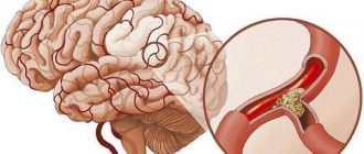
Since brain stem stroke develops identically to blockades of other localizations, they have common causes. The causes depend on the mechanism of damage to the arteries and neurons. Based on this, hemorrhagic and ischemic brainstem stroke are distinguished.
The first occurs due to a rupture of the artery supplying the brain, which leads to hemorrhage. Its cause is hypertension or congenital pathology of blood vessels, expressed in their thinning.
Causes of ischemic type
Ischemic stroke of the brain stem is accompanied by blockage of the arteries, due to vascular obstruction or the formation of a blood clot or atherosclerotic plaque. Among the risk factors for this form of pathology are arterial hypertension, diabetes mellitus, increased concentration of cholesterol in the blood, atherosclerosis, and diseases of the cardiovascular system.
Note! Increase in blood pressure to 140-90 mm. Hg Art. increases the possibility of cerebrovascular accident.
The causes of ischemic stroke of the brain stem are differentiated into the following groups.
- Atherothrombotic. Ischemic phenomena occur as a result of a gradually enlarging blood clot inside the vascular canal. This condition is preceded by a sharp change in mood, absent-mindedness, aggressiveness, and memory impairment. This type occurs more often in the morning.
- Embolic. Occurs suddenly, the victim feels sharp pain from lightning-fast blockage of a vessel with an embolus. Such a stroke usually develops against the background of cardiac pathologies. More often detected during the day after physical or emotional stress.
- Hemodynamic. It develops with low blood pressure, when the brain does not receive the required amount of nutrients.
- Lacunarnaya. Accompanied by damage to the smallest capillaries, which are localized in the deep layers of the brain. It often happens during the day due to increased blood pressure.
- Hemorheological. Develops as a result of increased blood viscosity. Rarely diagnosed.
For reference. After blockage of the vascular canal, ischemic phenomena occur, nerve cells die, losing their functions. Brain swelling develops.
What causes hemorrhagic form
Brainstem hemorrhagic stroke is diagnosed when a vascular canal ruptures, leading to cerebral hemorrhage. Based on the location of hemorrhage, the type is differentiated into parenchymal, subarachnoid, and epidural.
Diagnostics
The main methods of instrumental diagnostics are CT, MRI. In case of strokes of brainstem localization, carotid angiography gives an idea of the condition of the arteries supplying blood to the brain, the presence of malformations and other anomalies in the formation of the vascular bed, stenotic processes, occlusion of the vascular lumen, which is often detected in vascular atherosclerosis.
Computed tomography allows early differentiation of damage to the brainstem from damage to the substance of the hemispheres within the brain, which is associated with a better prognosis for recovery and a reduced likelihood of undesirable consequences. When diagnosing a stroke of the venous, hemorrhagic or ischemic type in the area of the trunk within the brain, ultrasound methods are used - duplex scanning, echoencephaloscopy, transcranial ultrasonography.
For example, during echoencephaloscopy, the nature and direction of dislocation (displacement) of the middle parts of the brain is revealed, which often occurs against the background of hydrocephalus or edema of brain tissue. Transcranial ultrasonography is also used for dislocation syndromes and signs of compression of the brain substance. Interpretation of the results of duplex scanning shows the presence of arterial stenosis or impaired patency of cerebral vessels.
Providing emergency assistance
After the first signs of a stroke occur, first aid should be started immediately. Only this can save a person from disability and death.
Necessary:
- lay the victim down, raising his head;
- provide a flow of fresh air;
- free the patient’s body from constrictive clothing and accessories;
- turn your head to the side for better evacuation of vomit;
- Apply a damp towel to the head area.
While waiting for the ambulance to arrive, you need to monitor your blood pressure, heartbeat, and breathing.
Consequences of brain stem damage
Trunk stroke is characterized by a high mortality rate. Even if the victim can survive, he will completely or partially lose certain functions. Timely treatment and rehabilitation do not guarantee the restoration of lost skills. The consequences of brainstem stroke depend on the form of the disease, location and extent of brain damage.
Death is caused by stem swelling with pinching of the area under the meninges. This causes heart failure, respiratory blockage, and epileptic seizures. With large-scale tissue damage, urinary tract infections, thrombosis, pneumonia, and bedsores appear. Myocardial infarction, sepsis, and gastric bleeding may develop.
Attention. Patients with mild cerebrovascular accidents who are able to walk often fall and break limbs, which also leads to death.
Swallowing dysfunction
Swallowing problems after a stroke are called dysphagia. The skill is difficult to restore; special techniques are used for this. Initially, the victim is taught the ability to swallow crushed food.
Such a swallowing disorder reduces the effectiveness of the entire rehabilitation program, since the patient receives food using gastrostomy tubes.
Speech disorders
Damage to the brain stem leads to speech impairment, which is accompanied by difficulties with pronunciation and understanding of some words. People begin to speak quietly, indistinctly, inarticulately. This pathological phenomenon has a favorable prognosis.
It is necessary to work with a speech therapist. After completing the course, the patient continues the exercises at home.
Deterioration in limb functionality
A brainstem stroke always leads to impaired motor functions. The patient begins to involuntarily wave and move his arms and legs. This is due to loss of control over the muscular system.
It is possible to restore motor skills only in the first months after the stroke. The more time has passed after the blockade, the worse the prognosis: it will be impossible to regain muscle strength.
Problems with coordination
The phenomena of ischemia and hemorrhage during a brainstem stroke provoke dizziness, which impairs coordination of movements. The painful sign must be eliminated as soon as possible.
Rehabilitation
An adequate set of rehabilitation measures directly affects the results of treatment and the severity of post-traumatic neurological deficit. Rehabilitation is carried out under the supervision of the attending physician and a group of specialized specialists. Usually this is: a neurologist, rehabilitation specialist, physiotherapist, occupational therapist, speech therapist and neuropsychologist.
Doctors strive to create favorable conditions for the patient to return to normal life and relieve neurological symptoms. For example, the efforts of a speech therapist are aimed at restoring speech function.
Rehabilitation methods
- Bobath therapy – stimulates motor activity by changing body position.
- Vojta therapy is based on encouraging the patient to make directed movements through irritation of certain areas of his body.
- Mulligan therapy is a type of manual therapy aimed at reducing muscle tone and relieving pain.
- Using the Exart design, which is a suspension system designed to develop hypotrophied muscles.
- Performing exercises on cardio equipment and a stabilization platform to improve coordination of movements.
- Occupational therapy is a set of techniques and skills that allow the patient to adapt to the social environment.
- Kinesio taping is a field of sports medicine that involves applying elastic adhesive tapes along the muscle fibers and increasing the effectiveness of muscle contractions.
- Psychotherapy is aimed at neuropsychological correction at the rehabilitation stage.
Physiotherapy:
- medicinal electrophoresis;
- laser therapy (has an anti-inflammatory and regeneration-stimulating effect);
- acupuncture.
Drug therapy based on intake:
- nootropic drugs (Picamilon, Phenotropil, Nimodipine), which improve metabolic processes in neurons;
- sedatives, hypnotics and tranquilizers to normalize the psycho-emotional background.
Diagnostic measures
To make a correct diagnosis, a detailed examination of the patient is required. The doctor can only assume the presence of a brain catastrophe based on the clinical picture.
Therefore, the victim undergoes a series of diagnostic procedures:
- Computed tomography, which provides three-dimensional visualization of the brain matter. The condition of the organ, the presence of damage, and ischemic phenomena are assessed.
- Magnetic resonance imaging, which allows you to study the organ in the smallest detail. The specialist evaluates the functioning of the brain and its blood circulation.
- Doppler ultrasound scan, which examines the carotid arteries.
- Angiography, which reveals the condition of the vascular channels and the presence of blood clots.
- An electrocardiogram that monitors the functioning of the heart muscle.
To obtain detailed information about the functioning of the heart, the victim is sent to an echocardiogram.
Therapy methods
If any type of stroke is suspected, a person is urgently hospitalized for emergency medical care. Only a third of patients with damage to the brain stem survive the stroke, and even for them the prognosis for recovery after a brain stem stroke is unfavorable.
Surgical intervention
The hemorrhagic form of the pathology often leads to death. Therefore, if this type of stroke is suspected, brain surgery is performed urgently.
Cranial trephination helps eliminate hemorrhage in the shortest possible time, this allows you to quickly reduce the pressure inside the skull.
When diagnosing an ischemic type of stroke, a thrombolytic is injected into the affected area. The manipulation is accompanied by drilling of the skull.
Local administration of the drug accelerates the resorption of blood clots. This procedure is not performed for vascular diseases and the presence of aneurysms.
Pharmacological treatment
For ischemic stroke, drug therapy is usually limited. The patient is prescribed drugs that combat cerebral swelling, normalize blood pressure, and restore nutrition to tissue structures.
In case of hemorrhage, the victim takes anticoagulants and thrombolytics. Intravenous medications to stop bleeding may be required. As symptomatic therapy, the patient is prescribed antibacterial medications. Such patients are fed through a special tube.
Five sick conditions
In neurotraumatology, there are five conditions for patients with TBI:
| State | Criteria | ||||
| Consciousness | Vital functions | Neurological symptoms | Threat to life | Forecast for recovery | |
| Satisfactory | Clear | Saved | Absent | No | Favorable |
| Moderate | Moderate stun | Preserved (possible bradycardia) | Hemispheric and craniobasal focal symptoms are pronounced | Minimum | Usually favorable |
| Heavy | Sopor | Moderately disturbed | Stem symptoms appear | Significant | Doubtful |
| Extremely severe | Coma | Grossly violated | Craniobasal, hemispheric and brainstem symptoms are severely expressed | Maximum | Adverse |
| Terminal | Terminal coma | Critical Violations | General cerebral and brainstem disorders dominate and overlap hemispheric and craniobasal | Survival is impossible | Absent |