"Split Brain"
The separation of the hemispheres doubles consciousness. Sperry said: “In other words, each hemisphere seems to have its own independent and private sensations, its own perceptions, its own concepts, and its own motivating impulses.”
How does a split-brain person function after surgery?
Having two "brains" in one body can create a serious dilemma. When one split-brain patient got dressed, he would sometimes pull his trousers down with one hand and pull them up with the other. One day this patient grabbed his wife with his left hand and shook her violently. His right hand gallantly came to the woman's aid and intercepted his militant left hand. Although such conflicts can occur, it is much more common for split-brain patients to behave completely normally. The reason here is that both sides of the brain have almost the same experience at the same point in time. Moreover, if conflict arises, one half usually prevails over the other.
Split-brain effects are most easily observed during special tests. For example, we could flash a dollar symbol to the right side of the brain of a patient named Tom. And direct the image of the question mark to the left half of his brain. Next, Tom is asked to draw what he sees, using his left hand blindly. The left hand draws the dollar symbol. If Tom is then asked to point with his right hand to a picture of what his “invisible” left hand has drawn, he will point to the question mark. In short, in a split-brain person, one hemisphere may not know what is happening in the other. This is an extreme case of the situation when “the right hand does not rise, what is the left doing?
Interhemispheric asymmetry (IA), according to modern concepts, is considered as a complex system of interaction between the cerebral hemispheres, being basic for the normal functioning of the central nervous system. It plays an important role in the regulation of motor acts, including in various forms of movement disorders [1, 2]. There is a large amount of data on the different contributions of the left and right hemispheres to the activity of the human brain, as well as various aspects of functional interhemispheric asymmetry (FIA) - ontogenetic [3], morphological [4], both normally and in various pathological processes [5] . However, despite a fairly long history of studying this problem and a huge number of publications devoted to various aspects and features of FMA, a complete theory explaining the functional asymmetry of the cerebral hemispheres does not yet exist [6].
The term “FMA of the brain” appeared after the work of the French anatomist and surgeon Paul Broca, who noted the connection between damage to the left hemisphere and the development of aphasia [7]. Ideas about FMA were formed under the influence of two groups of facts: studies of local brain lesions, which showed that damage to the symmetrical areas of the hemispheres is accompanied by various clinical symptoms, as well as from the quite obvious observation of motor asymmetry of the human hands [8]. This allowed us to assume the presence of a stable lateralization of functions in the human brain. These ideas are supported by morphological and partly neurochemical data on the presence of structural differences in the structure of the cerebral hemispheres [8]. There is a classical concept of FMA, which formulates the concept of the presence of one dominant hemisphere [7].
Development of FMA throughout life
It is believed that the main features of asymmetry are laid down at the time of birth of a child [9], and the formation of FMA occurs in the first years of life in the process of speech development and mastery of complex objective actions [2, 10]. The phenomenon of MA does not appear immediately, since the corpus callosum begins to fully function only by 2 years of age [11].
In the early stages of ontogenesis, the leading role belongs to the right hemisphere of the brain, and the development of interhemispheric relations occurs “from right to left” [10]. The prerequisites for the “advanced” functioning of the right hemisphere are anatomical and laid down from birth [10]. They are formed by such an organization of afferent pathways that ensures advanced arrival of information into the projection fields of the right hemisphere compared to the symmetrical zones of the left [10]. At the same time, the multimodal sensory systems of the right hemisphere are less differentiated and more closely connected with each other [12]. Thanks to such morphofunctional features, the afferent links of mental functions are formed in the child mainly in the right hemisphere. With the development of speech and complex voluntary movements (i.e., efferent parts of the psyche), the left hemisphere is already actively functioning [10]. According to various authors [2, 5, 6, 10], from 33 to 55% of the population are right-handed, in whom there is a clear lateralization of the afferent and efferent links of higher nervous activity. Moreover, FMA after the period of its formation in childhood is very stable [1, 7, 13].
The problem of age-related changes in interhemispheric interactions and restructuring of the functional activity of the right and left hemispheres of the brain during the aging process remains the subject of active study [8, 14]. With age, interhemispheric differences gradually smooth out, possibly being an expression of plasticity processes and a compensatory mechanism that prevents the development of degenerative changes associated with aging [15]. Some authors [8] suggest that the smoothing of MA occurs mainly due to a decrease in the activity of the left hemisphere, and note that in old age, in most cases, the activity of the right hemisphere predominates. Other researchers [16] postulate a predominant decrease in the functional activity of the right hemisphere with aging.
Structural and functional organization of interhemispheric asymmetry
There is still an opinion that the morphological substrate of FMA is the secondary (projection-associative) and tertiary (overlap zones) areas of the cortex [12]. The asymmetry is based on the different organization of the functional systems of the right and left hemispheres, which is determined by many factors, including anatomical, neurochemical, immunological, and electrophysiological [2, 6, 8, 17, 18].
Most studies focus on the asymmetry of speech motor and motor areas of the cerebral cortex [2, 6, 18-22]. In particular, the hypothesis about the structural and functional asymmetry of the motor cortex of both hemispheres is actively discussed, but the exact mechanisms that ensure this asymmetry are not fully known [21].
Differences in the anatomical structure of the cerebral hemispheres are considered. Thus, there are differences in the sizes of cytoarchitectonic fields in the temporal lobes of the right and left hemisphere [20]. The temporal region and occipital lobe are almost always greater in the left hemisphere, as is the severity of the sulci and cell density [23]. Asymmetry of the arcuate fasciculus connecting Broca's area and Wernicke's area has been described - in the left hemisphere of right-handed people, on average, it is 25% larger in diameter than in the right [24]. In general, the asymmetry of the fields that make up the motor and sensory speech areas is approximately 1.5 times higher than in other areas of the brain, which explains the presence of motor and sensory asymmetry of the brain [6, 18, 19]. In recent years, increasing attention of researchers [7, 25, 26] has been attracted by data on the asymmetry of the autonomic nervous system.
There are differences between the hemispheres of the brain. This concerns the analysis of stimuli, the method of processing incoming information [7] and movement control, depending on its complexity in the regulation of various types of activities [27]. The left hemisphere predetermines the initial trajectory [28] and amplitude characteristics of movement [29], and the right hemisphere determines the final position [28], taking into account spatial characteristics [29].
It is assumed that the hemispheres have different connections with the brain stem structures [31], for example, the predominant connection of the reticular formation of the brain stem with the left hemisphere, and the diencephalic structures with the right [32]. At the same time, the different nature of the interaction of cortical and subcortical structures in right-handers and left-handers is discussed: right-handers have a more complex and differentiated nature of interhemispheric interactions, reflecting the reciprocal influences of various regulatory systems of the brain compared to left-handers [5, 19]. The differentiated participation of hemispheres in the activities of various functional systems is discussed [8].
There are also neurobiochemical and metabolic asymmetries in the human brain [30]. Biochemical processes occur conjugately in symmetrical brain formations, while they differ in different hemispheres in both quantitative and qualitative characteristics [33]. It is these differences that influence the formation of the characteristics of intrahemispheric and intersystem relationships of electrogenesis of the cerebral hemispheres. In healthy subjects, there is an asymmetry in the α rhythm—the amplitude of α waves and the α index in the left hemisphere is lower than in the right [31]. MA was established based on the content of a number of neurotransmitters and metabolic activity, in particular the level of N-acetylaspartate, choline and inositol is higher in the right thalamus, and the content of norepinephrine is higher in the left thalamus and hypothalamus [23]. Biochemical asymmetry is also characteristic of the cortex as a whole. Thus, in the cortex of the right hemisphere there is more γ-aminobutyric acid, serotonin and higher activity of enzymes (catechol-O-methyltransferase, acetyltransferase, monoamine oxidase) [33, 34]. There is evidence of different sensitivity of the hemispheres to drugs [19].
Thus, FMA is a multi-level system, the main links of which have different “mobility”: from stationary cortical to more flexible, subcortical, associated with the work of nonspecific activating systems of the brain [7]. These structures can have an inhibitory or excitatory effect directly on the functional activity of specialized neurons or indirectly through changes in hemodynamics and metabolism [7].
Stationary and dynamic aspects of FMA
The presence of stable structural differences is a significant factor in the stability of the FMA, which is manifested by differences in functions in the symmetrical formations of the brain [8]. At the same time, MA is not something frozen; it is characterized by the ability to undergo dynamic changes [27]. The phenomenon of dynamic MA is observed when the functional state changes, which has been most fully studied in two models: “sleep-wakefulness” and “relaxation-stress”. Thus, under moderate stress, activity more often moves to the subdominant hemisphere, which can be regarded as a kind of rest for the dominant hemisphere [8]. Activation of diencephalic structures and the sympathetic nervous system occurs, which leads to an increase in the activity of the right hemisphere [7, 35]. In the morning there is greater activity in the left hemisphere, in the evening - in the right [7]. Manifestations of dynamic MA are observed both in normal and pathological conditions [7]. It is with the dynamic properties of FMA that adaptation processes are associated. However, with a number of diseases and, possibly, normal aging, switching between hemispheres becomes more difficult, which indicates a decrease in the quality of adaptation processes [8].
Dynamics of dominance of the cerebral hemispheres in the process of human life
The process of specialization of the cerebral hemispheres is complex and dynamic in nature: there is both high cooperation and disunity of their activity, which is determined by age characteristics and functional state [36]. In the learning process, a person uses both right- and left-hemisphere strategies, and they are successful to varying degrees depending on the type of learning. For example, in children, when mastering speed reading, right-hemisphere strategies are more successful, and when learning to navigate a maze, on the contrary, left-hemisphere ones are more successful [37].
The neurophysiological mechanisms of movement organization in right-handed people are based on a complex multifunctional system, and not only the leading left hemisphere can dominate, but also the right one, depending on which brain structures are involved in providing a motor task, which in turn is determined by its characteristics and features [38 , 39]. In the process of various types of activity, this role can move from the left to the right hemisphere and vice versa, i.e. alternating dominance of the cerebral hemispheres is observed [40].
In different functional states, neurophysiologically, there is a predominant activation of one of the hemispheres. Even a slight increase in activity in one of the hemispheres leads to inhibition of the work of neurons in the symmetrical region of the opposite hemisphere, which is ensured by commissural, predominantly inhibitory interhemispheric connections [7].
The right and left hemispheres are normal
For many years, ideas about the functioning of the brain were based on clinical observations and their comparison with pathomorphological data. The introduction of neuroimaging and functional research methods has made it possible to specifically study, in normal conditions and with damage to the central nervous system, the processes of structural and functional asymmetry of various brain systems, including their reorganization. For this purpose, functional magnetic resonance imaging (MRI), positron emission tomography, transcranial magnetic stimulation (TMS), electroencephalography and the study of motor potential associated with movement are used.
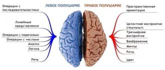
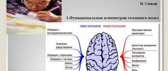
There are ontogenetic, morphological, and neurobiochemical differences between the hemispheres of the brain that determine the functional asymmetry of the hemispheres. In right-handed people, the potential activity and energy level of the left hemisphere is in most cases higher than that of the right [38]. It is assumed that it is the left hemisphere that organizes the integral work of the whole brain. Nonspecific activation of the brain or the principle of saving energy resources of its activation mechanisms are discussed as one of the options by which both hemispheres share control functions [38]. Everything that can be implemented with less activation costs occurs with the predominant participation of the right hemisphere, and more complex problems associated with the novelty of the situation, the subjective difficulty of the task, requiring significant costs and exceeding the activation and integrative capabilities of the right hemisphere, with the left hemisphere [38]. The left hemisphere is more analytical, responsible for motor attention, temporary organization of voluntary actions and the psychomotor sphere (for example, speech, logical thinking), and ultimately the choice of movement depends on it [2, 20, 38]. The right hemisphere determines holistic perception, including specific features and spatial characteristics (for example, complex forms of perception, creative thinking, non-verbal hearing, analysis of body diagrams and other, mainly afferent functions or components of the psychosensory sphere), and dominates the stage of anticipation and decision making [9 , 10, 38, 41]. The frontal and left hemisphere systems dominate in the organization of voluntary processes, while the parietal and right hemisphere systems dominate involuntary processes [42].
There are three mechanisms of interhemispheric interaction [9]: reciprocal interactions, when when one hemisphere is depressed, the functions of the other are reciprocally facilitated [43, 44], complementarity or a certain contribution of each hemisphere to the function or action performed [45] and superposition or correction of “distortions” of the space of the other hemisphere, which sees it in a mirror [46, 47]. Therefore, for the normal functioning of the central nervous system, including the implementation of movement, continuous interaction of both hemispheres is necessary [48-51]. Thus, normally, voluntary movement of one hand requires the participation of motor fields of both hemispheres [27]. It is assumed that the motor cortex of each hemisphere exercises motor control of both hands, and not the contralateral one, as was previously believed [28, 50, 52, 53], the opposite hemisphere is involved to a greater extent when moving one hand [22, 54, 55]. Moreover, the activity of different areas of the right and left hemispheres of the brain during movements with one hand, performed under conditions of different sensory afferentation, is not the same [27].
At the cortical level, there is a continuous interaction between the motor fields of the brain, controlled by sensory flow [27, 55]. In healthy right-handers, a study using paired-pulse TMS showed that the primary motor cortex (PMC) and the dorsal premotor area (dPMA) of the right hemisphere modulate the PMC of the left hemisphere during preparation for movement with the fingers of the leading right hand [27], and in the right hemisphere first exhibits activity in the dPMC and then in the PMC, influencing the PMC of the left hemisphere during the early and late phases of preparation for movement or only at the end of the phase, respectively. All this complements the existing ideas about the hierarchical model of the cortex for controlling a motor act and demonstrates the spatiotemporal interactions of these cortical fields of both hemispheres during preparation for movement [27].
Thus, normally the final motor program is formed in both hemispheres [56], while in the process of life there is a predominant activation of one of the cerebral hemispheres in different functional states, determined by current information or the need to solve a certain task [40]. However, data on the study of the functional organization of the brain in right-handers and left-handers at rest and during activity are ambiguous and contradictory for a number of indicators [32, 57-59], which is understandable, since the studies use not only a variety of types of tasks, but also different methods research, as well as methodological approaches to the analysis of experimental data. In addition, most of the research is based on clinical observations, which also confirm the fact that the hemispheres work together when implementing complex types of voluntary activities [11, 13, 38].
Right and left hemispheres in some pathological processes in the central nervous system
In various diseases of the brain, dysfunction of the right or left hemispheres, as well as disruption of interhemispheric interactions, can be observed. With local brain lesions, FMA changes depending on the location of the lesion, and, as a rule, its changes are accompanied by neurological deficits [7]. At the same time, many functions characteristic of the dominant and subdominant hemispheres are transformed due to neurological deficits and subsequent compensatory changes [7]. Psychoneurological symptoms in local brain lesions have been studied in some detail [19], however, the nature of the relationships that develop between the hemispheres in the process of recovery and rehabilitation still remains not entirely clear [7].
In some diseases, interhemispheric relationships can serve as a marker of the severity of the pathological process and be used as an indicator of the success of therapy. In particular, adolescents with minimal brain dysfunction experience a pronounced disruption of interhemispheric relations with a predominance of the level of constant potential in the right hemisphere. During a course of taking phenotropil, along with clinical improvement, there was also a normalization of interhemispheric relations [60]. In diseases of the brain and processes associated with aging, the indicators of interhemispheric relations depend on the type of pathological process, but in general, in each patient they are more stable than in healthy people, which is explained by focal pathology of one of the hemispheres [8].
Changes in FMA are one of the important pathogenetic mechanisms of depression [25], in which the functional activity of the prefrontal region of the left hemisphere decreases with an increase in the activity of the homologous zone of the right hemisphere [19, 25]. Ideas were formed about interhemispheric relationships in Parkinson's disease, cranial and cervical dystonia, brain tumors and psychogenic movement disorders [11].
The fundamental importance of MA in the regulation of motor skills in various forms of movement disorders is recognized by many researchers [9, 19, 36, 52, 54, 61]. The vast majority of research in this area has been conducted in stroke patients. In particular, when studying interhemispheric interaction associated with movement, a violation of MA was shown in patients with damage to the right hemisphere, in contrast to patients with damage to the left, in whom the direction of interhemispheric relationships was preserved [11]. It has been suggested that the cerebral hemispheres have a differentiated role in the regulation of different phases of the motor act: the right one in preparation, the left one in the implementation of movement [11]. The best restoration of interhemispheric relationships in case of damage to the left and intrahemispheric - right hemisphere is discussed [62], and differences in postural control after a lateralized stroke are considered [53].
There are various features of interhemispheric interactions in patients who have suffered a right- and left-hemisphere stroke [62, 63]. In patients with a lesion localized in the right hemisphere, disturbances in motor functions (especially precise control of the trajectory of movement [53]), parameters of autonomic processes, and changes in the bioelectrical activity of the brain have more pronounced manifestations and are observed not only in the affected hemisphere, but also in the intact hemisphere [64] , and the restoration of these impaired functions is much less active [31, 65].
In patients with a post-stroke focus in the left hemisphere, ceteris paribus, metabolic disorders are less pronounced, but the speed of information processing is more significantly reduced [7], and cognitive impairments are more common and more pronounced [31, 66], while the primary mnestic defect is restored more actively [62]. Using TMS and spectral analysis of EEG in patients after a left hemisphere stroke compared with healthy people, an increase in activation of the lateral PMC (Brodmann field 6) of both hemispheres and, to a lesser extent, in the PMC and the region of the parietal cortex of the unaffected hemisphere was shown [67]. According to functional MRI data, activation of the PMO in the unaffected hemisphere was higher compared to a similar motor area of the opposite hemisphere [30].
It is known that when one of the hemispheres is damaged during strokes, the functional deficit is compensated with the help of symmetrical structures of the other hemisphere [68]. This issue has been studied to a greater extent in left-hemisphere strokes. A number of reviews [28, 67, 69] discuss aspects of restoration of impaired functions after left hemisphere stroke. Significant recovery may be due to increased use of ipsilateral and contralateral motor fields not previously involved in the performance of a particular task. Using f-MRI and EEG coherence analysis, bilateral activation of the supplementary motor area and an increase in the activity of the PMC in the right and PMC in the left hemisphere were shown [67, 69]. It has been suggested that a functional shift towards the intact hemisphere probably facilitates the restoration of motor functions, being one of the manifestations of neuroplasticity, which is different from the predominantly bilateral processes in healthy people when performing complex and complex movements [67].
Thus, the phenomenon of FMA is complex, multifaceted and not fully understood. The dynamics of MA from a neurophysiological point of view are essentially associated with the involvement of various functional systems of the right and left hemispheres in a single activity, while the success of such activity both in normal conditions and in various pathological conditions depends on the balance of the systems of both hemispheres. Despite numerous studies in this area, the mechanisms of interhemispheric integration during the performance of complex voluntary movements remain poorly understood and not always explained, and in various diseases of the central nervous system, the FMA factor is often not taken into account, which is important for understanding the pathophysiological processes of the identified disorders and the rehabilitation treatment of these patients.
Difference between the left and right hemispheres of the brain
The functions of the brain are divided in a remarkable way. For example, approximately 95% of all adults use the left hemisphere for language (speaking, writing, and understanding). In addition, the left hemisphere has superiority in mathematics, timing and rhythm, and coordinating the order of complex movements such as those required for speech.
In contrast, the right hemisphere responds only to simple language and counting. Working with the right side of the brain is like talking to a child who only understands a dozen words. To answer questions, the right hemisphere must use nonverbal responses, such as pointing at objects.
Although the right side of the brain is weak in language, it has its own talents, especially perceptual skills. During the test, the right hemisphere is better at recognizing patterns, faces and melodies, solving design problems or drawing a picture. It is also involved in recognizing and expressing EMOTIONS.
Connections between hemispheres
There are some interesting facts about the connections that exist between the two halves of the brain. First, the left hemisphere is always slightly larger than the right. Secondly, in the right hemisphere there are long nerve fibers that connect it to the left. The left one, on the contrary, contains a large number of short fibers that create connections in limited areas. Brain asymmetry is a process that takes an average of ten to fifteen years to develop. Sometimes its speed can be determined by genetic characteristics. It is practically not observed in infants. Asymmetry is an acquired quality. In addition, it has been proven that it is less pronounced in illiterate people. That is, in the process of learning and acquiring new knowledge, the brain becomes more and more asymmetrical. Those who do not pay due attention to education slow down the development of many important functions.
Asymmetry in nature
It should be noted that the phenomenon under consideration is characteristic of many natural objects. Interhemispheric asymmetry is not only the prerogative of humans. If symmetry is represented in the structure of molecules and crystals, then asymmetry is in the arrangement of internal organs, the structure of the DNA helix. There is also hair asymmetry.
Research in this area leaves many mysteries. But the progress of science does not stand still. Perhaps the knowledge that now seems obvious will become completely outdated for future scientists. Perhaps scientists of the future will be able to finally unravel all the secrets of the highest product of evolution - the human brain.
Discovery of asymmetry
Asymmetry is a feature that has always interested scientists. But until a certain point, it remained a mysterious object that could only cause a lot of speculation even among the brightest minds of humanity. The history of the development of this area began with the discovery by Paul Broca of the relationship between human speech and the use of the right or left hand. This happened in 1861, when the scientist discovered that his patient, who suffered from loss of speech, had a lesion in the left hemisphere of the brain.
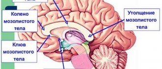
Also, the connection between the two parts is carried out using a special bundle of neurons - the corpus callosum. Thanks to him, they work harmoniously as a single whole. Some seriously ill patients underwent surgery to dissect the corpus callosum. This made it possible to study in more detail the characteristics of the right and left hemispheres.
Sperry's experiments
Split-brain studies have also shown that people with damage to the right hemisphere have very poor spatial orientation. Often such patients cannot find their way to the house in which they previously lived for decades. R. Sperry proved that when the corpus callosum is dissected, the following occurs: processes in the two hemispheres of the brain begin to occur independently. It's like two different people acting independently of each other. According to many scientists, asymmetry is a phenomenon that man acquired during evolution and is its acquisition.