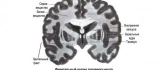
Anatomy of the brain
(c) Shutterstock
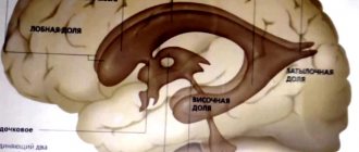
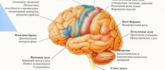
The brain consists of four lobes, each lobe has its own function.
The frontal lobe is located at the front and top of the brain. It is responsible for high levels of human thinking and behavior such as planning, judgment, decision making, control and attention.
The parietal lobe is located at the top of the brain, behind the frontal lobe. It is responsible for receiving sensory information. The parietal lobe of the brain is responsible for understanding someone's position in their environment.
The temporal lobe is located in the lower front part of the brain. It is associated with visual memory, language and emotions.
Finally, the occipital lobe is located at the back of the brain and processes what a person sees.
Along with the lobes, the brain includes the cerebellum and the brainstem.
The brain stem controls vital functions such as breathing, circulation, sleep, digestion and swallowing. These involuntary functions are under the control of the autonomic nervous system. The brain stem also controls reflexes.
The cerebellum is located in the lower back part of the brain, behind the brain stem.
Description of the hindbrain. Pons and cerebellum
The brain, with its surrounding membranes, is located in the cranial cavity. The upper ventral surface of the brain corresponds in shape to the inner concave surface of the cranial vault. The lower surface, the base of the brain, has a complex topography corresponding to the cranial fossae of the inner base of the skull.
The mass of the adult human brain ranges from 1100 to 2000. Over the course of 20 to 60 years, the mass and volume remain maximum and constant for each individual.
When examining a specimen of the brain, its three largest components are clearly visible. These are the paired cerebral hemispheres, the cerebellum and the brain stem.
The hindbrain and medulla oblongata were formed as a result of the division of the rhomboid vesicle.
The hindbrain, metencephalon, includes the pons, located anteriorly (ventrally), and the cerebellum, which is located behind the pons. The cavity of the hindbrain, and with it the medulla oblongata, is the IV ventricle.
In the deep transverse groove separating the pons from the pyramids of the medulla oblongata, the roots of the right and left abducens nerves emerge. In the lateral part of this groove the roots of the facial (VII pair) and vestibulocochlear (VIII pair) nerves are visible.
On the ventral surface of the bridge, which is adjacent to the clivus in the cranial cavity, a wide but shallow basilar (main) groove, sulcus basildris, is noticeable. The artery of the same name lies in this groove.
A cross-section of the bridge shows that the substance that forms it is heterogeneous. In the central sections of the bridge section, a thick bundle of fibers is noticeable, running transversely and belonging to the conduction path of the auditory analyzer - the trapezoidal body, cogrus trapezoideum. This formation divides the bridge into a posterior part, or tegmentum, and an anterior (basilar) part. Between the fibers of the trapezoidal body are located the anterior and posterior nuclei of the trapezoidal body, nuclei cogroris trapezoidei ventralis et dorsalis (anterior et posterior). In the anterior (basilar) part of the bridge (at the base), longitudinal and transverse fibers are visible. The longitudinal fibers of the bridge, fibrae pontis longitudindles, belong to the pyramidal tract (corticonuclear fibers, fibrae corticonucledres). There are also cortico-pontine fibers, fibrae corticopontinae, which end on the nuclei (proper) of the bridge, nuclei pontis, located between groups of fibers in the thickness of the bridge. The processes of the nerve cells of the pons nuclei form bundles of transverse fibers of the bridge, fibrae pontis transuersae. The latter are directed towards the cerebellum, forming the middle cerebellar peduncles.
In the posterior (dorsal) part (pons tegmentum), in addition to the ascending fibers, which are a continuation of the sensitive pathways of the medulla oblongata, there are focal accumulations of gray matter - the nuclei, V, VI, VII, VIII pairs of cranial nerves. Directly above the trapezoid body lie the fibers of the medial lemniscus, lemniscus medidlis, and lateral to them - the spinal lemniscus, lemniscus spindlis. Above the trapezoidal body, closer to the median plane, there is a reticular formation, as well as a higher-posterior longitudinal fasciculus, fasciculus longitundindlis dorsalis (posterior). Lateral and above the medial lemniscus lie the fibers of the lateral lemniscus.
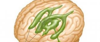
The cerebellum (small brain), cerebellum, is located posterior (dorsal) from the pons and from the upper (dorsal) part of the medulla oblongata. It lies in the posterior cranial fossa. Above the cerebellum hang the occipital lobes of the cerebral hemispheres, which are separated from the cerebellum by the transverse fissure of the cerebrum, fissura transversa cerebrdlis.
In the cerebellum, there are superior and inferior surfaces, the boundary between which is the posterior edge of the cerebellum, where there is a deep horizontal fissure, fissura homontalis.
It begins at the point where its middle peduncles enter the cerebellum. The superior and inferior surfaces of the cerebellum are convex. On the lower surface there is a wide depression - the cerebellar valley, vallecula cerebelli; The dorsal surface of the medulla oblongata is adjacent to this depression. In the cerebellum, there are two hemispheres, hemispheria cerebelli (neocerebellum, except for the flocculus), and an unpaired median part - the cerebellar vermis, vermis cerebelli (phylogenetically the oldest part). The upper and lower surfaces of the hemispheres and the vermis are cut by many transverse parallel fissures of the cerebellum, fissura cerebelli, between which there are long and narrow leaves (gyri) of the cerebellum, folia cerebelli. Groups of gyri, separated by deeper grooves, form the cerebellar lobules, lobuli cerebelli. The grooves of the cerebellum run, without interruption, through the hemispheres and through the vermis, and each lobe of the vermis corresponds to two (right and left) lobes of the hemispheres. The more isolated and phylogenetically older lobule of each hemisphere is the flocculus. It is adjacent to the ventral surface of the middle cerebellar peduncle. With the help of the long leg of the flocculus, pedunculus flocculi ffloccularis, the flocculus is connected to the cerebellar vermis, with its node, nodulus. The cerebellum is connected to neighboring parts of the brain by three pairs of peduncles. The lower cerebellar peduncles (rope bodies), pedunculi cerebellares cauddles (inferiores), are directed downward and connect the cerebellum to the medulla oblongata. The middle cerebellar peduncles, pedunculi cerebellares medii, are the thickest, they go anteriorly and pass into the bridge. The superior cerebellar peduncles, pedunculi cerebellares craniales (superiores), connect the cerebellum to the midbrain. The cerebellar peduncles contain fibers of the pathways that connect the cerebellum with other parts of the brain and the spinal cord.
The cerebellar hemispheres and the vermis consist of the intracerebral body, cogrus medullare, white matter and a thin plate of gray matter covering the white matter on the periphery of the cerebellar cortex, cortex cerebelli. In the thickness of the leaves of the cerebellum, the white matter has the appearance of thin white stripes (lamellas), laminae albae.
The white matter of the cerebellum contains paired cerebellar nuclei, nuclei cerebelli cerebellaris. The most significant of them is the dentate nucleus, nucleus dentatus. On a horizontal section of the cerebellum, this nucleus has the shape of a thin curved gray strip, which with its convex part faces laterally and backward. In the medial direction, the gray stripe is not closed; this place is called the gate of the dentate nucleus, hilum nuclei dentti, filled with white matter fibers forming the superior cerebellar peduncle. Inward from the dentate nucleus, in the white matter of the cerebellar hemisphere, there is a cork-shaped nucleus, nucleus emboliformis, and a spherical nucleus, nucleus globosus. Here, in the white matter of the worm, is the most medial nucleus - the nucleus of the tent, nucleus fastigii.
The white matter of the worm, bordered by bark and divided along the periphery by numerous deep and shallow grooves, on a sagittal section has a bizarre pattern reminiscent of a tree branch, hence its name “tree of life.”
The gray matter of the pons is represented by the nuclei of the V, VI, VII, VIII pairs of cranial nerves, which provide eye movements, facial expressions, and the activity of the auditory and vestibular apparatus; the nuclei of the reticular formation and the proper nuclei of the pons, which participate in the connections of the cerebral cortex with the cerebellum and transmit impulses from one part of the brain to another through the pons. In the dorsal sections of the bridge, ascending sensory pathways follow, and in the ventral sections, descending pyramidal and extrapyramidal pathways follow. There are also fiber systems here that provide bilateral communication between the cerebral cortex and the cerebellum. The cerebellum has nuclei (centers) that provide coordination of movements and maintain body balance. The activity of the cerebellum is under the regulatory influence of the cerebral cortex due to cortical-cerebellar connections, which are carried out through the pons nuclei.
Disturbances in the activity of the cerebellum are characterized by pronounced incoordination of movements and dissonances in the functioning of internal organs. Precise, targeted movements that require the coordinated work of many muscles and balance organs are especially affected. Speech and handwriting disturbances are also observed. The handwriting is uneven and large letters.
The reticular formation is a collection of nerve cells located in the central part of the brain stem. This is a kind of “accumulator” of brain energy. Impulses from the reticular formation rise to the cerebral cortex and are capable of suppressing or stimulating its activity (ascending influences). Descending influences of the reticular formation can change the intensity of motor reactions and autonomic functions of the body. Widespread changes in the body caused by the activity of the reticular formation gave rise to calling it a “nonspecific system” of the brain, i.e. a department of the brain, the influence of which does not have an “exact addressee” and is directed to all higher and subcortical nervous structures. However, studies in recent years have shown that in addition to nonspecific influences, the reticular formation can also have specific ones, i.e. directed to strictly localized nerve centers. The functions of the reticular formation are numerous: regulation of the state of sleep and wakefulness, simple and instinctive forms of behavior, participation in the course of reflex reactions, regulation of the conscious and unconscious state.
In the process of ontogenesis, the maturation of the structures of the brain stem occurs most intensively in the first two years of postnatal development. The final formation of these structures, and especially the diencephalon, is completed only at the age of 13-16, when the sexual development of adolescents ends.
Many features of lower and higher nervous activity in adolescent children are explained by the functional properties of the diencephalon and some other subcortical structures of the brain. Mental processes, according to modern concepts, are not localized in certain brain structures. They are formed on the basis of a systemic hierarchical organization of brain structures, each of which is specialized in the implementation of certain operations, and their interaction ensures the implementation of an integral function.
Functions of the cerebellum:
Movement coordination
. Most body movements require coordination of several muscle groups. The cerebellum allows the body to move smoothly.
Maintaining balance
. The cerebellum detects changes in movement balance. It sends signals to the body to adjust to movement.
Eye coordination
.
The cerebellum helps the body learn movements that require practice and fine tuning. For example, the cerebellum plays a role in learning the movements needed to ride a bicycle.
Researchers believe the cerebellum influences thinking and is linked to language and mood, but these functions are not yet well understood.
Brain mass
The mass of the human brain ranges from 1000 to more than 2000 grams, which on average represents approximately 2% of body weight. The brain of men weighs on average 100-150 grams more than the brain of women[2]. It is widely believed that a person’s mental abilities depend on the mass of the brain: the larger the brain mass, the more gifted the person. However, it is obvious that this is not always the case[3]. For example, the brain of I. S. Turgenev weighed 2012 g[4][5], and the brain of Anatole France - 1017 g[6]. The heaviest brain—2850 g—was found in an individual who suffered from epilepsy and idiocy[7][8]. His brain was functionally defective. Therefore, there is no direct relationship between brain mass and the mental abilities of an individual.
However, in large samples, numerous studies have found a positive correlation between brain mass and mental ability, as well as between the mass of certain brain regions and various indicators of cognitive ability [9][10]. A number of scientists [ who?
], however, cautions against using these studies to support inferences about low intelligence in some ethnic groups (such as Aboriginal Australians) who have smaller average brain sizes[11]. A number of studies indicate that brain size, which is almost entirely dependent on genetic factors, cannot explain most of the differences in IQ [12][13][14]. As an argument, researchers from the University of Amsterdam point to a significant difference in cultural level between the civilization of Mesopotamia and Ancient Egypt and their today's descendants in Iran and modern Egypt[15].
The degree of brain development can be assessed, in particular, by the ratio of the mass of the spinal cord to the brain. So, in cats it is 1:1, in dogs - 1:3, in lower monkeys - 1:16, in humans - 1:50. In Upper Paleolithic people, the brain was noticeably (10-12%) larger than the brain of modern humans [16] - 1:55-1:56.