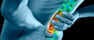
Vertebrogenic cervicalgia is a pain syndrome localized in the neck. It is characterized by a decrease in the mobility of the neck muscles, pain in their area, in addition, dizziness, decreased clarity of vision and autonomic dysfunction may be observed.
The sensations vary in nature: from mild tingling to severe throbbing pain. When moving, it intensifies and causes noticeable discomfort with sudden muscle tension during sneezing, laughing or coughing.
Causes of vertebrogenic cervicalgia
This pain syndrome does not always appear as a result of serious diseases of the spine; in some cases, the cause of pain may be more “modest” problems associated with improper or unsuccessful load on the spine and neck muscles. So, often the causes of this disease are:
- poor head position during sleep;
Causes of vertebrogenic cervicalgia - hypothermia;
- sedentary work and prolonged stay in one uncomfortable position;
- neck injuries;
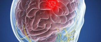
- tumors or infections;
- overload of the neck muscles as a result of heavy physical exertion at work or during physical training;
- diseases of the cervical spine (herniated intervertebral disc, osteochondrosis, etc.).
It is worth considering that the cervical region is very “delicate”, it is here that a huge number of nerve endings and blood vessels pass through the relatively narrow spinal canal, therefore almost all problems of the spine in this region result in at least severe pain, and at maximum – serious disruption of the functioning of internal organs .
Symptoms of the disease
The diagnosis is made in the presence of dystrophic or traumatic changes in the spine identified during examination (MSCT or MRI), as well as in the presence of the following symptoms:
- Pain in the neck, without radiating.
- Tension of the muscles of the neck and collar area.
- Crunch in the neck.
- “Wedging” of the neck.
Complaints from patients with this disease
With cervicalgia, pain can be of a very different nature - tingling, shooting, throbbing, etc. In this case, the pain can sharply intensify with any sudden movement or physical stress (sneezing or coughing, for example).
The most common complaints from patients are:
- inability to tilt or turn the head, so the patient is forced to turn the whole body or bend over to look to the side or down;
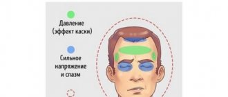
- dizziness;
- pain in the back of the head;
- numbness in the hands or back of the head;
- noise in ears.
In some cases, other neurological symptoms appear.
The presence of prolonged pain in the back of the head, shoulders and neck, as well as pain when turning the head, may indicate the initial stage of a chronic spinal disease. Most often in this case we are talking about the appearance of spondylosis or osteochondrosis in the cervical spine. In this case, the disease is diagnosed using radiography and magnetic resonance imaging, since in this case there are degenerative changes in the intervertebral discs and the vertebrae themselves.
Treatment
Treatment of hernias and protrusions is complex. Pain and inflammation are relieved with medications, then physiotherapeutic procedures, manual therapy, massage, balneology are prescribed (taking into account health status, age, contraindications). Surgery is indicated as a last resort when there are no other options to solve the problem.
Our clinic diagnoses and treats osteochondrosis, as well as other spinal pathologies. To eliminate painful processes in the cervical spine, highly qualified specialists are required. The clinic employs experienced doctors, uses advanced equipment, and applies the latest treatment methods. You can make an appointment by phone or online.
Diagnosis of the disease
The success of diagnosis largely depends on how competently the clinical analysis of pain was carried out and how carefully the anamnesis was collected. Particular attention should be paid to the conditions under which the pain first occurred. This may be sleeping in an uncomfortable position, or an anti-physiological position that a person is forced to take during professional activities. We can also talk about carrying heavy objects, making an unsuccessful turn, or hypothermia (general or local).
Subsequently, an X-ray examination of the cervical spine is carried out in a conventional projection and with various functional tests, magnetic resonance imaging and computed tomography. In some cases, panmyelography and computed tomography in combination with myelography may be needed - such studies are most important in postoperative diagnosis. Various methods of functional diagnostics (electroneurography, EMG, etc.) can also be used.
How is this pain syndrome treated?
Treatment of this pain syndrome in most cases is carried out using conservative methods. It is worth considering that in the case of vertebrogenic cervicalgia, we are dealing not only with pain, but also with the disease that causes this pain syndrome. Therefore, treatment in different cases can differ significantly, since, for example, a tumor and an intervertebral hernia are treated completely differently. But it is possible to identify common features of the conservative course of treatment, which are similar in the treatment of most diseases of the cervical spine.
Treatment of vertebrogenic cervicalgia is most often carried out using conservative methods.
In particular, conservative treatment includes:
- painkillers and non-steroidal anti-inflammatory drugs. It is worth noting that the main task of anti-inflammatory drugs in this case is also pain relief by reducing swelling and inflammation. But it is necessary to take into account that the course of treatment with non-steroidal anti-inflammatory drugs cannot be very long, otherwise there is a risk of complications;
- muscle relaxants. Such drugs are prescribed if the pain syndrome is caused by muscle spasm. But you should be careful when using muscle relaxants when treating diseases in the cervical spine;
- temporary use of a cervical collar. This collar is worn for 1 to 3 weeks, and it must be selected individually, otherwise excessive extension of the neck is possible, which will cause harm, not benefit;
- traction treatment (spinal traction) can also be used to relieve pain. But recently, this method has been losing its popularity, since with not very qualified treatment there is a risk of developing one or more intervertebral disc protrusions;
- physiotherapy. This element is practically key in the treatment of such diseases. It is necessary to start exercise therapy immediately after the pain syndrome has subsided;
- physiotherapeutic treatment methods and the use of a special orthopedic pillow.
Surgical treatment
It should be noted that surgical treatment and, in general, any operations in the cervical spine are quite risky for the reasons described above - a high concentration of nerve endings and blood vessels. Therefore, the decision on surgical intervention must be balanced and motivated. The main indications for surgical intervention are:
- subacute or acute damage at the cervical level of the spinal cord, which results in disruption of the functioning of internal organs;
- an increase in paresis at the site of innervation of the nerve root with the appearance of a threat of necrosis. At the same time, the intensity of pain often decreases, but weakness increases.
News
Neck pain is one of the fairly common pathological symptoms. Almost every person has experienced neck pain at least once during their life. Periodically recurring pain in the neck of insignificant duration (from 3 to 5 days) is observed, according to various authors, in 10-20% of the adult population. Chronic neck pain is observed in a significantly smaller number of people - about 2% of the population. Neck pain is an interdisciplinary problem, since pain of similar localization can be observed in the clinical picture of a number of somatic and neurological diseases. Neck pain can be a fairly benign and fleeting symptom and is observed in the vast majority of patients. However, neck pain may also be the first clinical manifestation of a serious organic disease. The main task of the doctor is to exclude the oncological nature of the pain syndrome (primary and metastatic tumors of the vertebrae, extraspinal tumors, multiple myeloma, lung cancer), traumatic nature (vertebral fractures, especially after whiplash), infectious nature (meningitis, purulent epiduritis, tuberculous spondylitis, acute and subacute thyroiditis, lymphadenitis), metabolic nature (hyperparathyroidism, osteoporosis) and referred pain in diseases of internal organs - heart, lungs, esophagus. Neck pain resulting from these diseases is very rare. In this article we will look at the most common causes of neck pain.
The vast majority of patients with neck pain are diagnosed with spinal osteochondrosis. Currently, there is an overdiagnosis of this cause of neck pain and, on the contrary, ignoring other more common causes of cervicalgia. Functional disorders of the musculoskeletal system with the formation of blocks in small joints of the spine with the appearance of reflex pain musculoskeletal syndromes are unreasonably rarely diagnosed, and the role of myofascial pain syndromes, in which the muscle suffers primarily, is underestimated. Neck pain is usually subdivided taking into account the localization of the main pain syndrome and the nature of pain irradiation. If the pain is localized only in the neck, it is called cervicalgia; when the pain radiates to the arm or head, the terms cervico-brachialgia and cervicocranialgia are used, respectively.
History and clinical analysis
The success of diagnosis depends primarily on a competent clinical analysis of pain manifestations and a thorough history taking. Particular attention should be paid to the analysis of the conditions under which pain first appeared - long-term anti-physiological posture, especially associated with professional activities (dental work, long-term driving, computer work) or after sleep, carrying heavy objects (shoulder bag, backpack), bad turn , after local or general cooling. A thorough interview must be conducted to identify past injuries, no matter how minor. Whiplash is of particular importance. It is necessary to ask about chronic somatic diseases. It is important to identify the connection between pain syndrome and exacerbation of diseases of the visceral organs, especially the heart and lungs. Information about the role of mental factors is essential - pain can debut or worsen after suffering emotional stress, against a background of anxiety, panic, fear, depressed mood. When analyzing the pain syndrome itself, special attention should be paid to the localization of pain, its irradiation, its occurrence during sleep or wakefulness, its connection with a certain body position in which the pain weakens or intensifies, the nature of the pain (sharp, shooting, tearing, i.e. the so-called radicular or deep, aching, dull, diffuse, which is most typical for muscle pain), symmetry or asymmetry of pain. When examining a patient, special attention should be paid to identifying developmental anomalies, body asymmetries, postural characteristics, the severity of cervical physiological lordosis, neck position, and the volume of active and passive movements in the neck. It is imperative that all muscles of the neck and shoulder girdle are examined sequentially by palpation. This is followed by a thorough neurological examination. The features of examining patients with neck pain are described in detail in the monographs of Ya.Yu. Popelyansky, 1989; K. Levita, J. Zahse, V. Janda, 1993; A.M. Veina, G.G. Voznesenskaya, A.B. Danilova et al., 1999; D.R.Shtulman, Ya.Yu.Popelyansky et al., 1995; G.Ya.Ivanicheva, 1997; D.G.Trevell, D.G.Simons, 1989. Data from additional research methods help make a diagnosis and verify the nature of the lesion. X-ray studies of the cervical spine in direct and lateral projection in states of extreme flexion and extension, computed tomography (more informative for visualizing bone structures), magnetic nuclear resonance (more informative for visualizing the spinal cord, intervertebral disc and ligamentous apparatus) are used. Data from additional examination methods play an important role in making a diagnosis, but the first place in diagnosis rightfully belongs to clinical symptoms. Underestimation of clinical manifestations and overestimation of radiographic research methods is a common cause of overdiagnosis of spinal osteochondrosis. The presence of signs of osteochondrosis on radiographs after the age of 25 is almost obligatory, but this does not mean that the patient’s pain is in all cases associated with these findings. Discogenic hernias detected on MRI in many cases remain clinically insignificant and acquire clinical significance only when clinical signs of radicular compression are identified. Thus, only a comparison of clinical manifestations with the results of special diagnostic methods and their logical correspondence to each other contribute to making the correct diagnosis. The most common causes of neck pain can be divided into two large groups: vertebrogenic and non-vertebrogenic. Vertebrogenic causes of neck pain: firstly, spinal osteochondrosis, which can be complicated by compression syndromes in the form of radiculopathy and reflex muscular-tonic syndromes and, secondly, vertebrogenic functional disorders with the formation of reversible blocking in the intervertebral joints. The most common cause of non-vertebral neck pain is myofascial pain syndromes.
Radiculopathy
Radiculopathy as a complication of spinal osteochondrosis occurs only in 5-8% of cases. In the vast majority of cases, complications of osteochondrosis are manifested by muscular-tonic reflex syndromes, which account for up to 95% of complications of spinal osteochondrosis. At the cervical level, the most clinically significant morphological manifestations of osteochondrosis are osteophytes and arthrosis of joints and uncovertebral joints. Disc herniations as a cause of root compression are less common at the cervical level than at the lumbar level. We should not forget that at the cervical level, along with the roots, the vertebral arteries can also be compressed. To make a diagnosis of radiculopathy, an appropriate clinical picture must be observed. Radiculopathy is characterized by specific shooting, fairly intense pain in the neck with distal spread of pain along the affected root down to the fingers. Radicular pain can intensify when coughing, sneezing, when moving the neck, during sleep, and is usually accompanied by spontaneously occurring sensations of numbness, tingling, and burning.
Paresthesia is usually more pronounced in the distal parts of the arm, and pain in the proximal parts. Such manifestations are combined with symptoms of loss of motor and sensory functions, which strictly correspond to the zone of innervation of the affected root. Symptoms of prolapse include: hypoesthesia, loss of reflexes, muscle weakness and muscle atrophy. It should be emphasized that the presence of isolated pain, even strictly in the area of a single root, is not enough to make a diagnosis of radiculopathy. A doctor has the right to make a diagnosis of radiculopathy only if symptoms of prolapse are simultaneously present. We can only speak conditionally about the symptoms of tension in radiculopathy at the cervical level. For example, with radiculopathy at the cervical level, turning and tilting the head towards the affected root is accompanied by severe pain resulting from narrowing of the intervertebral foramen and increased microtrauma to the root. In this case, the pain is shooting in nature, spreads from the neck to the arm and is localized in the zone of innervation of the affected root. If the pain intensifies on the affected side when the head is tilted in the opposite direction, then this does not indicate a radicular lesion, but the suffering of spasmodic muscles that react with pain when stretched. As a rule, radiculopathy is always combined with reflex muscle spasm. At the cervical level, the seventh cervical root is most often affected - 70% of cases and the sixth cervical root - 20% of cases of radiculopathies at the cervical level, the remaining 10% are affected by the fifth and eighth cervical roots. Lesions of the third and fourth cervical roots are extremely rare (R. Adams et al., 1997).
Reflex muscular-tonic syndromes
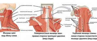
The role of isolated reflex muscular-tonic syndromes is the most significant and clearly dominates among the clinical manifestations of complications of spinal osteochondrosis. Muscular-tonic syndromes can form in any paravertebral or extravertebral muscles. Their favorite places to localize are the trapezius muscles, scalene muscles, and the sternocleidomastoid muscle. The pain is provoked by movements in the neck and arm, and significantly intensifies with movements in which the affected muscle is stretched. The pain is usually aching, deep, and pulling. On palpation, the muscles are tense and painful. Against the background of spasmed muscles, even larger painful muscle thickening or local muscle hypertonicity are determined. There are no symptoms of prolapse. The most common cause of vertebrogenic neck pain is associated not with morphological changes in the spine, as is observed with osteochondrosis, but with functional disorders. They are manifested by limited mobility in the vertebral motion segment, which is formed as a result of reversible blocking - a disorder localized in the intervertebral joint. Functional blocking in the intervertebral joint leads to irritation of the nociceptors of the synovial joint membranes, which is perceived by the patient as pain and starts a vicious circle: pain-muscle spasm-pain. The main causes of reversible functional blocking are long-term antiphysiological postural strain (usually associated with professional activities or observed during deep sleep), unsuccessful, sharp turns of the neck.
Myofascial pain syndromes
Myofascial pain syndromes are a very common cause of neck pain. To make a diagnosis of myofascial pain syndrome, it is necessary to identify the following signs: a spasmodic muscle that is painful on palpation, painful muscle thickening in it, active trigger points and an area of pain irradiation. Trigger points are usually located in painful muscle lumps. When you press an active trigger point, a sharp pain appears at the point itself and at a distance - in the area of referred pain. A characteristic “jumping symptom” is the patient’s flinching in response to palpation of an active trigger. All muscles of the neck and shoulder girdle can be involved in the process.
Treatment
The first necessary condition for treating neck pain is to create rest for the affected neck muscles, avoiding prolonged postural strain. For osteochondrosis of the spine with radiculopathy, immobilization of the cervical spine by wearing a cervical collar is recommended. During periods of acute spontaneous pain, light, dry heat is recommended. Novocaine blockades of the most tense muscles and painful muscle compactions can be used. Novocaine cocktails with analgin and hydrocortisone are used. Applications with dimexide are effective. Dimexide should be consumed in a 40% solution with the addition of novocaine, voltaren, hydrocortisone. Applications are applied for no more than one and a half hours a day. 10 applications are recommended per course. Pharmacological treatment of neck pain is very diverse. Non-steroidal anti-inflammatory drugs (NSAIDs) are used: ibuprofen, indomethacin, diclofenac, aspirin, etc. The form of administration is chosen based on the intensity of the pain syndrome. The course of treatment for radiculopathies is 1 or 1.5 months; for musculoskeletal syndromes, the use of drugs for 2 weeks or less is sufficient. Local administration of these agents in the form of gels is possible. It is not recommended to prescribe several NSAIDs at once, since in this case their side effects may be potentiated. Pain relief is one of the main goals of therapy. NSAIDs alone are not enough. It is possible to use analgesics, from the lightest (paracetamol) to the most powerful - strong narcotic analgesics. The anti-pain effect can be enhanced by adding small doses of carbamazepine to the analgesic. Effective pain management is impossible without the use of psychotropic drugs. In the acute period, tranquilizers with a relaxing effect (diazepam) should be prescribed, 1-2 tablets per day. For recurrent, chronic pain syndromes, antidepressants are prescribed: amitriptyline at a dose of 75 mg per day; mianserin 30 mg per day; fluoxetine 20 mg per day. Treatment with antidepressants should be long-term and last at least 6 weeks. The use of muscle relaxants is justified because they help break the vicious circle of pain-muscle spasm-pain. The most preferred is tizanidine, which has both a muscle relaxant and analgesic effect. It is prescribed in a dose of 4 to 8 mg per day for 2 weeks. It is recommended to prescribe vascular drugs that improve microcirculation and venous outflow. The drugs are prescribed for a month. The most significant role in the treatment of neck pain is played by non-pharmacological agents: post-isometric relaxation, physical therapy, manual therapy, massage, acupuncture, physical therapy (physical therapy). When prescribing manual therapy, it is recommended to use only gentle manual techniques. You should know the contraindications for manual therapy: the presence of verified radicular or radiculomyeloischemic lesions, age-related spondylosis, osteoporosis, instability of the spinal motion segment with spondylolisthesis. Manual therapy at the cervical level should not be prescribed if the neck is very long and the muscle corset is weak. Postisometric relaxation is a method that has virtually no contraindications and can be widely used. The effectiveness of this method for musculoskeletal syndromes in the neck area is superior to all other types of therapy. Post-isometric relaxation is facilitated by preliminary warming of the muscles and the use of the technique against the background of a course of analgesics and muscle relaxants. If there are trigger points identified in spasmodic muscles, the following methods are used: puncturing with a dry needle, kneading, introducing a solution of novocaine or lidocaine into the active trigger, applications with dimexide, wet warm compresses. However, the best method is post-isometric relaxation of the affected muscle. One of the most important methods of therapy is exercise therapy. It is advisable to start it as early as possible, as soon as the pain at rest disappears. The set of exercises must be selected individually, the loads are increased gradually. Exercise therapy is carried out constantly, without interruptions. Once every few months, the exercise therapy complex should be corrected. In the future, to avoid relapses, the patient should undergo courses of massage and physiotherapy twice a year, and teach autogenic training with the ability to relax the neck muscles. Correct posture, correct position at the desk when working with a computer, correct mode of work with periodic relaxation of the neck muscles every 30 minutes are the key to preventing relapses.
Literature:
1. Voznesenskaya T.G. Pain in the back and limbs.-In the book. — Pain syndromes in neurological practice. Ed. Veina A.M. M.-MEDpress.-1999; chapter 6, p. 217-84. 2. Ivanichev G.A. Manual therapy (Manual, atlas).-Kazan.-1997; 448. 3. Levit K., Zahse J., Janda V. Manual medicine. 1993; 510. 4. Popelyansky Ya.Yu. Diseases of the peripheral nervous system.-M.-Med. 1989; 462. 5. Travell D.G., Simons D.G. Myofascial pain.-M.-Med. 1989; vol.1-2. 6. Shtulman D.R., Popelyansky Ya.Yu. and others. Diseases of the peripheral nervous system. — In the book — Diseases of the nervous system. Ed. Yakhno N.N. and others - M.-Med. 1995; vol.1, chapter 6, p. 394-545. 7. Adams RD, Victor M., Ropper AN Pain in the back, neck and extremities.-In Principles of Neurology.-Sixth Ed. 1997; P.2, S.2, Ch.11, P.194-225.
Journal Consilium Medicum, volume 2, no. 12, 2000.
Similar articles:
News → Christian Georg Schmorl (German: Christian Georg Schmorl) is a German doctor and pathologist.
Articles → “Osteochondrosis” or “radiculitis”? (experience of approaching the terminological dilemma)
Glossary → Amossa symptom
Articles → Clinical and surgical parallels of compression forms of spinal osteochondrosis
Articles → Posterior epidural migration of a lumbar disc fragment: description of a series of six cases