What is the pituitary gland and where is it located?
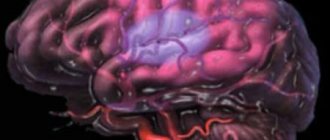
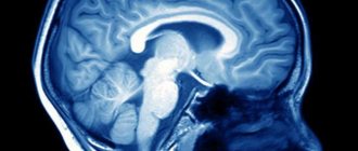
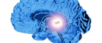
The pituitary gland is the main organ of the endocrine system, a small round gland. It is responsible for all other glands in the body. Therefore, it is very simple to answer the question of where the pituitary gland is located in humans. It is located in the brain on the lower part of the brain, in the sella turcica (bone pocket), where it connects to the hypothalamus (see photo below).
What is the pituitary gland responsible for?
The endocrine gland is responsible for the production of hormones in various organs:
- thyroid gland;
- adrenal glands;
- parathyroid gland;
- genitals;
- hypothalamus;
- pancreas.
It is important to distinguish the pituitary gland from another gland - the pineal gland. Located on opposite sides of the hypothalamus, these two formations have completely different functions. The pineal gland is responsible for circadian rhythms and melatonin production, but its function is not well understood.
Treatment of pathologies
A lack or excess of hormones supplied to the glands and organs leads to the occurrence of secondary diseases. Treatment for pituitary gland dysfunction in the brain is selected by an endocrinologist (oncologist) after conducting diagnostic research methods.
How to test the pituitary gland in the brain:
- Laboratory diagnostics (venous blood examination);
- Visualization of the gland (ultrasound, MRI, X-ray) – allows you to evaluate the parameters and changes in the structure of the pituitary gland.
After making a diagnosis, the doctor (or council) decides how to treat the pathology. The selection of therapy depends on the cause of the malfunction of the organ.
Treatment methods:
- Hormone therapy with drugs;
- Instrumental treatment (in the presence of neoplasms). Depending on the type of tumor, radiation therapy can be used as an independent method of treatment or in preparation for surgery.
To maintain brain functionality, neurometabolic stimulants and vitamin therapy are prescribed.
Tasks of the posterior pituitary gland
The secretion of the hormone (ADH) from the pituitary gland in the brain allows you to regulate the excretory function of the kidneys and maintain water and electrolyte balance.
The production of oxytocin allows you to maintain a labile emotional background. In women, uterine muscle contractions are regulated and lactation is stimulated during the postpartum period.
Function of the anterior pituitary gland
The adenohypophysis in the brain synthesizes most of the hormones that are responsible for the functionality of the entire body.
The tasks of hormones:
- ACTH – sends signals to the adrenal glands to produce cortisol;
- “Growth hormone” (somatotropin) – regulates metabolic processes, stimulates cell division and body growth;
- Thyrotropin – ensures proper functioning of the thyroid gland;
- Gonadotropin – regulates the functioning of the gonads and reproductive function;
- Melanin – regulates pigmentation.
The hormone prolactin is important for women. With its help, lactation is regulated.
The structure of the pituitary gland
The pituitary gland is a small appendage of the brain. Its length is 10 mm and width is 12 mm. Its weight in men is 0.5 grams, in women – 0.6 grams, and in pregnant women it can reach up to 1 gram
How is the pituitary gland supplied with blood? Blood enters it through two pituitary arteries (branching from the internal carotid): superior and inferior. For the most part, blood flows to the lobes of the gland through the anterior (superior) artery. Entering the infundibulum of the hypothalamus, this artery penetrates the brain and forms a capillary network, which passes into the portal veins heading to the adenohypophysis, where they branch again, forming a secondary network. Further, dividing into sinusoids, the veins supply the organs with blood, which is enriched with hormones. The posterior part is supplied with blood flow using the posterior artery.
All irritations of the sympathetic nerves enter the pituitary gland, and many small neurosecretory cells are concentrated in the posterior lobe.
Small neurosecretory cells are relatively small neurons located in several nuclei of the hypothalamus and form a small-cell neurosecretory system that regulates the release of pituitary hormones.
The anatomical structure of the pituitary gland includes three sections (lobes):
- adenohypophysis (anterior lobe);
- intermediate share;
- neurohypophysis (posterior lobe).
Let's look at the anatomy and histology of each part of the pituitary gland.
Adenohypophysis: features, what hormones it secretes
The adenohypophysis is the largest lobe of the pituitary gland: its size is 80% of the volume of the organ. The bulk of the cells of the adenohypophysis are glandular cells - adenocytes, which secrete hormones.
Interesting fact! In pregnant women, the adenohypophysis increases slightly, but after childbirth it returns to its normal size. And in people aged 40-60 years it decreases slightly.
The adenohypophysis consists of three parts, the basis of which are different types of glandular cells:
- distal segment. Self is basic;
- tubular segment. Consists of tissue that forms the shell;
- intermediate segment. It is located between the two previous segments.
The main task of the adenohypophysis is the regulation of many organs in the body. The main functions of the anterior pituitary gland:
- increased production of gastric juice;
- decrease in heart rate;
- coordination of heat exchange processes;
- improving motility of the digestive tract;
- pressure regulation;
- influence on sexual development;
- increased sensitivity of cell tissue to insulin;
- regulation of pupil size.
Homones secreted by the adenohypophysis are called tropins or tropic hormones, as they act on independent glands. The anterior pituitary gland secretes many different hormones:
- somatropin – a hormone responsible for growth;
- adrenocorticotropin - a hormone responsible for the proper functioning of the adrenal glands;
- folliculotropin is a hormone responsible for the formation of sperm in men, and follicles in the ovaries in women;
- luteotropin - a hormone responsible for the production of androgens and estrogens;
- prolactin is a hormone responsible for the formation of breast milk;
- thyrotropin is a hormone that controls the activity of the thyroid gland;
Neurohypophysis: structure and functions
The neurohypophysis consists of two parts: the nervous and the infundibulum. The funnel-shaped part connects the pituitary gland with the hypothalamus, due to which releasing hormones (releasing factors, liberins) enter all lobes.
Histology of the neurohypophysis:
The tissue of the posterior lobe contains ependymal cells (pituicytes), as well as the endings of the processes of nerve cells of the hypothalamus. The axons form the hypothalamic-pituitary tract. Through these nerve fibers, the hormones vasopressin and oxytocin enter the pituitary gland from the hypothalamus.
Functions of the posterior lobe:
- blood pressure regulation;
- control of water exchange in the body;
- regulation of sexual development;
- decreased motility of the digestive tract;
- heart rate regulation;
- dilated pupils;
- increased levels of stress hormones;
- increased resistance to stress;
- decreased sensitivity of cells to insulin.
Hormones in the posterior lobe of the pituitary gland are produced by ependymal cells and the endings of neurons, which are the basis of the neurohypophysis:
- oxytocin;
- vasopressin;
- vasotocin;
- asparotocin;
- mesotocin;
- valitocin;
- isotocin;
- Glumitacin.
The most important hormones of the posterior lobe are oxytocin and vasopressin. They accumulate in the depot and are then released into the blood. The first is responsible for contractions of the walls of the uterus and the release of milk from the breast. The second is for the accumulation of fluid in the kidneys and contraction of the walls of blood vessels.
Intermediate lobe of the pituitary gland
The intermediate part of the pituitary gland is located between the adenohypophysis and the neurohypophysis and is responsible for skin pigmentation and fat metabolism. This section produces melanocyte-stimulating hormones and lipotropocytes. The intermediate part in humans is less developed than in animals and has not been fully studied.
Table Functions of the pituitary gland
| Share (department) | Hormone | Functions |
| Adenohypophysis (anterior lobe) | Follicle stimulating hormone | Maturation of follicles and sperm |
| Luteinizing hormone | Growth and development of the corpus luteum | |
| Prolactin | Stimulation of lactation | |
| Thyroid-stimulating hormone | Formation of thyroid hormones in the thyroid gland | |
| Somatotropic hormone | Growth and maturation of all cells | |
| Adrenocorticotropic hormone | Regulates the production of hormones by the adrenal glands (mineralocorticoids, glucocorticoids), as well as androgens. | |
| Neurohypophysis (posterior lobe) | Antidiuretic hormone (vasopressin) | Stimulates the contraction of vascular smooth muscle, including in the kidneys, reducing filtration and secretion of urine. |
| Oxytocin | Stimulates uterine contractions during and after childbirth. “Attachment hormone” of mother to child. |
Adrenocorticotropic
Speaking about what hormones the pituitary gland produces, one cannot fail to note adrenocorticotropic hormone, which regulates the activity of the adrenal glands (they produce and accumulate cortisone, cortisol, adrenocorticosterone). The action of an adrenocorticotropic biologically active substance is aimed at a person’s fight against stressful situations, control in the field of sexual development, and regulation of the reproductive organs.
An increase in the content of adrenocorticotropic hormone is observed during inflammatory processes in the pituitary gland, with pathologies of the adrenal glands, taking certain potent drugs, and in the period after a major operation. When the functioning of the pituitary gland, adrenal cortex is suppressed, and with the development of a tumor process in the adrenal glands, the levels of adrenocorticotropic hormone decrease significantly.
Development of the pituitary gland in the body
The pituitary gland begins to develop in the embryo only at 4-5 weeks and continues after the birth of the child. In a newborn, the mass of the pituitary gland is 0.125-0.25 grams, and by puberty it approximately doubles.
The anterior lobe of the pituitary gland begins to develop first. It is formed from the epithelium, which is located in the oral cavity. This tissue forms Rathke's pouch (epithelial protrusion), in which the adenohypophysis is an exocrine gland. Next, the anterior lobe develops into an endocrine gland, and its size will increase until the age of 16.
A little later, the neurohypophysis begins to develop. For it, the building material is brain tissue.
Interesting fact! The adenohypophysis and neurohypophysis develop separately from each other, but ultimately, after coming into contact, they begin to perform a single function and are regulated by the hypothalamus.
Pancreatic hormone
When the functions of the anterior part of the adenohypophysis are impaired, changes occur in the pancreas. However, the connection between these glands is very complex and not well understood. From the extracts of the anterior part of the adenohypophysis, a pancreatic hormone was obtained, which causes the proliferation and increase in the number of islets of Langerhans, and a diabetogenic factor, which causes the destruction of the islets of Langerhans, while the secretion of insulin decreases and diabetes mellitus occurs. These facts have not yet found a satisfactory explanation.
Hormones of all endocrine glands influence the functions of the pituitary gland. This mutual influence can be both exciting and inhibitory.
What pituitary hormones are used to treat various diseases?
- Some pituitary hormones are used in the treatment of various diseases and conditions:
- oxytocin. It is used for weak labor, as it promotes uterine contractions;
- prolactin. Will help women who have given birth by stimulating milk production;
- gonadotropin. It improves the functioning of the female and male reproductive system.
vasopressin. It has almost the same properties as oxytocin. Their difference is that vasopressin acts primarily on the muscles of the vascular wall, and only then on the smooth muscles of the uterus and intestines. It lowers blood pressure by dilating blood vessels;
antigonadotropin. Used to suppress gonadotropic hormones.
Diagnosis of the pituitary gland
There is not yet a technique that can immediately diagnose and identify all disorders in the functioning of this multifunctional gland. This is due to the huge range of systems that pituitary hormones act on.
Attention! All procedures necessary for the diagnosis and treatment of disorders should be prescribed only by the attending physician.
If there are symptoms of gland dysfunction, a differential diagnosis is prescribed, including:
- blood test for the presence of hormones. You can check the content of almost any compound in the blood. It must be remembered that the production of some substances is associated with circadian rhythms, day of the cycle (in women), age and some other factors.
- conducting computed tomography or magnetic resonance imaging with contrast. This study makes it possible to identify voluminous formations of the gland due to their increased blood supply.
No research can be viewed in a vacuum. It is imperative to carry out a comprehensive diagnosis, as well as evaluate the clinical manifestations of a possible pathology.
Fat metabolism hormone
When extracts from the anterior part of the adenohypophysis are administered to rats fed a diet rich in fat, ketone bodies (acetone, acetoacetic and beta-hydroxybutyric acids) accumulate in the blood and urine. They appear as intermediate products of increased fat breakdown caused by the pituitary ketogenic hormone. Obesity is associated with decreased pituitary secretion. With excess fat nutrition, you can detect the appearance of large amounts of ketogenic hormone in the blood and urine. Ketogenic hormone stimulates the breakdown of fats in the liver; after shutting down the liver, it does not cause the formation of ketone bodies. Insulin reduces the formation of ketone bodies by the liver, therefore, it acts in the opposite direction to the ketogenic hormone.
Pituitary gland diseases: causes and symptoms
When a malfunction of an organ occurs, its cells begin to be destroyed. The cells secreting somatotropic hormones are the first to be destroyed, then the gonadotropic ones, and the last to die are the cells producing adrenocorticotropins.
There are many causes of pituitary gland diseases:
- a consequence of an operation during which the pituitary gland was damaged;
- impaired blood circulation in this area (acute or chronic);
- traumatic brain injuries;
- an infection or virus affecting the brain;
- taking hormonal medications;
- innate nature;
- a tumor that compresses the organ;
- consequences of radiation in the treatment of cancer;
Symptoms of the disorder may not appear for several years. The patient may be bothered by constant fatigue, a sharp deterioration in vision, headaches or fatigue. But these signs can indicate many other diseases.
Dysfunction of the pituitary gland consists of either excessive production of hormones or their deficiency.
With hyperfunction of the gland, diseases such as:
- gigantism. This disease is caused by an excess of somatotropic hormones, which is accompanied by intensive human growth. The body grows not only outside, but also inside, which leads to multiple heart problems and neurological diseases with severe complications. The disease also affects people's life expectancy;
- acromegaly. This disease also appears when there is an excess of the hormone somatotropin. But, unlike gigantism, it causes abnormal growth of individual parts of the body;
- Itsenko-Cushing's disease. This disease is associated with an excess of adrenocorticotropic hormone. It is accompanied by obesity, osteoporosis, diabetes mellitus and arterial hypertension;
- hyperprolactinemia. This disease is associated with excess prolactin and causes infertility, decreased sex drive and milk production from the mammary glands in both parties. More often it occurs in women.
Hypofunction of the pituitary gland is also quite common. It can be either the result of a pathological process or artificially caused, for example, after surgery on the gland. With insufficient production of hormones, the following diseases occur:
- dwarfism This is the opposite of gigantism. It is quite rare: 1-3 people out of 10 suffer from this disease. Dwarfism is diagnosed at 2-3 years of age, and is more common in boys;
- diabetes insipidus. This disease is associated with a lack of antidiuretic hormone. It is accompanied by constant thirst, frequent urination and dehydration.
- hypothyroidism A disease associated with secondary hypofunction of the thyroid gland. It is accompanied by a constant loss of strength, decreased intellectual level and dry skin. If hypothyroidism is not treated, all development in children stops, and adults fall into a coma with a fatal outcome.
Pituitary tumors
Pituitary tumors can be benign or malignant. They are called adenomas because they arise from glandular tissue. It is still unknown exactly why they appear. Tumors can form after injury, long-term use of hormonal drugs, due to abnormal growth of pituitary cells and due to genetic predisposition.
There are several classifications of pituitary tumors.
Based on tumor size, they are classified as:
- microadenomas (less than 10 mm);
- macroadenomas (more than 10 mm).
According to localization they distinguish:
- formation of the adenohypophysis;
- formation of the neurohypophysis.
By distribution relative to the sella turcica:
- endosellar (extends beyond the sella);
- intrasellar (does not extend beyond the saddle).
According to functional activity, they are distinguished:
- hormonally inactive;
- hormonally active.
There are also many adenomas associated with the work of hormones: somatotropinoma, prolactinoma, corticotropinoma, thyrotropinoma.
The symptoms of pituitary tumors are similar to the symptoms of diseases caused by gland dysfunction.
A pituitary tumor can only be diagnosed by a thorough ophthalmological and hormonal examination. This will help determine the type of tumor and its activity.
Today, pituitary adenomas are treated with surgical, radiation and drug methods. Each type of tumor has its own treatment, which can be prescribed by an endocrinologist and neurosurgeon. The most effective way to remove tumors is surgically, but this is not always possible due to the location of the tumors and their prevalence. Sometimes, under the influence of radiation and chemotherapy, the tumor can decrease in size, becoming operable. In general, pituitary surgery has been studied quite well and such operations are not uncommon in neurosurgical hospitals.
The pituitary gland is a very small but very important organ in the human body, as it is responsible for the production of almost all hormones. But, like any other organ, it can experience dysfunction. The difficulty is that often disturbances in the functioning of the pituitary gland manifest themselves as problems in completely different organs and systems, so it can be difficult for a doctor to find the true cause of the patient’s problems. Well, ordinary people should be attentive to themselves, avoid head injuries and not use hormonal drugs without a doctor’s prescription. You need to carefully monitor your body and pay attention to even the most minor symptoms.
Prolactin
Among the pituitary hormones and their functions, prolactin (luteotropic, lactotropic hormone) stands out, which is one of the most important in the woman’s body. It directly affects a woman’s sexual development, regulates lactation processes (for example, it is prolactin that prevents pregnancy with continued breastfeeding), is responsible for the formation of maternal instinct, and maintains normal progesterone levels. Prolactin is also important for men - in the male body it is directly involved in the regulation of sexual functions.
The pituitary gland cannot produce a sufficient amount of prolactin in the presence of tumors in the gland, with tuberculosis, or with trauma to the skull and brain structures (if the gland has a depressing negative effect).
The level of prolactin in the human body can increase against the background of anorexia, prolactinoma, hypothyroidism, and polycystic ovary syndrome.


