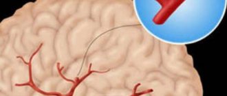
Angiography of the brain (angiogram or arteriography) is a study that allows you to examine the condition of the blood vessels. A diagnostic test is prescribed before complex operations, in the presence of symptoms such as headache, tinnitus, fainting, and dizziness. Based on the results of the examination, it is possible to identify the presence of congenital anomalies and pathological areas in the cerebral region.
Angiography of the vessels of the brain and neck is divided into 3 types: X-ray, MRI angiography and CT angiography.
X-ray method
X-ray angiography of the vessels of the neck (upper and lower spine) and head is not performed if the following contraindications are present:
- hypersensitivity to the substance used for contrast (in most cases iodine is used) and the components of the anesthetic drug;
- failure of organs such as kidneys, liver, heart;
- disorders of the hemostasis system;
- dysfunction of the endocrine system;
- acute inflammatory and infectious disease;
- mental health disorders.
Before performing X-ray angiography of the arteries and veins of the brain, you need to undergo fluorography and an electrocardiogram.
Venography of the brain requires special training. So, 14 days before the diagnostic test, you need to avoid drinking alcohol. To protect the kidneys from the introduction of a large volume of contrast agent, hydration is carried out before diagnosis, saturating the body with fluid. This will dilute the contrast, further facilitating its removal.
To eliminate the risk of symptoms caused by an allergic reaction, you must take an antihistamine before the test. 4 hours before the diagnosis you need to stop eating and drinking water.
Before undergoing X-ray angiography, a person is placed on a table intended for examination, the position of the torso is fixed, and connected to a cardiac monitor. An injection catheter is installed into the vein cavity. Before diagnosis, premedication is carried out by administering an antihistamine through a catheter to prevent an allergic reaction, a tranquilizer, and an analgesic.
The diagnostic procedure involves performing a puncture or puncture of a vessel with further catheterization to introduce a contrast agent (usually iodine). In most cases, the catheter is placed in the femoral artery. Every action that is carried out inside the vessel is monitored by the doctor using X-ray television. After the end of the event, a pressure bandage is applied to the place where the puncture was performed for 1 day.
After the diagnostic procedure, if there are no contraindications, you need to drink a lot to speed up the removal of the contrast agent from the body.
MRI angiography
Magnetic resonance angiography of the brain or MRI of cerebral vessels in angiography mode involves exposing the required area to magnetic fields and radiofrequency radiation. The method is non-contrast, that is, angiography does not require the introduction of a contrast agent, which is different from the previous research method. However, sometimes MRI angiography of cerebral vessels is performed using a special gadolinium-based contrast agent to obtain a clearer image and enhance diagnostic efficiency.
MRI angiography of blood vessels is performed for the following indications:
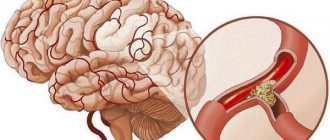
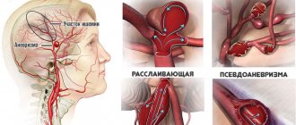
- if the presence of an aneurysm is suspected - local expansion of the vascular wall;
- in case of aneurysm dissection;
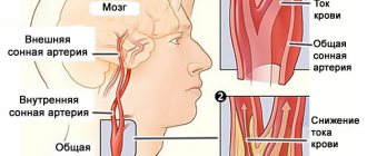
- with vascular stenosis;
- with an inflammatory process in the wall of a vessel (vasculitis);
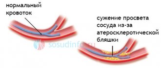
- with atherosclerosis.
Despite the informative value of the diagnostic measure in studying the condition of blood vessels, it can be harmful if contraindications are not taken into account. Relative limitations in the presence of which MRI angiography is possible, but only after excluding the provoking factor, include:
- the presence of an insulin pump, a nerve stimulator;
- the presence of a non-ferromagnetic implant in the inner ear;
- the presence of a prosthetic heart valve (in a high field if there is a suspicion of malfunction);
- development of decompensated heart failure;
- pregnancy period (research on the possibility of using the method in pregnant women does not exist today);
- claustrophobia - a panic attack that occurs when being in a confined space, including in the tunnel of a diagnostic apparatus.
There are also absolute restrictions, in the presence of which magnetic resonance angiography is prohibited:
- built-in pacemaker, in which the heart rhythm is simulated due to changes in the magnetic field;
- the presence of a ferromagnetic or electronic implant in the middle ear;
- the presence of a large metal implant or ferromagnetic fragment in the body;
In addition, contraindications include the presence of a hemostatic clip in a cerebral vessel, because MRI can cause intracerebral or subarachnoid bleeding.
Types of research
The methodology for performing the procedure determines two types of examinations:
- puncture (the contrast is poured exactly into the vessel using a puncture needle);
- catheterization (the agent is injected using a catheter connected to the local vascular bed).
MRI
Based on the area examined, brain scans with contrast can be presented as:
- general angiography (visualization of vessels of different calibers is performed);
- selective angiography (means scanning of the vertebrobasilar, carotid region);
- superselective technique (a vessel with a caliber that does not correspond to any blood pool is examined).
The vascular system imaging pathway determines the following scan types:
- classical angiography - taking x-rays with preliminary introduction of contrast into the vessels of the head;
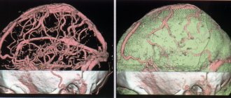
- CT angiography - scanning of the vascular system in the brain using an x-ray method with preliminary contrast and further 3D modeling of the display of the blood supply system;
- MR angiography involves examination using magnetic resonance imaging, often without prior contrast.
Computed tomography (MSCT) MRI
CT angiography
CT angiography or computed tomography angiography shows pathologies in blood vessels and allows one to study the pattern of blood movement through their internal cavity.
Indications for the use of CT angiography are:
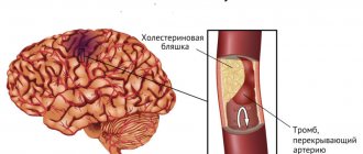
- the presence of stenosis or thrombosis of the vessel;
- the presence of an aneurysm in the vessel;
- suspicion of another vascular disease or congenital pathology.
Before undergoing a diagnostic procedure, it is necessary to exclude contraindications, which differ in some ways from the limitations inherent in other angiography methods. Among them:
- hypersensitivity to substances that are included in the contrast;
- development of renal failure;
- development of diabetes mellitus in severe form;
- pregnancy period (due to possible teratogenic effects);
- presence of severe general condition;
- overweight and obesity;
- disruption of the endocrine system;
- development of multiple myeloma;
- presence of acute heart failure.
The technique requires special training. Thus, before the study, possible contraindications are excluded, in particular, an allergic predisposition to the injected contrast agent. In order to reduce the risk of a corresponding reaction, take an antihistamine before the test.
The essence of the diagnostic procedure is as follows:
- The patient is placed on a special table.
- A catheter is inserted into the cubital vein, through which an iodine-based contrast agent is passed.
- Next, multiplanar and three-dimensional computer reconstruction is carried out with interpretation of the resulting images.
In some cases, computed angiography causes complications, including contrast extravasation. This negative consequence is the penetration of the substance into soft tissues that are located outside the vessel. As a rule, the volume of contrast entering the tissue does not exceed 10 ml. If it spreads to a larger extent, it causes severe damage to the subcutaneous tissue.
Factors that increase the risk of extravasation include a history of multiple punctures of one vessel and a weakened immune system. Characteristic symptoms are pain and swelling in the area where the needle was inserted. Treatment consists of ensuring an elevated position of the injured area and applying cold compresses.
Other negative consequences of computed angiography include intolerance to the contrast agent, the symptoms of which in most cases appear suddenly. Clinical manifestations of allergies are rash, itching, burning, hyperemia of the skin, swelling, feeling of lack of air.
Decoding the results
The interpretation of angiography results is carried out by a specialist. Based on the obtained angiogram, he evaluates the structural and functional state of the vascular bed. Depending on the density of organic tissues, X-ray radiation passes through them differently.
Because of this, radiographs look like:
- bones - white;
- liquor – black;
- the medulla is gray.
For all types of vessels, the so-called “branching, like in trees” is considered normal: smooth contours, uniform narrowing of the lumen. Various deviations from this norm may indicate various diseases that have already been mentioned above. Thus, angiography shows what pathologies are present in the patient and how exactly they can be cured.
Advantages and disadvantages
Normally, after an angiography is performed, the person is immediately released from the hospital to go home. Compliance with the regimen after the study is not required. No unpleasant symptoms or discomfort should occur after the procedure. Therefore, the diagnostic procedure is considered safe and can be done to identify pathologies in a child. The disadvantage of angiography is that it has a large list of contraindications that act as an obstacle to the study. If you neglect them, the risk of internal bleeding and other complications increases.
Other disadvantages include the possibility of an allergic reaction to the injected contrast agent. The severity of this varies depending on the level of sensitivity of the body and can be limited to redness or be more serious, for example, with the onset of anaphylactic shock. Therefore, to prevent an allergic reaction, it is recommended to conduct a sensitivity test before the study. People at risk include people who have a history of similar reactions to contrast agents and who suffer from asthma.
Both plain radiography and angiography require prior confirmation of pregnancy. This can be explained by the fact that the image is obtained using x-ray radiation, which has a negative effect on the fetus. If possible, it is recommended to postpone the study until after delivery. Alternatively, other imaging modalities that are not radiation-based, such as ultrasound, can be used.
Contraindications
The main contraindications are:
- allergic reaction (intolerance) to iodine preparations and other radiopaque substances;
- pregnancy (due to ionizing radiation during the procedure). In this case, MR angiography may be performed;
- mental illnesses that do not allow you to comply with all the conditions for the procedure (for example, a person will not be able to help but move during the photo);
- acute infectious and inflammatory diseases (as the risk of complications increases);
- violation of the blood coagulation system (both downward and upward);
- the general condition of the patient, regarded as severe (this may be stage III heart failure, end-stage renal and liver failure, coma, and so on). Essentially, this subgroup of contraindications is relative.
Alternative diagnostic methods
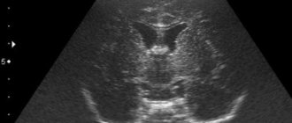
If for one reason or another it is impossible to perform angiography, alternative diagnostic methods are used. One of these is Doppler ultrasound of blood vessels. In this case, it is possible to identify circulatory disorders, changes in the structure and tone of the walls of arteries and veins. Used in the diagnosis of the head, neck, vessels near the spinal cord, and other organs. No special preparation is required for diagnosis.
The second alternative method is color duplex scanning. This is a type of Doppler ultrasound, which is used to obtain an informative picture of blood flow and the structure of the arteriovenous plexus of the head. In this case, stenosis, congestion, and congenital anomalies are detected.
There is no need to be afraid of angiography of cerebral vessels. The diagnostic procedure is absolutely safe, informative, and rarely causes negative consequences. Therefore, if alarming symptoms such as headache, dizziness, or tinnitus appear, you should immediately consult a doctor. Perhaps just such a study will be needed.