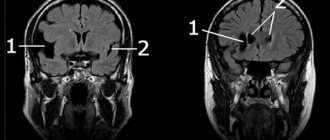
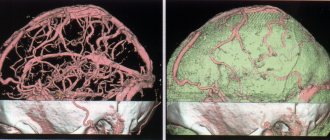
MSCT (multispiral computed tomography) is a relatively new method for studying human internal organs. Today it is considered one of the most accurate and effective. Three-dimensional models of the examined tissues, which are created with its help, help to identify pathology and its location in just a few seconds. MSCT of the brain allows you to visualize and examine in great detail the internal structures of this complex organ.
The image is so detailed and accurate that even the most minor changes in the structure of the nervous tissue are revealed.
- What is the basis of MSCT?
- When is MSCT of the brain performed?
- Advantages of the method
- How is multi-slice tomography performed?
- Contraindications and risks
- MSCT and other diagnostic methods
- How often can MSCT be done?
Options
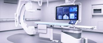
During the examination, the X-ray tube rotates around the patient's body with continuous movement of the table.
As a result, the study is carried out in turns - in a spiral, which is why CT is called “spiral”.
The prefix “multi-” means the presence of several rows of detectors in the tomograph (4, 8, 16 or more).
MSCT of the head - performed without contrast or with contrast.
In patients with head injuries, contrast administration is not indicated. In this case, a scan is performed with reconstructions in the bone and soft tissue window:
- in three planes;
- with three-dimensional reformatting.
Patients with head tumors and vascular abnormalities are advised to administer contrast.
CT perfusion is also used.
- The purpose of this study is to time-by-time assess the blood flow in a certain area after the injection of contrast into a vein.
- Volumetric blood flow, its peak velocity, etc. are assessed.
- The method detects foci of ischemia at an extremely early stage, when no changes are yet detected on the images.
- Perfusion is also indicated to assess tumor vascularization.
Recently, perfusion studies are performed less frequently due to the proliferation of DWI MRI. This method:
- allows you to do without contrasting;
- fast enough;
- specific
An area of diffusion restriction in a patient with neurological symptoms can be regarded as a manifestation of ischemia.
Contrast study
To obtain a clearer image, in some cases, the patient is injected with a colored liquid. Vascular examination is carried out only with contrast. The liquid entering the vessels stains them and on the monitor the doctor clearly sees the state of the vascular system.
Examination of brain tissue can be carried out either with or without a colored substance. For a complete examination of the brain, the patient may be injected with a contrast agent. The decision about the need for such a step is made by the attending physician and the radiation therapy doctor.
The colored substance is a nonionic composition with iodine. In addition to examining blood vessels, it is usually used to detect brain tumors. MSCT with contrast makes it possible to see malignant tumors - to determine the boundaries of neoplasms, the stage of development of the pathology, and the presence of damage to parenchymal tissues.
The contrast agent is excreted from the body within 24 hours by the kidneys. After administering the liquid, a person may experience a metallic taste in the mouth and a sensation of heat spreading throughout the body.
Advantages of the method
- Speed of execution. Computed tomography of the brain is a rapid imaging method. Data collection takes 2-3 minutes.
- Low dependence on the patient's condition. CT is less sensitive to head movements in contrast to MRI, incl. due to the short procedure time. Even if the patient is unconscious and makes head movements, the likelihood of obtaining informative images is higher than with MRI.
- High spatial resolution. The method shows bones with high resolution and allows you to evaluate the smallest traumatic changes.
Resolution for soft tissue is lower, but sufficient for visualizing gross pathology.
CT scan
What does it show
An informative method helps the doctor recognize any brain pathology. The session allows you to identify:
- brain diseases;
- stroke;
- hemorrhage;
- fractures of the cranial bones;
- aneurysm;
- bleeding;
- hematoma spot;
- cyst;
- tumors;
- inflammation.
Using a tomograph, the structures of the cerebellum, pons, hypothalamus, and thalamus are visualized. Both hemispheres of the brain are thoroughly examined.
The brain is the main element of the entire central nervous system. Its structures contain a huge number of neurons that create certain electrical impulses. Special connections are formed in the brain; the organ exercises control over all systems of the body. It controls the functions of all internal organs, ensures the necessary reactions and the formation of connections.
When the disease occurs, natural control and communication mechanisms are disrupted. A detailed examination using MSCT helps to identify the causes of disorders and the development of pathology. Some pathologies are congenital. The procedure shows the localization and parameters of the pathological focus, all the smallest changes in nearby tissues.
It is especially important to perform a session after a stroke. The results of the procedure will help the doctor prescribe the correct therapy to restore vital functions. The image clearly shows all pathological changes in organic structures and in the walls of blood vessels.
Nakhodki
| Nakhodki | What is being assessed |
| Fractures | Fracture line, number, nature, size, extent, which bones are involved (vault or base of the skull). Displacement of fragments (direction, extent). Impact of fragments on surrounding structures |
| Intracranial hemorrhages | Localization (sub- or epidural, or parenchymal hematoma, or subarachnoid hemorrhage). Character, structure (homogeneous or layered, hypo- or hyperdense). Size, impact on surrounding structures. Displacement of intracerebral structures. Breakthrough of blood into the ventricles of the brain |
| Concussion lesions | Localization, size, structure and nature of the lesion, its type (according to Kornienko), impact on neighboring structures, connection with the fracture line and fragments |
| Foreign bodies | Location, size, nature of objects (exogenous - bone fragment or endogenous - bullet, fragment, piece of wood, metal), connection with vital systems, direction of the wound canal, its size |
| Displacements and dislocations | The direction and type of displacement (for example, a shift of the temporal lobe under the tentorium of the cerebellum or vice versa - the cerebellum is above the tentorium). Moving the midline structures to the right and left. The most dangerous type is herniation of the cerebellum into the foramen magnum with compression of the brain stem and disruption of vital functions. |
| Ischemic stroke | Hypodense foci with blurred edges, smoothness and swelling of the brain convolutions, narrowing of the sulci, hyperdense artery in the ischemic zone due to thrombosis (in the acute phase). Cystic lesions, atrophic changes in the brain (in the chronic phase) |
| Hemorrhagic stroke | Size and localization of intracerebral hematoma, volumetric impact on neighboring structures, their dislocation. Connection with the ventricular system of the brain. Ventricular breakthrough is an unfavorable prognostic factor |
| Subarachnoid hemorrhage | Localization, severity of hemorrhage. Possible association with trauma or aneurysm. Subarachnoid hemorrhage often occurs due to a ruptured aneurysm and may require angiography to locate it. |
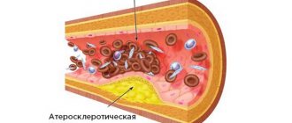
| Vascular pathology | Type (aneurysm or malformation), localization, extent, structure (thrombosis, calcifications), presence of complications (hemorrhages). Atherosclerosis of blood vessels (degree of narrowing, atherosclerotic plaques). Vascular anomalies (circle of Willis). Intravenous contrast is required for diagnosis |
| Intracranial tumors | Probable type of tumor (cerebral - glioma, astrocytoma; extracerebral - meningioma, schwannoma of large cranial nerves), metastases. Localization, dimensional values, structure, vascularization, damage to neighboring tissues (invasion, displacement) |
| Developmental anomalies | Type of anomaly (for example, holoprosencephaly or schizencephaly), location, severity, associated disorders |
| Post-operative changes | The nature of alterations (burr holes, milling holes), their size, type (covered or not). Presence of drainages, shunts |
| Increased intracranial pressure | Sharp balloon-like dilation of the cerebral ventricles, acute angle between the lateral ventricles on coronal sections (110 degrees or less), narrowing of the external cerebrospinal fluid spaces, the presence of an obstructive lesion (for example, a tumor) |
| Age-related changes | Symmetrical decrease in the volume of brain tissue, expansion of cerebrospinal fluid spaces, decrease in tissue density around the lateral ventricles, atherosclerosis |
Contrasting
Contrast is used for:
- assessment of tumor vascularization;
- visualization of blood vessels.
A contrast containing iodine in non-ionic form is used, for example:
- omnipack;
- ultravist;
- yopamiro.
The contrast is injected into the vein with an injector at high speed - 3-5 ml/sec. If necessary, a study of the vessels of the neck is also performed.
How often can MSCT be done?
The radiation dose depends on which organ is being studied. In any case, it is significantly less than the radiation dose during a conventional x-ray examination. MSCT of the brain provides radiation exposure of only 2 millisieverts (m3v), which is 3 times less than a similar examination of the spine and 5 times less than that of internal organs. It can be considered insignificant, since this dose is approximately equal to natural radiation exposure for 1 year.
Experts do not recommend doing multislice tomography more than once a year.
Medical practice allows it to be carried out several times a month, if there are compelling reasons for this, since the method is significantly superior in speed, accuracy and efficiency to other types of examinations.
Preparation
If the patient is in a state of motor agitation, for example, as a result of a stroke or severe injury, sedation is indicated. In stationary conditions the following are introduced:
- or a tranquilizer;
- or an anesthetic;
- or a muscle relaxant.
This is necessary to limit mobility and obtain high-quality images. In outpatient practice, special preparation is usually not needed except for contrast studies of vessels and tumors.
In this case, you need to evaluate the level:
- creatinine;
- urea in the blood;
- kidney function.
You must come to the clinic in advance, taking with you:
- direction;
- test results;
- data from previously performed studies, incl. MRI and X-ray, CT, expert opinions.
Please note: you must take the results of previous studies with you.
Contraindications for examination
During the diagnostic period, the patient is exposed to x-rays and intravenous contrast, a substance that contains iodine.
Therefore, MSCT is contraindicated for:
- pregnancy and lactation (if diagnostics are necessary, breastfeeding should be avoided for at least 2-3 days);
- identifying an allergy to iodine or a vascular contrast agent;
- hyperthyroidism or thyrotoxicosis;
- insufficiency of kidney or liver function;
- bronchial asthma;
- decompensated diabetes mellitus;
- severe heart failure
Relative contraindications include severe pain, motor or mental agitation, as they can reduce image clarity and complicate diagnosis due to the patient’s inability to maintain a stationary position.
Methodology
You must first remove all metal jewelry and dentures.
- The patient lies down on the table with his head resting on the headrest.
- If contrast is performed, a catheter is installed in the vein of the elbow.
- The table slides into the tomograph and scanning begins.
- The collected data is transferred to the workstation, where it is converted into an easy-to-view format.
Duration
The examination takes several tens of seconds; most of the time is spent on preparation. On average, a study without contrast takes 2-3 minutes, with contrast - up to a quarter of an hour.
Alternatives
| Method | Note |
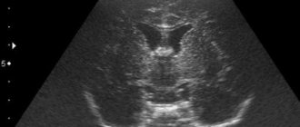
| Ultrasound | In small children with open fontanelles |
| Radiography | Approximate method for searching for bone trauma |
| MRI | Detailed brain assessment in outpatients, acute phase stroke assessment using DWI |
| Nuclear medicine | Search and evaluation of neoplastic lesions |
Indications
MSCT of all cerebral vessels is a procedure necessary to identify the condition after a stroke and for a number of other ailments. An examination of the organ should be performed if it is necessary to identify the causes of confusion and numbness. The study is necessary to undergo in case of injury, constant dizziness and fainting.
The diagnostic method is prescribed by doctors:
- to detect vascular pathology;
- for diagnosing arterial hypertension;
- to localize a cyst or tumor;
- before surgery;
- for diagnosing hearing loss;
- to determine the causes of dizziness and fainting.
The procedure is necessary for cancer therapy. It allows you to clearly monitor the condition of the affected areas and see the results of the prescribed drug therapy. Tomography has been used by doctors for a very long time. When performing a scan, all the nuances of the pathology are revealed. Preparing and conducting the session does not have any difficulties.
