The functions of the spinal cord - reflex and conduction - are very important. It is thanks to these functions that a person can carry out his life activities. Let's talk more about the work and physiology of this part of the nervous system.
Breathing, heartbeat and even sexual desire all depend on the functioning of the spinal cord. There are two important functions of the spinal cord: conduction and reflex. Let's look at each of them in more detail.
Reflex function
It is thanks to the reflex function of the spinal cord that we withdraw our hand from something hot and cough if something gets into our respiratory tract. A person’s reaction to stimuli - this is how we can simply characterize this function. That is, all reflexes are carried out due to it. The importance of this function is beyond doubt, because all our actions are based on reflexes.
In short, this happens due to the reflex arc. It looks like this: the receptor senses the stimulus - the impulse goes to the SC - there the impulse switches to the neuron responsible for movement, and we react to the stimulus (we withdraw our hand, sneeze).
Each such arc has three links:
- afferent – responsible for transmitting impulses from the organ to the SM;
- intercalary – connecting the previous link directly with the neuron responsible for executing the command;
- efferent, through which the impulse travels from the brain to the organ.
Reflexes, as is known, are congenital and acquired. They close at a certain level. This is why the neurologist checks our knee reflex: he wants to make sure the spinal cord is working.
The spinal cord performs two functions: reflex and conduction.
The reflex function is carried out by the nerve centers of the spinal cord, which are the segmental working centers of unconditioned reflexes. Their neurons are directly connected to receptors and working organs. It has been established that each segment of the spinal cord innervates three metameres (transverse segments) of the body through its roots and receives sensitive information also from three metameres. Due to this overlap, each metamer of the body is innervated by three segments and transmits signals (impulses) to three segments of the spinal cord (safety factor). The spinal cord receives afferentation from receptors in the skin, motor system, blood vessels, digestive tract, excretory and genital organs. Efferent impulses from the spinal cord go to the skeletal muscles, including the respiratory muscles - intercostal muscles and the diaphragm, internal organs, blood vessels, sweat glands, etc. The overlying parts of the central nervous system, without having a direct connection with the periphery, control it through the segmental centers of the spinal cord.
The conductive function of the spinal cord is carried out through ascending and descending pathways. Ascending pathways transmit information from tactile, pain, temperature receptors of the skin and from proprioceptors of skeletal muscles through neurons of the spinal cord and other parts of the central nervous system to the cerebellum and cerebral cortex. Ascending paths include:
1) the anterior spinothalamic tract is the afferent pathway of touch and pressure (tactile sensitivity);
2) the lateral spinothalamic tract is the path of pain and temperature sensitivity;
3) the anterior and posterior spinocerebellar pathways (V. Goversa and P. Flexig) are afferent pathways of the muscular-articular (proprioceptive) sensitivity of the cerebellar direction;
4) the thin (gentle) fascicle of F. Gaulle and the wedge-shaped fascicle of K. Burdach are the afferent pathways of the muscular-articular (proprioceptive) sensitivity of the cortical direction from the lower extremities and the lower half of the body and, respectively, from the upper extremities and the upper half of the body.
Descending pathways connect the cerebral cortex, subcortical nuclei and brainstem formations with motor neurons of the spinal cord. They provide the influence of the higher parts of the central nervous system on the activity of skeletal muscles. The descending pyramidal tracts include: the anterior corticospinal (pyramidal) and lateral corticospinal (pyramidal) tracts - conduct impulses of voluntary motor reactions from the cerebral cortex to the anterior horns of the spinal cord (control of conscious movements).
The descending extrapyramidal tracts that control involuntary movements include: reticular-spinal cord (reticulospinal), tectospinal (tectospinal), pre-door-spinal (vestibulospinal) and red-nucleus-spinal cord (rubrospinal) tracts.
A person has 31 pairs of spinal nerves, corresponding to 31 segments of the spinal cord: 8 pairs of cervical, 12 pairs of thoracic, 5 pairs of lumbar, 5 pairs of sacral and a pair of coccygeal nerves. Each spinal nerve is formed by connecting the anterior (motor) and posterior (sensory) roots and is a relatively short trunk. Upon exiting the intervertebral foramen, the nerve is divided into two main branches: anterior and posterior, both of which are mixed in function. In addition, the following branches depart from the spinal nerve: the meningeal branch (it goes into the spinal canal to the hard shell of the spinal cord) and the white connecting branch to the nodes of the sympathetic trunk.
Through the spinal nerves, the spinal cord carries out the following innervation: sensitive - to the trunk, limbs and part of the neck, motor - to all muscles of the trunk, limbs and part of the neck muscles; sympathetic innervation - of all organs that have it, and parasympathetic - of the pelvic organs.
The posterior branches of all spinal nerves have a segmental arrangement. They go to the back surface of the body, where they are divided into cutaneous and muscular branches that innervate the skin and muscles of the back of the head, neck, back, lumbar region and pelvis. These branches are named after the corresponding nerves (for example, the posterior branch of the I thoracic nerve, ... II, etc.). Only some of them additionally have special names. For example, the posterior branch of the first cervical nerve is called the suboccipital nerve, and the second branch of the cervical nerve is called the greater occipital nerve.
The anterior branches are much thicker than the posterior ones. Of these, only 12 pairs of thoracic spinal nerves have a segmental (metameric) location. These nerves are called intercostal because they run in the intercostal spaces on the inner surface along the lower edge of the corresponding rib. They innervate the skin and muscles of the anterior and lateral walls of the chest and abdomen. The anterior branches of the remaining spinal nerves form plexuses before going to the corresponding area of the body
There are cervical, brachial, lumbar and sacral plexuses. from the plexus, each of which has its own name and innervates a specific area.
The cervical plexus is formed by the anterior branches of the four superior cervical nerves. It is located in the area of the four upper cervical vertebrae on the deep muscles of the neck. In front and on the side it is covered by the sternocleidomastoid muscle. Sensory (cutaneous), motor (muscle) and mixed nerves (branches) depart from this plexus.
1) Sensory nerves: the lesser occipital nerve, the greater auricular nerve, the transverse nerve of the neck, the supraclavicular nerves respectively innervate the skin of the lateral part of the back of the head, the auricle, the external auditory canal, the anterolateral region of the neck, the skin in the area of the clavicle and below it.
2) The muscle branches innervate the deep muscles of the neck (scalenes, etc.), as well as the trapezius, sternocleidomastoid muscles, and the infrahyoid muscles receive innervation from the cervical loop.
3) The phrenic nerve is a mixed nerve and the largest nerve of the cervical plexus. Its motor fibers innervate the diaphragm, and its sensory fibers innervate the pericardium and pleura.
The brachial plexus is formed by the anterior branches of the four lower cervical, part of the anterior branch of the IV cervical and I thoracic spinal nerves. In the plexus, there are supraclavicular (short) branches, extending mainly from the trunks of the supraclavicular part: upper, middle and lower, and subclavian (long) branches, extending from three bundles: medial, lateral and posterior subclavian part, surrounding the axillary artery on three sides.
The short branches of the brachial plexus innervate the muscles and skin of the chest, all the muscles of the shoulder girdle and the muscles of the back. The shortest branch of the infraclavicular part of the brachial plexus is the axillary nerve, which innervates the deltoid, teres minor muscles and the capsule of the shoulder joint.
The long branches of the brachial plexus innervate the skin and muscles of the free upper limb. These include the following nerves:
1) the medial cutaneous nerve of the shoulder innervates the skin of the medial surface of the shoulder;
2) the medial cutaneous nerve of the forearm innervates the skin of the anteromedial surface of the forearm;
3) the musculocutaneous nerve innervates the shoulder flexor muscles: biceps, brachialis, coracobrachialis and the skin of the anterolateral surface of the forearm;
4) the median nerve on the shoulder does not give branches, innervates the anterior group of muscles of the forearm, except for the flexor carpi ulnaris and the medial part of the deep flexor digitorum, on the hand - the muscles of the eminence of the thumb (except for the adductor muscle), two lumbrical muscles, the skin of the lateral part of the palm, the palmar surface of 3.5 fingers, starting with the thumb, and partially the dorsal surface of these fingers;
5) the ulnar nerve on the shoulder also does not give branches; it innervates the flexor carpi ulnaris, the medial part of the deep flexor digitorum, the muscles of the eminence of the little finger, all interosseous, two lumbrical muscles, the muscle that adducts the pollicis, the skin of the medial parts of the hand, palmar and dorsum 1 .5 and 2.5 fingers, starting with the little finger;
6) radial nerve - the thickest nerve of the brachial plexus, innervates the extensor muscles of the shoulder and forearm, the skin of the posterior surface of the shoulder, forearm, the skin of the lateral parts of the dorsum of the hand and the dorsal surface of 2.5 fingers, starting with the thumb.
The lumbar plexus is formed by the anterior branches of the upper three lumbar nerves and partially by the anterior branches of the XII thoracic and IV lumbar nerves. It is located next to the lumbar vertebrae in the thickness of the psoas major muscle. Short branches of the lumbar plexus innervate the quadratus lumborum muscle, the iliopsoas muscle, the abdominal muscles, as well as the skin of the lower abdominal wall and external genitalia (muscle branches, iliohypogastric, ilioinguinal and genital femoral nerves). The long branches of this plexus innervate mainly the free lower limb. The largest branches of the lumbar plexus are:
1) the lateral cutaneous nerve of the thigh innervates the skin of the lateral surface of the thigh to the knee joint;
2) the femoral nerve innervates the anterior group of thigh muscles and the skin above it. It is the thickest nerve of the lumbar plexus. The longest subcutaneous branch of this nerve, the saphenous nerve, descends along the medial surface of the leg and foot, where it innervates the skin of the anteromedial surface of the leg and the medial edge of the foot to the big toe;
3) the obturator nerve from the plexus descends into the small pelvis, and from there, through the obturator canal, it enters the medial surface of the thigh and innervates the medial group of muscles that adduct the thigh, the skin above them, as well as the hip joint.
The sacral plexus is formed by the anterior branches of the IV (partial) and V lumbar nerves and the upper four sacral nerves. Located in the pelvic cavity on the anterior surface of the piriformis muscle. Short and long branches extend from it. Short branches include the superior and inferior gluteal nerves, the pudendal nerve, the obturator internus, the piriformis nerve, and the quadratus femoris nerve. The pudendal nerve innervates the muscles and skin of the perineum and external genitalia, the remaining nerves innervate the adjacent muscles of the pelvis and gluteal region.
The long branches of the sacral plexus are represented by the posterior cutaneous nerve of the thigh and the sciatic nerve. Both nerves enter the posterior surface of the thigh through the infrapiriformis foramen, where the posterior cutaneous nerve of the thigh innervates the skin of the perineum, gluteal region and posterior thigh, and the sciatic nerve (the largest nerve in the human body) innervates the entire posterior group of thigh muscles. Next, the sciatic nerve descends into the popliteal fossa and divides into two branches: the tibial and common peroneal nerves. The tibial nerve runs along the back of the leg between the superficial and deep muscles (flexors of the leg and foot), innervating them. It then passes behind the medial malleolus to the plantar surface of the foot and divides into the medial and lateral plantar nerves, which innervate the skin and muscles of the sole of the foot. The common peroneal nerve in the thickness of the long peroneal muscle is divided into the superficial and deep peroneal nerves, both passing to the dorsum of the foot. The first innervates the long and short peroneus muscles, the skin of the dorsum of the foot and fingers, the second innervates the anterior group of leg muscles (extensors of the foot and fingers), the muscles of the dorsum of the foot, the capsule of the ankle joint and the skin of the first interdigital space of the dorsum of the foot. The cutaneous branches of the tibial and common peroneal nerves, connecting on the posterior surface of the leg, form the sural nerve, which innervates the skin of the lateral edge of the foot. Thus, in the lower leg and foot, the tibial and common peroneal nerves provide innervation to all muscles and skin of these areas, with the exception of the skin of the medial surface of the lower leg and foot (innervated by the saphenous nerve of the thigh).
Inflammation of a nerve is called neuritis
(mononeuritis
), spinal roots - radiculitis (Latin radix - root), nerve plexus - plexitis (Latin plexus - plexus). Multiple inflammation or degenerative damage to the nerves is polyneuritis.
Pain along the nerve, not accompanied by a significant impairment of the function of the organ or muscle, is called
neuralgia. Burning pain, intensifying in attacks, is called causalgia
(
Greek kausis - burning, algos - pain), observed after damage (wound, burn) to nerve trunks rich in fibers of the sympathetic nervous system. Pain that acutely occurs in the lumbar region during physical exertion, especially lifting weights, is called lumbago
(lumbago).
Painful, motor and autonomic disorders caused by damage to the spinal cord roots due to spinal osteochondrosis are discogenic radiculopathies
(banal radiculitis).
Inflammation of the spinal cord is called myelitis. Purulent inflammation of the tissue in the epidural space of the spinal cord is epiduritis. A disease characterized by the formation of cavities in the center of the gray matter of the spinal cord is called syringomyelia. An acute viral disease caused by damage to the cells of the anterior horns of the spinal cord and the motor nuclei of the cranial nerves is called polio.
Conductor function
The conductor function of the SC is as follows: the gray matter directs impulses from the peripheral nerves located in the organs to other parts of the central nervous system. The conductors that make up the white matter transmit signals from skin receptors, muscles, and internal organs. The impulses are then transmitted along short paths to other segments of the spinal cord, and along long paths to the brain.
This function is performed by descending and ascending pathways, which are located in the white matter. They communicate not only between different parts of the spinal cord. They also keep in touch with the brain. In addition to the motor centers responsible for the functioning of skeletal muscles, there are also sympathetic and parasympathetic autonomic centers. In the lateral horns of the cervical and thoracic regions there are also nerve centers that innervate almost all internal organs: the heart, gastrointestinal tract, blood vessels, and others.
In the sacral region there are centers responsible for the innervation of the pelvic organs. It is their lesions that can lead to uncontrolled urination and defecation.
When the spinal cord is damaged, a condition called spinal shock occurs. The physiology of SM is designed in such a way that the activity of all human reflex centers located below the site of injury is inhibited. This condition lasts for about six months, although, depending on the type of injury, complete recovery may not occur. This leads to the fact that the white matter cannot perform its conductive function.
Brain stem
The brain stem includes:
- medulla,
- bridge,
- midbrain.
Brain stem functions
- responsible for primitive forms of behavior,
- supports vital functions.
Did you like the site? Support us by subscribing on social networks!
- Site group in VK
- Website profile on Twitter
- Site community on Facebook
Medulla
The medulla oblongata (medulla oblongata) is 2.5–3 cm, located between the pons and the origin of the C1 root of the spinal cord.
Centers of the medulla oblongata
- Vital autonomic centers: respiration, vascular-motor center, digestion.
- Protective reflexes: sneezing, coughing, vomiting, blinking, sucking, chewing, swallowing.
- Centers that control the muscles of the limbs and trunk (lateral reticulospinal tract).
The medulla oblongata contains the nuclei of the IX, X, XI, XII pairs of cranial nerves, which are involved in the innervation of the head and neck.
The X pair innervates the internal organs of the thoracic and abdominal cavities.
Pons
The pons (discovered by the scientist Varolio in 1560) is located between the midbrain and medulla oblongata.
Reflex function:
- Within the bridge are located the nuclei of the V, VI, VII and VIII pairs of cranial nerves innervating the head.
- The intrinsic neurons of the pons form its reticular formation.
- Pontine RF affects the cerebral cortex, causing its activation or inhibition.
Among these neurons, a group of nuclei is localized, forming a pneumotaxic center that regulates the change of inhalation and exhalation.
Vestibular nuclei:
- ankylosing spondylitis's upper nucleus
- Roller's inferior vestibular nucleus
- medial nucleus of Schwalbe,
- lateral nucleus of Deiters.
The nuclei, together with the cerebellum, take part in maintaining the balance and tone of skeletal muscles.
From Deiters' nucleus comes the lateral vestibulospinal tract.
Excitatory effect on extensor motor neurons and inhibitory effect on flexor motor neurons.
When the vestibular apparatus is irritated, muscle tone is redistributed in such a way as to maintain balance.
Organ characteristics
The morphofunctional characteristics of the spinal organ are as follows:
The spinal cord consists of two symmetrical halves, separated from each other by a deep central fissure. At the back they are separated by a connective tissue septum.
Inside the organ there is a dark area called gray matter. In the periphery of the spinal cord there is light white matter.
From the cross-sectional side, the organ has an H-shaped gray matter. The places where the gray matter protrudes are called horns. They are anterior (ventral), posterior (dorsal) and lateral (lateral).
Gray matter includes neuronal cell bodies, unmyelinated and thin myelinated fibers, and neuroglia. It differs from white matter in that it contains multipolar neurons.
Longitudinal myelinated fibers make up the white matter. The spinal cord pathways are made up of nerve fibers that connect different parts of the nervous system.
The neurons of the organ are divided into neurites, radicular cells, as well as internal and tufted neurons.
Each dorsal horn includes a spongy layer, a gelatinous substance, a horn nucleus, and a thoracic nucleus.
The posterior horn contains a gelatinous substance that inhibits the functioning of the spinal cord.
The anterior horns are equipped with large neurons of the spinal cord, which form nuclei - somatic centers; they also have medial and lateral groups of motor cells. The medial cells are responsible for the functioning of the muscles of the human torso, and the lateral cells are responsible for the muscles of the arms and legs.
Functioning of the conduction system
How does the conduction system of the heart work?
Due to irritation of the ACS, an electrical impulse is generated in it. It spreads through three conduction bundles to both atria and reaches the AV node. Here there is a delay of the impulse, which ensures the sequence of contractions of the atria and ventricles.
Next, the impulse passes to the His bundle and Purkinje fibers, which approach the contractile cells. Here the electrical impulse fades away. The coordinated activity of all elements is called cardiac automatism. The conduction system of the heart can be clearly seen in the video in this article.
What happens if the functions are disrupted?
The spinal cord may rupture or atrophy. In any situation, the patient must be urgently taken to the hospital.
Organ rupture
When an organ ruptures, very unpleasant, serious and unpredictable consequences can occur.
When the brain ruptures, a person’s sensitivity and activity are lost, and partial or complete paralysis of the body may occur. The consequences of a spinal cord rupture manifest themselves in a person receiving partial or complete disability, due to which he is unable to independently care for himself and live as before. Such a rupture occurs when you receive a domestic injury, fall from a height, or get into a car accident. If the entire body refuses to work, spinal shock occurs, which often leads to the death of a person.
What does the human spinal cord look like?
Experts distinguish five sections of the spinal cord, each of which includes a certain number of vertebrae:
- The neck consists of the cervical spine (8 vertebrae).
- The chest consists of the thoracic region (12 vertebrae).
- Loin from the lumbar region (5 vertebrae).
- Sacrum from the sacral region (5 vertebrae).
- Coccyx from the coccygeal region (one vertebra).
What does the organ consist of?
The spinal cord has round sections, but its structure also includes thickened areas that are flattened from front to back.
The cervical thickening can be seen near the third cervical and first thoracic vertebrae. In the area of 10–12 thoracic vertebrae there is a lumbosacral thickening. In the area of somatic neurons, which are located in the thickenings of the organ, there are many roots with a large number of nerve fibers. The thickness of somatic neurons is greatest, since they are larger than other neurons.
The spinal cord supplies the internal organs and skeletal muscles of a person with nerve fibers that communicate with the central nervous system. The valuable organ of the spine consists of 31 segments, which are responsible for different organs and parts of the body:
Eight cervical segments are responsible for the head, neck, chest cavity, heart, lungs and arms.
12 thoracic and 5 vertebral segments are responsible for the abdominal cavity and trunk muscles.
The leg muscles and lower abdominal cavity control the 5 sacral and coccygeal segments.
Cerebrospinal fluid can be seen in the canal, which is located in the center of the spinal organ.
External structure of the spinal cord
External structure of the spinal cord and features: this organ, and not the brain, of the nervous system externally resembles a cylindrical cord, which is covered with membranes. The diameter of the organ is 1 centimeter, but the diameter changes throughout its entire length.
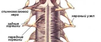
Along the entire length of the brain, not the brain, 124 roots extend, which form 31 pairs of spinal nerves, which consist of 2 roots (anterior and posterior). The anterior median fissure runs in the middle part of the anterior surface, the longitudinal superficial groove stretches along the posterior surface. The spinal cord is visually divided into symmetrical parts due to the formation of these grooves and fissures. The posterior lateral sulcus runs along the sides of the longitudinal sulcus. The posterior radicular filament enters it.
Outside the anterior longitudinal fissure there is an anterior lateral groove. In the cervical and thoracic parts, a longitudinal groove is closed between the grooves. The anterior roots are formed due to the exit of the radicular filaments from the anterior lateral groove. The posterior bundles are formed from the posterior radicular filaments, which are arranged in a single row.
The anterior and posterior bundles follow to their intervertebral foramen, where the spinal ganglion is formed. Afterwards, both nerves merge and form a mixed spinal nerve, not the brain. Later the nerve splits into two branches.
The structure and direction of the radicular branches depends on their exit from the foramen; due to the rapid growth of a person, the lumbar and sacral branches run downwards parallel to the spinal cord of the nervous system. In the sacral region, the conus medullaris lies among the nerve roots; this formation is called the cauda equina. White.