General information
In the middle of the dura mater and the arachnoid membrane there is a subdural cavity of insignificant width filled with a small amount of interlayer fluid - cerebrospinal fluid.
In some fragments, the dura mater grows in the form of processes into the narrow spaces of the brain. In areas where the processes grow, the membrane bifurcates, forming triangular sinuses also covered with endothelium - the dura mater sinuses. The plates of these tanks are very tightly stretched and do not move, even when cutting.
These tanks are designed to contain venous blood, which gradually drains from the veins supplying nutrition and oxygen to the brain in the skull. From the sinuses, blood flows into the internal jugular veins; in addition, there is communication between these recesses and the arteries of the outer surface of the head thanks to spare arterial outlets.
Meninges: processes of the dura mater. cisterns of the subarachnoid space.
The membranes of the brain. Dura mater encephali.
The membranes of the brain, meninges, are a direct continuation of the membranes of the spinal cord - hard, arachnoid and soft.
The dura mater encephali is a dense whitish connective tissue membrane lying outside the other membranes. Its outer surface is directly adjacent to the cranial bones, for which the hard shell serves as the periosteum, which is how it differs from the same shell of the spinal cord. The inner surface facing the brain is covered with endothelium and, as a result, is smooth and shiny. Between it and the arachnoid membrane of the brain there is a narrow slit-like space, spatium subdurale, filled with a small amount of fluid. In some places the hard shell splits into two sheets. This splitting occurs in the area of the venous sinuses (see below), as well as in the area of the fossa at the apex of the pyramid of the temporal bone (impressio trigemini), where the trigeminal ganglion lies.
The hard shell gives off several processes from its inner side, which, penetrating between the parts of the brain, separate them from each other.
Sickle cerebri, falx cerebri. Tentorium cerebellum, tentorium cerebelli.
Falx cerebri, the falx cerebri, is located in the sagittal direction between both hemispheres of the cerebrum. Attached along the midline of the cranial vault to the edges of the sulcus sinus sagittalis superioris, its anterior narrow end grows into the crista galli, and its wide posterior end fuses with the upper surface of the cerebellar tentorium.
Tentorium cerebelli, the tentorium of the cerebellum, is a horizontally stretched plate, slightly convex upward, like a gable roof. This plate is attached along the edges of the sulcus sinus transversa of the occipital bone and along the upper edge of the pyramid of the temporal bone on both sides to the processus clinoideus posterior of the sphenoid bone. The tentorium separates the occipital lobes of the cerebrum from the underlying cerebellum.
Falx cerebellum, falx cerebelli. Diaphragm sellae, diaphragma sellae.
Falx cerebelli, the falx of the cerebellum, is located, like the falx cerebellum, in the midline along the crista occipitalis interna to the foramen magnum of the occipital bone, covering the latter on the sides with two legs; this low process projects into the posterior notch of the cerebellum.
Diaphragma sellae, sellae diaphragm, a plate that bounds above the receptacle for the pituitary gland at the bottom of the sella turcica. In the middle it is pierced by a hole for the passage of a funnel, infundibulum, to which the hypophysis is attached.
Arachnoidea encephali. Tanks, cisterna. Granulations of the arachnoid membrane, granulationes arachnoideales.
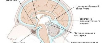
The arachnoid membrane, arachnoidea encephali, as in the spinal cord, is separated from the dura mater by a capillary gap in the subdural space. The arachnoid membrane does not go into the depths of the grooves and recesses of the brain, like the pia mater, but spreads over them in the form of bridges, as a result of which between it and the soft membrane there is a subarachnoid space, cavitas subarachnoidealis, which is filled with a transparent liquid. In some places, mainly at the base of the brain, the subarachnoid spaces are especially strongly developed, forming wide and deep containers of cerebrospinal fluid, called cisterns.
The following tanks are available:
1. Cisterna cerebellomedullaris (largest) between the posterior edge of the cerebellum and the medulla oblongata.
2. Cisterna interpeduncularis between pedunculi cerebri.
3. Cisterna chiasmatis in front of chiasma opticum.
4. Cisterna fossae lateralis cerebri in the pit of the same name.
All subarachnoid spaces communicate widely with each other and, at the foramen magnum of the occipital bone, directly continue into the subarachnoid space of the spinal cord. In addition, they are in direct communication with the ventricles of the brain through openings in the region of the posterior wall of the IV ventricle: apertura mediana ventriculi quarti, opening into the cisterna cerebellomedullaris, and apertura lateralis ventriculi IV. In the subarachnoid spaces lie cerebral vessels, which are protected from compression by connective tissue bars, trabeculae arachnoideales, and the surrounding fluid.
A feature of the structure of the arachnoid membrane is the so-called granulation of the arachnoid membrane, granulationes arachnoideales, which are outgrowths of the arachnoid membrane in the form of roundish bodies of gray-pink color, protruding into the cavity of the venous sinuses or into nearby blood lakes. They are present in children and adults, but reach their greatest size and abundance in old age. Increasing in size, the granulations, by their pressure on the cranial bones, form depressions on the inner surface of the latter, known in osteology as foveolae granulares. Granulations serve to drain cerebrospinal fluid into the bloodstream through filtration.
Soft shell, pia mater encephali.
The soft shell, pia mater encephali, is closely adjacent to the brain, extending into all the grooves and crevices of its surface, and contains blood vessels and choroid plexuses. Between the membrane and the vessels there is a perivascular gap that communicates with the subarachnoid space.
Structure
The hard shell is a fibrous type protective plate that penetrates from the inside to the bone tissue of the skull. Forms processes that grow into the cranial space: a crescent-shaped continuation of the cerebrum, a continuation of the cerebellum in the form of a sickle, tentorium, sellar plate, etc.
Between the dura mater and the bone tissue of the skull there is an epidural cavity of the brain, which essentially means the union of multiple spaces separated by connective tissue bases (tractions). These areas develop after birth, during the closure of pulsating fontanelles. At the arch, these spaces expand, since there are not so many cartilaginous foundations here. On the vault of the skull, and in the direction of the venous sinuses and cranial joints, the mentioned cavities become narrower and the interweaving of the cords is very dense. All connecting cavities are provided with endothelium and filled with fluid. Using experiments, it has been scientifically proven that epidural fluid flows into the external network of small vessels of the dura mater.
The dura mater of the brain is divided into two more or less reinforced plates, of which the outer one is the periosteum of the skull. Each of the plates is delaminated. Without exception, all layers are equipped with fibrillar protein, essentially the basis of the connecting material. They are connected into bundles, placed equally horizontally in each layer. In neighboring layers, the beams intersect, forming a cross.
Pathology of the dura mater of the spinal cord
External pachymeningitis often develops. During its development, inflammation occurs, affecting the epidural tissue, after which inflammation spreads to the entire surface of the dura mater of the spinal cord.
Diagnosing the disease is quite difficult. But the incidence of spinal pachymeningitis is higher than the development of pathologies associated with inflammation in the dura mater of the brain. To identify it, it is necessary to build on the patient’s complaints, medical history, as well as laboratory tests of cerebrospinal fluid and blood.
Sinuses and processes of the dura mater of the brain
The following are considered to be the processes of the dura mater:
- A large crescent-shaped continuation, or the crescent-shaped process of the largest hemispheres of the brain, is located between both large parts of the brain;
- A small falciform process, or a falciform process near the cerebellum, extends into the cavity between the cerebellar hemispheres, joining the bone tissue of the occiput from the internal occipital indentation to the large foramen of the occiput;
- Cerebellar tentorium - located between the parts of the cerebral hemispheres at the back of the head and the cerebellum;
- The plate of the sella turcica is located above the sella turcica; in the middle there is a hole through which a funnel runs.
Sinuses (lacunae) of the dura mater of the brain, formed due to the splitting of the dura mater into two strands, are essentially channels through which blood from the veins is drained from the head into the internal dual veins.
The hard shell plates that form the lacunae are tightly reinforced and do not move. Therefore, these sinuses are visible in the section. They are not equipped with valves. This structure of these tanks allows venous blood to flow freely from the brain, completely independent of pressure surges inside the skull. On the inner walls of the bone tissue of the skull, in the areas where these recesses of the hard shell are located, there are proper tentories. In medical practice, the following names of the dura sinuses are used:
- The superior vertically dividing sinus is located longitudinally along the entire upper-outer border of the falx of the cerebral hemispheres, from the edge resembling the cockscomb of the ethmoid bone to the indentation of the occiput inside. In the anterior parts, this tank is equipped with anastomoses with veins of the paranasal space. Its termination at the rear is included in the transverse manifold.
- The lower vertically dividing lacuna is located inside the lower spacious border of the falx of the cerebral hemisphere. It is much smaller than the top one.
- The straight sinus is located vertically in the splitting of the cerebellar membrane in the direction of the attachment of the falx of the cerebral hemisphere to it. This collector combines the posterior ends of the superior and inferior sagittal sinuses.
- The transverse cistern is located in the part of the separation of the cerebellar plate from the dura mater of the brain. On the inner side of the scales of the bone tissue of the back of the head, an extensive groove of the transverse sinus is related to this depression.
- The occipital lacuna lies at the bottom of the cerebellar falx. Descending longitudinally from the inside of the occipital border, this tank is located to the posterior border of the large opening of the occiput, where it diverges into two grooves that frame this opening at the back and on both sides.
- The sigmoid collector is double, located in the sigmoid branch on the inside of the skull, characterized by an S-shaped appearance. In the area of the opening of the great veins, this tank flows into the jugular vein.
- The cavernous sinus is double and lies on the vault of the cranium away from the sella turcica. The carotid artery and some intracranial nerve fibers pass through this tank. The recess has a very intricate structure in the form of interconnected caves, which is why it got its name.
- The sphenoparietal lacuna is double, refers to the spacious posterior border of a small wedge-shaped bone fragment, in the cleft it connects in this place with the dura mater of the brain.
- The upper and lower stony recesses are double and lie longitudinally along the upper and lower borders of the triangle of bone tissue in the temporal region.
In some areas, all these cisterns form connections - anastomoses - with the external veins of the skull through connections of vessels. In addition, the sinuses of the TO connect to the diploic arteries, which are located in the spongy structure of the bones of the base of the skull and are included in the superficial vessels of the head. Thus, blood from the veins of the brain flows through the branches of its vessels located on the surface and in the depths into the sinuses of the brain and then into both internal large veins.
Comparison with soft shell
The most basic difference between the dura mater and the soft mater is the presence of double layers, a large number of veins and capillaries in the second. In addition, the pia mater is located closest to the gyri, glia and barbules, separated only by the glial diaphragm. In specific areas, the soft membrane penetrates into the spaces of the ventricles of the brain and forms vascular weaves that synthesize cerebrospinal fluid. Whereas the dura mater has sinuses, and has a slightly different structure and functional tasks.