General information about the cisterns of the brain
The meninges have a three-layer structure:
- hard, which is located directly next to the cranial bones;
- arachnoid;
- soft, which covers the brain.
Let's look at each of the layers in more detail:
- In the structure of the dura mater there are small processes that are designed to separate different parts of the brain. This layer fits tightly to the skull. The largest process is considered to be the one that divides the human brain into two equal hemispheres; in appearance it resembles a crescent. A special diaphragm is located at the top of the hard layer; it protects the brain from external damage.
- After the hard layer comes the arachnoid (arachnoid). It is very thin, but at the same time provides sufficient strength. Simultaneously connects to the hard and soft shell. This layer is intermediate.
- The soft shell, or as it is also called the soft leaf, envelops the brain itself.
Between the soft and arachnoid layers there is a subarachnoid cavity in which cerebrospinal fluid circulates. The spaces between the convolutions of the brain contain cerebrospinal fluid.
Cisterns are structures that are formed from depressions above the interarachnoid space.
Tanks
Brain cisterns are small hollow formations located between the arachnoid and pia mater and containing spinal cerebrospinal fluid. All tanks are connected to each other through various holes. These sacs also communicate with the fourth ventricle of the brain.
Anatomy
The anatomical features of the cisterns are that they completely repeat the surface relief of the telencephalon - the gyri and sulci. These formations are narrow and almost flat oblong passages. In some areas they expand and turn into full-fledged containers of cerebrospinal fluid.
Types of tanks
There are the following types of tanks:
- Cerebellar. This tank is the largest among all the others. It is located between the cerebellum and the medulla oblongata. The posterior wall of this cavity is limited by an arachnoid membrane.
- Basal. Represented in the form of a pentagon.
- Prepontinnaya. It lies in front of the bridge. The basilar artery passes through it, giving its branches to the cerebellum.
- Quadrigeminal cistern. It is located between the cerebellum and the corpus callosum.
- Bypass or enveloping cistern of the brain. This tank looks like a canal running along the sides of the cerebellar peduncles. It is subsequently connected to the previous cavity.
Types of tanks, their characteristics, what they are responsible for
Let's look at the main types of tanks:
- The largest is considered to be the one located between the cerebellum and the medulla oblongata; it is called the magnum occipital;
- interpeduncular fills the area between the processes of the midbrain;
- the visual chiasm is surrounded by Cisterna chiasmatis, which runs along its frontal parts;
- the bypass is located in the space between the upper part of the cerebellum and the occipital lobes;
- The prepontine is located between the interpeduncular and cerebellocerebral. Located on the border of the subarachnoid region in the spinal cord;
- the basal cisterns include the interpeduncular and cross cisterns and form a pentagon;
- the bypass tank is located on the border of the interpeduncular, caudal and quadrigeminal (posterior part), has an unclear shape;
- The quadrigeminal cistern is located in the area of the corpus callosum and cerebellum. In its structure it has archanoid cystic formations, which cause dysfunction of the cranial nerve endings and pressure inside the skull;
- the superior cerebellar cistern covers the top and front of the cerebellum;
- The cistern of the lateral fossa is located in the lateral region of the cerebrum.
It should be noted that the cisterns are mainly located in front of the brain. They are connected to each other by the openings of Manaji and Lushka, the spatial openings are completely filled with cerebrospinal fluid.
If we consider the arachnoid layer using the example of a child’s body, we can say that it has a more delicate structure.
In newborn babies, the volume of the interarachnoid region is very large; it decreases as the child grows.
What does an MRI of the cerebellopontine angle show?
As a result of magnetic resonance imaging, a series of images of scanned sections is obtained in increments of 1 mm. The study is carried out in axial, sagittal and frontal projections. Using complex algorithms, a doctor can reconstruct a three-dimensional image of the brain, reflecting the topography of structural elements, nerve trunks, and blood vessels. The 3D model shows the slightest changes in the structure of the cerebral substance and allows us to evaluate the interaction of the pathological area and surrounding tissues.
MMU lipoma (indicated by arrows) on an MRI image of the brain in axial projection
The area of the cerebellopontine angle is a limited space next to the medulla oblongata, open anteriorly, towards the base of the skull and towards the posterior cranial fossa. The arachnoid membrane is located in the lower part of the area. The formation is filled with cerebrospinal fluid (lateral cistern).
The narrow space is limited by the medulla oblongata, cerebellum and pons. The roots of the vestibulocochlear, facial, trigeminal and abducens nerves are located in this area. The anterior inferior cerebellar and labyrinthine arteries and veins flowing into the superior petrosal sinus pass nearby.
MRI shows:
- the state of the cerebral structures that form the space of the region of interest;
- features of the location and structure of blood vessels;
- condition of the roots of the cranial nerves (from V to XI pairs);
- inflammatory, neoplastic and other pathological processes;
- abnormalities in the structure of this part of the brain.
Magnetic resonance imaging is more informative in relation to the study of tissues whose cells contain a sufficient amount of fluid. When scanning with a targeted examination of the cerebellopontine angle, detailed images of cerebral structures are obtained. To improve efficiency, contrast-enhanced MRI is used.
MRI scan of the brain with a targeted study of the cerebellopontine angle, frontal section
The “staining” solution fills the lumen of the veins and arteries, the intercellular space in the area under study. The images show a clear picture of the network of blood vessels, structural anomalies (loops, anastomoses, aneurysms, etc.) are clearly visible, and the area of compression of the cranial nerve root during vasoneural conflict is determined.
When using contrast, MRI visualizes tumors whose diameter does not exceed 3 mm. Tumor tissues accumulate an enhancing drug; the vascular network, depending on the degree of malignancy (malignancy), has a characteristic pattern. Based on the images obtained, the radiologist can guess the nature of the process, and the final diagnosis is made by the attending physician.
Magnetic resonance imaging shows cysts, abscesses, hematomas and other formations with liquid contents. The method is highly informative regarding foci of demyelination of nerve fibers.
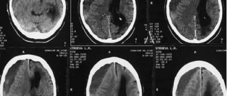
Signs of an arachnoid cyst (indicated by arrows) on MMU tomograms
The importance of proper formation and movement of cerebrospinal fluid for brain function
In a healthy person, the circulation of cerebrospinal fluid (CSF) occurs continuously. It is located not only in the cisterns of the brain, but also in its central cavities. These sections are called the cerebral ventricles. There are several varieties:
- lateral;
- third and fourth (connected by the Sylvian aqueduct).
It is important to note that it is the fourth ventricle that is directly connected to the human spinal cord. Cerebrospinal fluid performs the following functions:
- washes the outer surface of the cortex;
- circulates in the cerebral ventricles;
- penetrates deep into the brain tissue through the cavities around the vessels.
These areas are not only the main area of cerebrospinal fluid circulation, but also its storage. The cerebrospinal fluid itself begins its formation at the junction of the blood vessels of the ventricles. These are small processes that have a velvety surface and are located directly on the walls of the ventricles. There is an inextricable connection between the tank and the cavity around it. When special slits are used, the main tank interacts with the fourth ventricle of the brain. Thus, cerebrospinal fluid is synthesized, which is transported through these gaps to the subarachnoid region.
Among the features of the movement of cerebrospinal fluid are:
- movement in different directions;
- circulation occurs slowly;
- it is influenced by cerebral pulsation and respiratory movements;
- the main amount of cerebrospinal fluid enters the venous bed, the remainder into the lymphatic system;
- directly takes part in metabolic processes between brain tissues and organs.
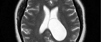
Malformation of the great cerebral vein
Malformation of the vein of Galen is a change in the vascular system in which a plexus is found in the form of a tangle, consisting of embryonic-type vessels and, necessarily, an enlarged great vein (aneurysm). That is, malformation always includes an aneurysmal change in the vein.
Malformations are characterized by underdevelopment of the elastic and muscle fibers of the middle tunic of the vein, so even a slight increase in pressure is accompanied by an expansion of the lumen of the vessels, which only worsens over time. An aneurysm may have the appearance of a sac or a diffuse increase in the lumen of the vessel.
Arteriovenous malformation of the vein of Galen can be mural, when the brain arteries approach the aneurysmally dilated vein and flow into it directly, and choroidal - pathologically altered tangles of vessels feeding the malformation, flowing into the dilated vein of Galen.
The malformation together with the aneurysmally dilated vein of Galen puts pressure on the surrounding tissues, promoting their atrophic changes, displacement, blockade of liquor and venous outflow, which causes hydrocephalus to increase.
Video: Vein of Galen malformation
Symptoms of deformation
The main signs of changes in the size of the tanks are: headache, nausea, blurred vision. As symptoms progress, serious complications develop.
When a large volume of fluid accumulates, the patient is diagnosed with hydrocephalus. It comes in two types:
- internal (cerebrospinal fluid accumulates in the cerebral ventricles);
- external (accumulation is observed in the subarachnoid region).
The main symptoms include morning swelling under the eyes. In this case, an urgent examination by a doctor is required to make an accurate diagnosis. During pregnancy, to exclude disorders of brain development in the child, a mandatory ultrasound examination is performed in the first trimester.
Treatment of diseases associated with deformities
If deformation processes are detected early, drug therapy is carried out. If the amount of accumulated fluid is very large, the patient may require urgent surgery. To do this, a small hole is made in the patient's skull into which a tube is placed. With its help, excess liquid is pumped out. Today, neuroendoscopy is becoming an increasingly popular method, which is performed without the use of additional excretory tubes and does not cause harm to the patient.
Consequences of the disease
In case of chronic hydrocephalus, the patient is registered with a neurologist and regularly undergoes the necessary tests. If treatment is not started on time, hydrocephalus leads to disability in the child. His development is inhibited, he speaks poorly, and his vision functions may be impaired. With timely treatment, doctors note a high recovery rate. If deformations in the cisterns of the brain are diagnosed during intrauterine development, then most likely such a child will be born defective.
Prevention of violations
Most brain development disorders occur during fetal development. You must adhere to the following recommendations:
- try to avoid infectious diseases, especially in the first trimester of pregnancy;
- Take medications with caution.
To prevent the development of hydrocephalus in children, it is necessary to avoid traumatic brain injuries and infectious diseases of the nervous system, since these factors are considered to provoke the development of hydrocephalus.
To maintain the viability of a patient with cistern deformities, doctors prescribe medications and regular examinations. If a deterioration of the condition is suspected, urgent surgical intervention is performed.
Diagnostics
In case of disturbances in the cerebrospinal fluid system, the following diagnostics are performed: MRI, CT, fundus examination, examination of brain cisterns using radionuclide cisternography, as well as neurosonography.
It is very important to know how the liquor system works, how its pathology arises and manifests itself. In order to undergo full treatment in case of detection of pathologies, it is necessary to consult a specialist in time. In addition, ultrasound results at different stages of pregnancy make it possible to study the development of the fetal brain in order to make a correct prognosis and plan treatment in the future.