Not every patient knows for what purpose REG of the head is performed or what it is. This is one of the examination methods that helps determine how normal the blood flow of large and small vessels of the head and neck is. Rheoencephalography is used when it is necessary to determine the presence of pathologies in the vascular bed of the brain, to assess the state of cerebral circulation after injuries, strokes, and surgical interventions.
How is data read during REG?
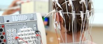
To carry out the examination, a polygraph is required, which will produce results on a paper tape. This is a special device that supplies electrical impulses to sensors connected to the patient’s head.
Paired electrodes connected to a rheograph are applied to the subject's head. One of the electrodes emits a signal, which, after passing through the brain tissue, is recorded by the opposite electrode. Since body tissues and blood flow have different electrical conductivities, pulse fluctuations in the vascular bed cause certain changes in the data. The data is transferred to a computer, which not only records it, but also sends it to a polygraph.
The graphs are recorded on paper; two results are given if the activity of blood flow in the left and right hemispheres is compared. Also, in parallel with the REG of the head, an electrocardiogram can be recorded to track the correspondence of vascular filling and cardiac output.
When is rheoencephalography indicated for a person?
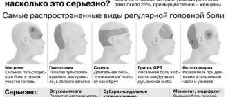
Using REG, you can obtain information about the functionality of the vascular bed without visualizing the object. The doctor does not see the arteries, but only receives data on how much they allow blood to pass through, how they narrow and expand as the pulse passes blood through them. The examination is prescribed for patients who complain of headaches, dizziness, fainting, poor sleep, and impaired mental abilities. Rheoencephalography is also performed for patients suffering from the following pathologies:
- Vegetative-vascular dystonia.
- Arterial hypertension.
- Hypotension.
- Cerebral atherosclerosis.
- Vertero-basilar insufficiency.
- Migraine.
- Neurocirculatory dystonia.
- Diabetes mellitus.
- Osteochondrosis of the cervical spine.
- Encephalopathy.
- Parkinson's disease.
- Epilepsy.
The examination is indicated for post-stroke patients who have suffered ischemia of cerebral tissue or hemorrhage into the head cavity. Also, REG can be performed after head or neck injuries, concussion, to determine whether there is a blood flow disorder. Rheoencephalography may be prescribed to monitor the effectiveness of the prescribed treatment, as this method of studying blood flow helps to obtain dynamic data for comparison.
If the question arises about why REG of cerebral vessels is performed, what it is, we can say that this is a narrowly focused examination that allows you to assess the condition of the vessels, but not determine the disease.
Rheovasography: features of the study
Rheovasography (RVG) is a rheographic method for studying the vascular bed of the upper and lower extremities. Such a study provides information about the state of hemodynamics in the bloodstream of the extremities, the main indicators of which are peripheral vascular resistance, vascular elasticity, and the amount of blood outflow. Based on the data obtained, a rheographic index is calculated, characterizing the intensity of blood flow to an organ (arm or leg).
Direct indications for RVG are:
atherosclerotic lesions of the arteries of the extremities or suspicion of it;- thrombosis or thrombophlebitis, as well as suspicion of one of these processes;
- varicose veins;
- Raynaud's syndrome;
- diabetes mellitus (diagnosis of diabetic macroangiopathies).
In each of these cases, rheovasography acts as an objective research method that confirms or rejects the diagnosis with high accuracy. In addition, the method is used to monitor patient treatment. This procedure has no contraindications.
Contraindications to REG
Rheoencephalography can be performed on literally every person. The procedure does not require special preparation and does not cause any discomfort. The high-frequency current supplied to the electrodes is so weak that it is not felt at all. The procedure can be performed even on children of any age; it is harmless.
REG can be canceled only if the patient’s skin is damaged in the place where the electrodes need to be attached. Also, the examination is not carried out for patients with skin diseases of the scalp: seborrheic dermatitis, lichen, purulent eczema, etc.
The difference between REG and MRI, CT, Dopplerography
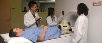
Some experts believe that the REG examination has already become obsolete, since it does not reach the information content of magnetic resonance or computed tomography. But not every head physician can install a bulky tomograph tube in a clinic. This equipment is quite expensive and requires special maintenance.
But even with a magnetic resonance imaging scanner, a neurologist cannot always send a patient for a tomography. In such situations, when the patient has a pacemaker, he cannot undergo an MRI examination. Head CT scanning is contraindicated for pregnant women due to radiation exposure.
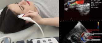
That is why it is advisable to use rheoencephalography. This procedure can act not only as a separate examination method, but also as a complement to Doppler sonography in an adult patient and neurosonography in a one-year-old child. A comprehensive examination helps to accurately determine the cause of the pathology and select the most effective drugs for treatment.
Characteristic
The doctor writes a mysterious abbreviation in the direction and, without going into too much detail, sends home the patient who forgot to ask what it is - REG.
REG, rheoencephalography, is a non-invasive type of study of cerebral vessels, which is based on measuring the electrical resistance of tissues at the moment a weak high-frequency impulse passes through them. Blood is a conductor of electric current and, filling the vessels, reduces their resistance, as can be seen on the rheoencephalogram.
Clinical rheography is used to show cerebral blood supply. Another type of study, integral, is aimed directly at assessing vascular resistance.
The information obtained from REG allows you to see:
- tone of the walls of blood vessels;
- time of change in their resistance;
- elasticity;
- condition of arteries and veins;
- blood volume;
- viscosity;
- speed of pulse wave propagation.
The rheoencephalogram shows signs of impaired blood flow, but this is not enough to reliably say about the presence of a particular pathology and, moreover, to differentiate between diseases. In this regard, recently there has been a lot of debate about the advisability of conducting REG and replacing it with more informative research methods. So, if REG records a weakening of blood flow and an increase in resistance, then magnetic resonance imaging allows you to see the exact location of a blood clot or a damaged area of a vessel.
However, it is too early to write off rheoencephalography. It is indispensable in cases where MRI and CT are contraindicated: during pregnancy, lactation, the presence of pacemakers or other implants, obesity, claustrophobia, contrast agent intolerance, and the need to avoid radiation. Sometimes, to clarify the diagnosis, this procedure is complemented by echoencephalography.
REG is often confused with EEG. To carry out these studies, electrodes are placed on the head. The main difference is in the purpose: electroencephalography examines brain activity, and REG examines the state of blood vessels.
Preparing for the examination
It is advisable to get a good night's sleep before undergoing the procedure. Two hours before the diagnostic examination, it is not recommended to drink tea, coffee, or Coca-Cola; smoking and taking medications containing alcohol are strictly prohibited. If the patient is taking drugs that affect the vascular bed (for example, vasodilators, antispasmodics, drugs to lower blood pressure), he should coordinate their use before the examination with the attending physician.
Immediately before the procedure, the patient needs to rest. It is advisable to sit quietly for 15-20 minutes in silence, do not talk to anyone, do not read, do not play games on a mobile phone or tablet, and do not enter into discussions.
How does the procedure proceed?
Usually, to conduct an REG of the head, the patient needs to sit in a special chair. Sometimes the patient is asked to lie down on the couch and is examined in a lying position. The examination is carried out on a multi-channel rheograph: from 2 channels to 6. The extent of the study depends on the number of channels; the more channels on the device, the larger part of the brain can be studied in one procedure.
At the site where the electrodes are connected, the skin is treated with alcohol. The electrodes themselves are coated with a special gel that increases signal conductivity. The sensors can be attached to the slots of a wide elastic band or placed on a special “cap”.
The location of the electrodes on the patient's head depends on which area of the brain needs to be examined:
- When studying the internal carotid artery basin, sensors are attached above the inside of the eyebrow and on the mastoid area (behind the ear).
- If the basin of the vertebral artery is examined (practised for osteochondrosis of the cervical spine), sensors are attached to the mastoid process and to the occipital protuberance.
- When it is necessary to assess blood flow in the external carotid artery, sensors are attached in front of the auricular tragus and above the outer edge of the eyebrow.
If it is necessary to simultaneously perform an ECG, sensors are also attached to the patient's wrists. The examination of the head lasts 10-15 minutes, while the patient does not feel anything. Data from the electrodes is sent to a computer; there are also older devices that send the result directly to paper.
Functional tests for REG
The patient may be asked to close his eyes at the very beginning of the procedure so that nothing distracts him. In addition, to clarify the data, the examination is carried out with functional tests:
- Test with vasodilating drugs. Nitroglycerin is most often used. But, if necessary, the subject may be asked to take aminophylline or caffeine.
- Test with vasoconstrictor drugs.
- To determine whether there is vascular spasm in the brain, a glycerol test is performed.
- The orthostatic test involves tilting the head to the sides, back and forth. The patient may be asked to stand up abruptly or bend over.
- Test with hyperventilation - when the patient needs to breathe frequently, hold his breath at a signal, breathe either through the nose or through the mouth.
During the examination, the doctor may press on some blood vessels with his finger. This is required in order to obtain the most complete data on the blood supply to the peripheral parts of the brain.
Patients with epilepsy should be examined with rheoencelography with caution, as the procedure may provoke an attack in them. It is functional tests that affect blood flow and neuron function that can have a negative impact.
How is the research conducted?
REG is performed with the patient sitting. Blood pressure is measured. An elastic band is placed on the subject's head, passing over the eyebrows, over the ears and along the back of the head. In this case, it is better if the hair is removed, because it will get under the tape and interfere with the diagnosis. In addition, it is quite painful.
Then small round electrodes are attached using tape: two above the eyebrows, two behind the ears and two in the occipital region. Sometimes small damp gauze pads are placed under the electrodes. After this, registration of the rheoencephalogram begins. This usually takes a few minutes.
After the main recording, various functional tests can be carried out. Most often, the patient is asked to take half or a whole tablet of nitroglycerin under the tongue. However, in case of low blood pressure, glaucoma, or intolerance to nitroglycerin, this test is not performed. The subject may refuse to undergo it. After taking nitroglycerin, a rheoencephalogram is recorded again. In some cases, tests are carried out with changes in the position of the body and head (tilts, turns), with holding the breath or hyperventilation, temperature tests, with physical activity and others. The examination itself takes up to 10 minutes. The results of the study are processed by a functional diagnostics doctor, and this procedure is performed by a nurse. This is associated with a possible delay in the readiness of the medical report.
What do the survey results show?
You should first draw the patient’s attention once again to what a head REG is. The data obtained from the rheograph are not a definite diagnosis; they only indicate whether the functionality of the vessels corresponds to the norm or deviations from it.
When deciphering the results, the doctor takes into account not only the general health of the patient, the presence of chronic diseases, but also age. REG data differs in children, young, mature people and the elderly.
On paper, the device produces lines of curves that are arranged in the form of graphs. The medical professional notes:
- Anacrotes are ascending wave lines, sharply rising, with slightly rounded tops.
- Cataktrots are smoothly descending descending lines.
- Incensoring – located in the middle third of the wave.
For the result, it matters how regular the waves are, whether there are curves at the tops, their height, how the incensor is located, whether there are additional waves, teeth. The norm for a 14-year-old patient is completely different from the blood flow norm for a 50-year-old patient. If in the first case the waves are uneven and interspersed with high peaks, then in the second case the doctor will see a smoother line, with evenly spaced anacrotes.
When deciphering the results of the REG of the head, the doctor receives information about:
- Tone of arteries, veins, capillaries.
- The degree of blood filling of a certain area of the vascular bed.
- Blood flow speeds in accordance with the movement of the pulse wave.
- Blood viscosity.
Based on the results of the rheogram, three types of functionality of the vascular bed can be identified, which indicate a deviation from the norm:
- Dystonic. With this type of disorder, vascular tone constantly changes, hypertonicity occurs, followed by low pulse pressure, which indicates a violation of venous outflow, stagnation.
- Angiodystonic. It is characterized by unstable tone, which is caused by stagnation of venous blood in a certain area of the head. It is observed when there is a defect in the walls of blood vessels, leading to loss of flexibility of the vessel, decreased elasticity and, as a result, disruption of the blood supply to brain tissue.
- Hypertensive. With this type of pathology, the outflow of venous blood is difficult, and persistent hypertonicity of the vascular wall is noted.
For the patient, the results given by the rheograph are of no information value. Only a doctor can “read” the rheogram, determine whether there are pathological changes in the blood flow, and prescribe the necessary treatment.
Interpretation of the results of a rheoencephalographic study
The rheograph records cyclic waves recorded in one heartbeat.
Designation of curve segments:
- anacrota - rising wave;
- katacrota - descending;
- incisura followed by a dicrotic tooth - a smooth transition between them.
When deciphering a rheoencephalogram, many factors are taken into account - the patient’s age, amplitude, wave inclination, curve indices. They determine the tone of blood vessels or veins, resistance and force of blood outflow.
The recording is transcribed by a specialist who has undergone qualified training and certification.
Today there is a free and paid service for deciphering the REG result online, which is carried out by specialists in this profile. However, it is still difficult to assess the effectiveness of the innovation.
Angiodystonic type REG
Most often, angiodystonia is diagnosed in adolescence and working age. Its appearance is explained by the presence of a defect in the structure of the walls of blood vessels.
This defect leads to a decrease in elasticity and disruption of the tone of small vessels in a certain pool. As a result, other organs and systems suffer: eyes, heart, gastrointestinal tract, etc.
Dystonic
Little different from the previous one. It is characterized by a tendency to regular changes in vascular tone, often with a predominance of hypotonicity.
The dystonic type with signs of venous dysfunction is accompanied by low pulse filling. Venous stagnation often forms and venous outflow becomes difficult.
Normotonic
The normotonic type does not have a separate category. Usually used when concluding a diagnosis, when it is necessary to indicate a mixture of different types with different disorders.
Hypertensive
The hypertensive type has a clear difference from those described above. When the venous outflow is impaired, this anomaly is determined by a persistent increased tone of the vascular network.
It is worth considering that these are not separate diseases, but the symptoms that accompany them.
Is rheoencephalography performed for children?
Since diagnosing the head using REG is absolutely safe, this procedure can be performed on children of any age. This type of examination is not carried out on newborns, since neurosonography is sufficient to obtain data on blood flow in the infant’s brain.
In some cases, a problem arises with data collection, since it is advisable to conduct REG when the patient is completely immobile. Not every child will be able to sit quietly, even if the procedure only takes 5 minutes. Therefore, increased mobility of a small patient can also be a reason for refusal to conduct an examination. Anesthesia is not used for a rheoencephalogram, since it is impractical to use it for such a short period of time.
The child undergoes REG together with one of the parents. To make the baby feel confident, to sit quietly, you can take him in your arms. If the patient cries, is afraid, or screams, the procedure is canceled.
When it is not possible to undergo an MRI or CT scan due to contraindications, but it is necessary to check the condition of the blood flow, it makes sense to agree to an REG. Examination of the head using rheoencephalography is a simple procedure available to many patients and does not require special training.
Prices
| Name of service (price list incomplete) | Price |
| Appointment with a neurologist, therapeutic and diagnostic, primary, outpatient | 1750 rub. |
| Consultation (interpretation) with analyzes from third parties | 2250 rub. |
| Consultation with prescription of a treatment regimen (for up to 1 month) | 1800 rub. |
| Consultation with prescription of a treatment regimen (for a period of more than 1 month) | 2700 rub. |
| Consultation with a candidate of medical sciences | 2500 rub. |
| Transcranial duplex scanning (TCDS) of cerebral vessels | 3600 rub. |
| Electroencephalography | 3100 rub. |
| MRI of the brain | 4200 rub. |
| CT scan of the head (brain structure) | 3300 rub. |
| Corporal acupuncture (session) | 1200 rub. |
| Superficial acupuncture | 1200 rub. |