Ultrasound of cerebral vessels or, in other words, transcrian Dopplerography, is a non-invasive method for studying the main blood veins and arteries using special ultrasound equipment, the operation of which is based on the use of the Doppler effect. To obtain detailed information, this method can be combined with duplex scanning of the great vessels of the neck and head.
The use of ultrasound (ultrasound) allows you to quickly obtain reliable information about the state of the blood vessels in the brain, which simplifies the diagnosis of the underlying disease, and also allows you to monitor the course of the disease during and after treatment.
What does an ultrasound scan of the brain show?
In order to understand what ultrasound of cerebral vascular structures is and what it can show, first you should familiarize yourself with the main methods of ultrasound examination of this organ:
- Echoencephalography allows you to evaluate the physical features of the structure of the brain matter, as well as identify developmental pathologies in parts of the brain.
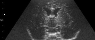
- Neurosonography (NSG) - using this ultrasound research method, the characteristic features of the structure of the brain are studied in children of the first year of life, or until the large fontanel is open.
- Doppler ultrasound (USD) is a combined method that combines the capabilities of ultrasound and Dopplerography. There are two types:
1. Transcranial ultrasound Doppler allows you to assess the condition of the arterial and venous circulatory system located in the brain. To perform a study in this way, access to acoustic windows in the temple, occiput, eyes and the area of the articulation of the cervical spine and occipital bone is required.
2. Extracranial ultrasound. Makes it possible to examine large cervical blood vessels outside the skull.
Duplex and triplex ultrasound scanning are modern methods for studying the vascular system of the brain. They allow you to interpret the characteristic features of the structure of each artery separately and estimate the speed of blood flow in them. This test also evaluates the tissues and joints surrounding the blood vessels.
In the first case, the specialist receives a two-dimensional display along and across the arteries, and in the second, more extensive information about the state of the vessels, containing, in addition to standard data, indicators of the direction and speed of blood flow, which are colored in different colors.
Let us consider in more detail the capabilities of the listed methods.
Ultrasound of the brain (echoencephalography) helps to diagnose and identify the following pathological processes in this organ in real time:
- destruction of brain tissue due to inflammatory infectious diseases (for example: meningitis or encephalitis);
- formation of tumors and cysts of various nature;
- ischemic disease;
- swelling of the brain;
- hemorrhagic stroke;
- atherosclerotic changes;
- stenosis of head vessels;
- violation of the patency of the liquor passages;
- the presence of structural changes in the brain matter due to traumatic brain injury.
Also, using this research method, a medical specialist can determine where the hemorrhage is located and assume the degree of destruction it caused.
Despite the obvious advantages, a final diagnosis cannot be made based on the results of a brain ultrasound, since this examination method is screening, that is, it makes it possible to predict the development of certain complications due to impaired blood supply. Therefore, to make an accurate diagnosis, more accurate methods of studying this organ using MRI or CT are required.
Often, to determine the functional abilities of blood vessels, a Doppler ultrasound of the brain is required. This research method is based on the fact that the sound wave sent by the sensor is reflected only from a moving object, that is, blood cells.
Carrying out this procedure is no different from a standard ultrasound examination: the difference lies in the principle of processing incoming information: the Doppler sensor sends ultrasound waves at a certain speed and receives them back at another. That is, the quality of the blood circulation of the vessel depends on how quickly the ultrasound wave returns. Based on the data obtained, a corresponding image is displayed on the monitor screen in the form of a diagram, the indicators of which will change and indicate the presence of vascular pathology.
Using Doppler ultrasound, only large vessels of the cervical spine are examined, which are located outside the cranium and in areas where the bones of the skull have minimal thickness. It allows you to evaluate the following characteristics of blood vessels in an adult:
The picture is clickable
- degree of passability;
- the presence of deposits in the form of atherosclerotic plaques;
- the presence of blood clots, their physical parameters;
- blood flow speed, valve condition;
- presence of aneurysms, anastomosis.
Since in children under one year of age there are several non-ossified areas (fontanelles) in the skull, they are prescribed an ultrasound of the brain (Neurosonography), which allows us to identify structural pathologies of this organ. Using this method, the following indicators are assessed:
- symmetry of the cerebral hemispheres;
- map of the location of the convolutions of the cerebral cortex;
- ventricle size;
- crescent shape;
- cerebellar contours;
- the presence or absence of the 5th fontanel between the hemispheres;
- the presence of cysts and other neoplasms in the medulla;
- indicators of homogeneity of the vascular plexus.
Since neurosonography is a diagnostic method for studying brain structures, it is done only when indicated, to refute or confirm a preliminary diagnosis. After decoding, the obtained data should be provided to a pediatric neurologist.
Ultrasound examination of brain structures, or echoencephalography as it is also called, is a non-invasive examination method based on measuring the displayed echo signal from the deep structures of the brain.
The information received by a special sensor is processed and displayed on the monitor in the form of a two-dimensional black and white image. Due to the fact that ultrasound is the most bladeless examination method and has virtually no contraindications, it is prescribed to patients of any age, including children.
What is this type of ultrasound?
Ultrasound of the brain (echoencephalography) is an intracranial ultrasound examination. It is based on the principle of reflection of an echo signal from the midline structures of the brain. Ultrasound, thanks to which such diagnostics is in principle possible, is reflected from brain structures at different angles, and this gives grounds to judge the location of the reflecting structure, its size, and shape. The reflected rays produce an image on the monitor, which is analyzed by a diagnostic specialist.
In the presence of what symptoms is it recommended to carry out such a diagnosis?
The patient should be concerned about the state of his health and contact his doctor as soon as possible for a referral for an ultrasound if the following symptoms are detected:
- Headaches of varying duration and intensity. Particular concern should be caused by pain, the origin of which is difficult for the patient to explain.
- Uncoordinated movements, disorders of the musculoskeletal system.
- Noises in the ears.
- Dizziness and confusion.
- Numbness of the limbs, characteristic weakness in the fingers.
- Constant weakness.
- "Heaviness" in the head.
- Loss of consciousness for no apparent reason.
- Memory impairment.
- Nausea for no clear reason.
- Various neurological abnormalities.
What medical indications are there for undergoing this examination?
In the following cases, doctors themselves usually insist on performing an ultrasound of the brain both for diagnostic purposes to identify pathologies and for the prevention of diseases of various kinds:
- Osteochondrosis of the cervical spine.
- High blood pressure (hypertension).
- History of stroke.
- Diabetes.
- Pathological level of cholesterol in the blood.
- Obesity.
- Cardiac ischemia.
- Suspicions of infectious diseases of the brain.
Is it necessary to do an ultrasound of the brain?
Ultrasound of the brain is a fairly objective and, despite the development of medical technologies, still a very effective diagnostic method. Such a study allows you to quickly, in real time, assess the state of the brain and identify abnormalities. Also, such research is quite accessible to the general population due to its relative cheapness. There are no contraindications for such an examination.
Since ultrasound has absolutely no negative impact on the patient’s body, it can be performed as often as needed. To summarize, we can come to the conclusion that ultrasound is quite indicative in diagnosing the brain and has a number of undeniable advantages that give it priority over other methods of monitoring the state of the brain. Every person should undergo it at least once a year, and if there are certain (and often serious) suspicions, there is no reason to delay going to a diagnostic specialist.
Who is the procedure indicated for?
An ultrasound examination of the brain and neck may be prescribed to a patient with the following symptoms of insufficient blood supply to this organ:
- periodic attacks of headache;
- apathy, weakness;
- frequent attacks of dizziness;
- impaired clarity of hearing, vision and memory;
- arterial hypertension;
- numbness of the limbs.
For example, if a patient complains of headaches, tinnitus and frequent fainting, this may indicate a circulatory disorder in the blood vessels of the head and neck. In this case, he should be prescribed an ultrasound outside the cranial brachiocephalic vessels (BCVs - large blood vessels that carry blood to the head). During this study, the specialist receives information about the structural features of the arteries and veins of the cervical spine, the thickness of their walls and the speed of blood flow in them.
Transcranial duplex scanning helps to identify many pathologies and find out the causes of headaches, nausea and other symptoms that can be precursors to serious diseases of the internal organs:
- diabetes;
- increased cholesterol levels in the blood;
- ischemic disease;
- hemorrhagic stroke;
- cardiovascular pathologies;
- obesity;
- ischemic attacks;
- cervical osteochondrosis;
- cerebrovascular disease.
This type of ultrasound is done to refute or confirm Parkinson's disease, which is characterized by neurological abnormalities - hand tremors, stiff back muscles and insufficient motor activity of the body.
If the above symptoms are present, the study evaluates the echogenicity of the substantia nigra of the brain - if it excessively reflects the signal from the sensor at high speed, then this indicates the presence of the disease in the patient. This research method is most informative in people over 60 years of age, but if there are obvious signs, it can be done at any age.
In addition, ultrasound of the structures and vessels of the brain can be prescribed to monitor the patient’s condition after traumatic brain injury to diagnose pathologies of the brain substance.
Many parents are interested in the following questions: why is it necessary to do an ultrasound of the brain of a newborn, and up to what age is it informative? The answer to it is quite simple - this method allows you to study the characteristic features of the structure of this organ, until the large fontanel is overgrown, without resorting to more complex diagnostic methods.
What does echoencephalography show?
After completion of the procedure, an examination protocol is filled out, which is deciphered by a neurologist.
During an ultrasound examination of the brain, the following indicators are assessed:
- size, structure of the liquor system;
- volume, type of cerebral hemispheres;
- state of the subarachnoid space;
- vascular patency;
- vascular wall thickness;
- the presence of space-occupying formations, their size, structure, localization.
Normally, echoencephalography reveals:
- symmetry of brain structures;
- homogeneous, clear, anechoic (black) ventricles;
- subcortical structures have clear boundaries and moderate echogenicity (gray);
- no M-Echo bias;
- lack of fluid in the interhemispheric fissure;
- vascular wall with smooth and clear contours;
- vessels passable throughout.
In addition to normal indicators, various abnormalities may be detected during the examination. An ultrasound of the brain can show: inflammatory processes, neoplasms, vascular pathologies, atherosclerotic changes.
The most characteristic sign for neoplasms is M-Echo displacement. At the same time, the structure of the brain loses homogeneity. Ultrasound visualizes a space-occupying formation of varying degrees of echogenicity (depending on the type of tumor). The contours can be either clear or blurry (a blurred contour indicates a malignant process).
- Cerebral edema is characterized by the presence of dark fluid in the ventricles and cisterns of the brain. In this case, the cavities are overstretched and expanded. A similar picture can be observed with intracranial hypertension. Often these conditions are combined with each other.
- When an abscess occurs, an ultrasound of the brain visualizes a round formation with a clear outer and inner contour. In a vertical position inside the cavity, you can see the liquid level.
In the video, puncture under ultrasound control:
- Intracranial hematomas are a hypoechoic (dark) spot without clear boundaries. Foci of ischemia are often visible along the periphery of the hemorrhage.
- Cerebral ischemia often does not produce any symptoms for a long time. During examination, necrotic or depleted light areas can be noted. They are spots of irregular shape, without clear boundaries.
- With atherosclerosis of cerebral vessels, a light plaque attached to the wall is detected. Often it can block up to 75% of the cavity, thereby preventing normal blood flow.
- An aneurysm is a sac-like deformation of the vascular wall. At the same time, it lacks a muscle layer. This explains the fragility of the vessel and the frequent occurrence of strokes.
Preparing for the study
Before you do an ultrasound of the blood vessels of the brain and neck, you should familiarize yourself with information on how to prepare for it.
Since during the examination the specialist will evaluate the functional abilities of the blood flow, 24 hours before the procedure the following rules should be followed:
- Do not drink energy drinks and drugs that have a stimulating effect on the nervous system, since the substances they contain affect blood circulation parameters - for this reason, the patient may exhibit deviations from accepted norms;
- Do not engage in active sports or other physical activity immediately before the study;
- You should not take sedatives (unless taking them is necessary);
- Also, 3-4 hours before the ultrasound, you should stop smoking and drinking tea or coffee.
Immediately before the procedure, it is necessary to remove jewelry that interferes with the examination from the lower part of the head and neck.
If the patient knows in advance about his fear of ultrasound, then he should spend some time on psychological preparation, or choose a different examination method.
How to do an ultrasound of the brain
Carrying out an ultrasound scan of the brain in adults is no different from the process of undergoing an ultrasound scan of any other organ: the patient lies down on a couch with his head on a special pillow. Next, the specialist examines the head using his fingers to identify areas of skull deformation, hematomas, and the presence of asymmetry.
The examined area is lubricated with a special gel to improve the conductivity of the ultrasound signal, special sensors are placed on the sides, and the received data is recorded on the monitor. If necessary, the specialist may also ask the patient to change his position, hold his breath or blink. After the study, the sensors are removed and the gel is wiped off with a paper towel.
If it is necessary to perform an ultrasound examination of the arteries supplying the brain, then in this case the patient is prescribed transcranial Doppler ultrasound (TCDG).
The difference between this procedure and standard ultrasound lies in the way the data obtained is recorded - changes in the displayed sound waves from moving blood cells are recorded. This research method is based on the use of the Doppler effect, that is, changes in the frequency and wavelength of ultrasound perceived by the receiver due to the movement of the source of ultrasound radiation. To obtain more detailed information, the patient may also be asked to undergo a duplex scan.
To assess blood flow using the Doppler method, you will need access to the temples, back of the head, forehead and eye sockets. Then everything is carried out as usual: the gel is applied, the specialist presses and moves the sensor over the examined area. The result is presented on a computer monitor in the form of a scale colored in different colors.
Using transcranial Doppler sonography, specialists diagnose almost all diseases caused by impaired blood flow in the brain vessels, such as atherosclerosis and hypertension. Also, the use of this method significantly reduces the time for diagnosing acute conditions - hemorrhagic stroke or ischemic attack.
Unlike Doppler ultrasound (USDG), duplex scanning combines two research functions: with the help of Doppler ultrasound, the blood flow of vessels is characterized, and B-mode makes it possible to visualize the structural features of arteries and veins.
Echo EG in children differs from ultrasound of the brain in that in the first case, the sensors are attached directly to the head, then an ultrasound signal is applied and an image is displayed on the screen in the form of a graph, which determines the echogenicity of the area under study, and in the second case, the ultrasound is generated by the sensor, which is leaned directly against the fontanel, and the ultrasound image familiar to everyone appears on the monitor. Parents should not worry - an EG echo is as painless as a regular ultrasound, but may take a little longer.
After the study, the doctor compares the results with normal values, draws up a conclusion and, if necessary, takes pictures of possible pathology.
How the procedure works
This diagnostic method is absolutely painless and does not require general or local anesthesia. The patient is examined standing, lying or sitting, depending on which organ needs to be examined.
The surface of the skin is lubricated with a special gel, which facilitates signal transmission. The ultrasound technician then directs the probe to the desired area.
During duplex scanning of the brain, the sensor is placed in places called “ultrasonic windows” - parts of the skull where the bones are thinner or where there are physiological openings in them.
Through such zones, the ultrasound beam easily enters the cranial cavity.
During the procedure, the patient may be required to adopt a more comfortable position for the examination. The doctor may also ask you to hold your breath or do other similar actions. The manipulation lasts approximately half an hour and causes absolutely no discomfort.
Decoding the results
The diagnostician deciphers the data obtained as a result of ultrasound of the structures and vessels of the head, so you should not try to independently interpret the information presented in the conclusion. After conducting the study, the following data is usually assessed and deciphered:
- size and structure of the ventricles;
- physical characteristics of the cerebral hemispheres;
- the presence of tumors, cysts, their structural features, size, location;
- patency of the blood vessels of the head and neck, their diameter, lumen, presence of inclusions on the walls, aneurysms, anastomosis, blood flow speed;
- The amount of space between the soft and arachnoid membranes of the brain and spinal cord, within which cerebrospinal fluid circulates, is also calculated.
The following indicators are considered the norm:
- cerebral hemispheres - symmetrical, without changes;
- the ventricles are of a homogeneous structure, with clear contours, anechoic;
- the basal ganglia should have moderate echogenicity;
- There should be no shift in the M-echo signal, since volumetric processes (tumors) cause its change;
- the volume of cerebrospinal fluid in the subarachnoid space should not exceed the permissible value at this age;
- absence of foreign inclusions, cysts.
Blood vessels should have clear contours, and their walls should be free of aneurysms and other structural pathologies. The lumen should be free of blood clots and there should be no anastomosis. The thickness of the vertebral arteries should normally be more than 2 mm in diameter.
Ultrasound of the structures and vessels of the brain is widely used in diagnosing cerebral hemorrhages as a result of traumatic brain injury, as well as after a stroke. Also, if necessary, using this research method, specialists ascertain physiological brain death.
Based on the results obtained during neurosonography (NSG), a pediatric neurologist makes a final diagnosis and prescribes the necessary treatment. Since such an examination is safe and takes a minimum of time, it is often prescribed when monitoring brain defects in children in the first year of life, as well as to confirm and remove the diagnosis.
It is important to understand that the ultrasound examination method is not a final diagnosis. For this reason, after undergoing the study, the patient must contact his attending physician for an accurate diagnosis and appropriate treatment of vascular pathologies.


