Electroencephalography, also called EEG, is a method that is used to study the state of the human brain and is based on recording its electrical activity. This examination allows you to detect the spread of pathological processes and signs of epilepsy.
The average duration of the procedure is about an hour, but it is highly informative and makes it possible to track any changes in the brain, the dynamics of the disease, and evaluate the impact of the therapy.
Since the procedure does not cause any pain or discomfort, EEG can be called not only the most accurate, but also the most gentle method of examining the brain.
How EEG works
The human brain consists of millions of special cells called neurons. Each of them generates its own electrical impulse. Within individual areas of the brain, impulses must be consistent. They can also strengthen each other or make each other weaker. Their strength and amplitude are not stable and are constantly changing.
Content:
- How EEG works
- Historical reference
- Significance of EEG
- Benefits of an Electroencephalogram
- Indications for the procedure
- Contraindications
- How to prepare for an EEG
- Performing an EEG
- Purpose of EEG video monitoring
- Conclusion of electroencephalography
This is the bioelectrical activity of the brain. To register it, special electrodes can be applied to the intact scalp, which will pick up vibrations, amplify them and record them in the form of special curves, so-called waves. The latter, depending on their shape, frequency and amplitude, are divided into five types: α- (alpha), β- (beta), δ- (delta), θ- (theta) and μ- (mu) waves. Each of the waves reflects the work of a specific part of the brain and is named by the first letter of its Latin name.
Their registration in real time is the essence of encephalography.
VideoEEG
Recently, according to standards, long-term video-EEG monitoring is indicated for patients with complex diagnostic situations. This type of study involves recording the bioelectrical activity of the brain for 8-24 hours with simultaneous video recording. The study time should capture the sleep state for a more accurate assessment of brain activity, because it is during sleep that foci of epileptiform and pathological activity often arise.
Historical reference
One of the founders of the electroencephalography method is considered to be the physiologist and psychiatrist from Germany Hans Berger. In 1924, using a device for measuring small currents called a galvanometer, he was the first to perform a procedure resembling an EEG recording.
Later, a special device called an encephalograph was created to conduct an electroencephalogram. Today, there are stationary encephalographs, which make it possible to conduct research exclusively in a special room, and portable ones, which can be moved.
It is noteworthy that initially EEG was considered exclusively as a method for identifying mental disorders in a person. Only over time, scientists found out that the technique also makes it possible to detect deviations not related to psychiatry.
Significance of EEG
EEG is a highly informative diagnostic method that performs a huge number of functions. An electroencephalogram provides the ability to:
- Assess the nature of brain dysfunction and how severe they are.
- Identify the side of the pathological focus.
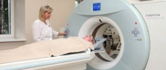
- Clarify information obtained during other diagnostic procedures (for example, computed tomography).
- Monitor how effective the therapy is.
- Identify areas of the brain in which epileptic activity is present.
- Assess brain function in the periods between attacks.
- Identify the causes of panic attacks and fainting.
- Study the sleep-wake cycle.
It should be taken into account that if a person has seizures, the study will be informative only if it is carried out after about a week.
Benefits of an Electroencephalogram
Today, electroencephalography is widely used in neuropathological practice. It makes it possible to clarify a huge number of problematic situations that are associated with the diagnosis and differentiation of neurological diseases. One of the undeniable advantages of encephalography is the fact that it not only helps to identify certain problems, but also helps to distinguish true disorders from hysterical manifestations or simulation.
In addition, the procedure is not as expensive as examination using a tomograph or other similar devices. EEG equipment is available in most hospitals.
The procedure does not have any negative impact on human health and condition. The patient remains fully functional. At the same time, the study can be carried out even on patients in serious condition, children and adults of any age, since it does not cause deterioration of the condition, discomfort or pain.
Indications for the procedure
Today, electroencephalography continues to be widely used in the practice of neurologists to solve a number of problems.
Thus, an EEG is recommended:
- For prolonged insomnia and other sleep disorders, including apnea, sleep walking and talking.
- For convulsive attacks.
- For diseases of the thyroid gland.
- If there are signs of developmental disorders in children or signs of mental disorders in adults.
- After recently experiencing traumatic brain injuries.
- If pathological changes in the vessels of the neck and head, brain tumors are detected during ultrasound.
- For frequent migraines, complaints of regular dizziness, persistent fatigue.
- For panic attacks, autism, Asperger's syndrome, stuttering, nervous tics, delayed speech and mental development in children.
- For meningitis, encephalitis, after a stroke and micro-stroke.
- After neurosurgical operations.
In what cases does EEG help make a diagnosis?
Electroencephalography, revealing the functionality and reserves of the central nervous system, has become the standard for brain research; doctors consider its implementation advisable in many cases and for various conditions:
- To assess the degree of functional immaturity of the brain in young patients (in a child under one year old, the study is always carried out during sleep, in older children - depending on the situation);
- For various sleep disorders (insomnia, drowsiness, frequent awakenings at night, etc.);
- In the presence of convulsions and epileptic attacks;
- To confirm or exclude complications of inflammatory processes caused by neuroinfection;
- For vascular lesions of the brain;
- After a TBI (brain contusion, concussion) – the EEG shows the depth of the GM’s suffering;
- To assess the severity of the effects of exposure to neurotoxic poisons;
- In case of development of an oncological process affecting the central nervous system;
- For mental disorders of various kinds;
- EEG monitoring is carried out when assessing the effectiveness of anticonvulsant therapy and selecting optimal dosages of medications;
- The reason for doing an EEG may be signs of dysfunction of brain structures in children and suspicion of degenerative changes in the nervous tissue of the brain in older people (dementia, Parkinson's disease, Alzheimer's disease);
- Patients in a coma need to have their brain assessed;
- In some cases, the study requires surgical operations (determining the depth of anesthesia);
- Encephalography will help to recognize how far neuropsychic disorders have gone in hepatic cellular failure (hepatic encephalopathy), as well as in other forms of metabolic encephalopathies (renal, hypoxic);
- All drivers (future and current), when undergoing a medical examination to obtain/replace a license, are asked to undergo an EEG for a certificate provided by the traffic police. The examination is easy to use and easily identifies those who are completely unfit to drive vehicles, which is why it was adopted;
- Electroencephalography is prescribed to conscripts who have a history of convulsive syndrome (based on medical card data) or in case of complaints of attacks with loss of consciousness accompanied by convulsions;
- In some cases, a study such as EEG is used to ascertain the death of a significant part of the nerve cells, that is, brain death (we are talking about situations when they say that “a person has most likely turned into a plant”).
Video: EEG and detection of epilepsy
Contraindications
It is noteworthy that there are no absolute contraindications for electroencephalography. If a person suffers from convulsive attacks, is diagnosed with coronary heart disease, hypertension, or mental disorders, an anesthesiologist must be present during the diagnosis.
The procedure should be postponed if there are open wounds, traumatic injuries, postoperative sutures or any signs of an inflammatory process in the area where it is necessary to install electrodes. Also, the study is not carried out for patients with ARVI.
Indications and contraindications
Electroencephalography makes it possible to clarify many situations related to the diagnosis and differential diagnosis of neurological diseases, therefore this research method is widely used and positively assessed by neurologists.
So, EEG is prescribed for:
- disorders of falling asleep and sleep (insomnia, somnambulism, obstructive sleep apnea syndrome, frequent awakenings during sleep);
- seizures;
- traumatic brain injuries;
- neuro-circulatory dystonia;
- frequent headaches and dizziness;
- diseases of the lining of the brain: meningitis, encephalitis;
- acute cerebrovascular accidents;
- brain tumors;
- recovery after neurosurgical operations;
- fainting (more than 1 episode in history);
- panic attacks;
- constant feeling of fatigue;
- diencephalic crises;
- autism;
- delayed speech development;
- mental retardation;
- stuttering;
- tics in children;
- Down syndrome;
- cerebral palsy;
- suspected brain death.
There are no contraindications to electroencephalography as such. Diagnostics are limited by the presence of skin defects (open wounds), traumatic injuries, recently applied, unhealed postoperative sutures, rashes, and infectious processes in the area where electrodes are supposed to be installed.
CM. SEE ALSO: Epilepsy in adults: diagnosis and treatment
The study should be carried out with caution in people with mental illnesses, since they cannot always correctly follow the doctor’s instructions (in particular, to remain with their eyes closed and not move during the procedure), as well as violent patients, since they have both the device itself and A cap with electrodes can even cause a feeling of rage. If it is necessary to conduct an EEG in such patients, they are first administered sedatives, which at the same time distort the results of the study, that is, make it less informative.
Not every diagnostic department has a portable electroencephalograph in its arsenal, so in such a situation, patients with cardiovascular pathology in its later stages, as well as patients with limited mobility, may be a contraindication for the study. Transporting them to the diagnostic department may have a higher risk than refusing this research method when making a neurological diagnosis.
How to prepare for an EEG
It should be noted that there are no special restrictions that must precede the procedure. However, there are a number of rules that are recommended to be followed to ensure that the examination is successful and informative.
First of all, inform your doctor if you are taking any medications. You may need to stop taking them for a while or change the dosage.
At least twelve hours before the procedure, or even better, a day before, eliminate from your diet foods that contain caffeine, carbonated drinks, chocolate and cocoa, as well as foods that contain them, foods with energy components, for example, taurine. You should also avoid products with sedative effects.
Wash your hair before the procedure. Additional styling products (oils, gels, balm, varnishes, etc.) should not be used, as this may affect the quality of contact of the electrodes with the skin.
If the main purpose of the procedure is to detect seizure activity, you need to sleep before the study.
In order for the result to be as reliable as possible, the patient should not be nervous or worried. In addition, you should refrain from driving for twelve hours before the procedure.
Eating is recommended two hours before the procedure.
If the patient is prescribed EEG sleep monitoring, the night before should be sleepless. Immediately before the procedure, the subject will receive a special sedative, which will enable him to fall asleep during the electroencephalogram.
If a child is to undergo an examination, parents should first psychologically prepare him for the manipulations, explaining that there will be no pain or discomfort. You can practice putting on a pool cap under the pretext of playing pilots or astronauts, teach your child to breathe deeply, showing him how to do it by personal example. On the eve of the procedure, the child should wash his hair without using any additional styling products. Before leaving the house, the baby should be fed and calmed down. Just in case, parents are advised to take with them tasty food and drink, and a favorite toy that will help calm and distract the baby.
Please note that if the above rules are not followed, the EEG result may not be very accurate or very informative. In this case, the procedure will have to be repeated.
Performing an EEG
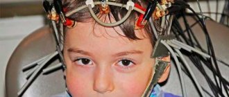
Electroencephalography is performed in a special room, which is completely isolated from light and sound. The patient sits in a chair or is asked to lie on the couch. A special cap with electrodes is first placed on his head. During the procedure, the patient is alone in the room, contact with doctors is maintained using a camera and microphone. If the diagnosis is performed on a child, one of the parents remains in the office.
Before starting the procedure, the patient is asked to close and open his eyes several times to adjust the equipment. During the diagnosis, the eyes must be closed. If during the procedure the patient needs to change position or visit the restroom, he can inform the doctors about this, after which the diagnosis will be suspended.
It is extremely important that the patient lies with his eyes closed and does not move during the procedure. In the event that a person opens his eyes slightly or moves, the doctor makes a corresponding note, since these actions must be taken into account when deciphering the electroencephalogram.
After the resting EEG is recorded, so-called “stress tests” are performed. Their goal is to test how the brain will react to situations that are stressful for it.
So, a hyperventilation test can be performed. The patient is asked to breathe frequently and deeply for three minutes. Photostimulation with a stroboscopic light source can also be used. It flashes frequently, and this allows you to evaluate how the brain reacts to bright light.
Stress tests can trigger convulsions or an epileptic seizure. The doctors who conduct the study have the appropriate skills to provide emergency care to the patient if necessary.
After the study is completed, the physician should remind the patient to resume taking medications that were stopped the day before the EEG.
The total duration of the procedure is from forty minutes to two hours.
What does EEG show in adults?
The doctor may ask for an EEG to find out what is happening in the brain (its bioelectrical activity), if there are problems with memory, abnormal movements, episodes of falling, fragments of unusual behavior, if there have been convulsions, epileptic attacks, or problems sleeping.
How long does an EEG last?
- A short (routine) EEG takes from 30 minutes to 2 hours.
- Long recordings can take several days.
What types of EEG are there for an adult?
- Night or day.
- Wakefulness with the inclusion of sleep, wakefulness without sleep, only sleep.
How to prepare for an EEG
- Wash your hair the night before the EEG. An adult should have clean, dry hair, washed with the simplest shampoo without the use of hair nourishing or styling products.
- Ideally, you should be calm while recording, but quiet activities will not interfere with the study (for example, you can read a book, watch TV, write, play on the phone...
- If an adult has special needs or you think they would not be able to perform the procedure without them, please let the EEG provider know in advance.
Before EEG
If an adult experiences seizures during certain situations, be sure to report this before the test. Before testing, the EEG technologist will ask several questions, collecting the adult patient's complaints.
During the EEG
- The accompanying person, if desired, can be in the same room with the adult being studied. It is important that no one interferes with the electroencephalogram.
- The patient should sit in a comfortable chair, preferably with a backrest, or, if desired, can lie down on the bed.
- After collecting the necessary information, the technologist usually measures the head and determines the size of the cap. We use very high quality caps with comfortable built-in electrodes.
- The hat is filled with a water-based gel, which is easily washed off from the hair after the test is completed with water.
How to do an EEG:
- You will need to open and close your eyes while recording.
- Sit with your eyes closed against a background of controlled flashing light of different frequencies.
- Hyperventilate (take long, deep breaths).
Decoding the EEG of an adult
At the end of the conclusion you can usually see:
- normal EEG or pathological;
- a brief description of the main background activity;
- reaction to the tests;
- presence or absence of epileptiform activity.
The presence of a theta rhythm on the EEG of an adult does not necessarily indicate pathology.
Purpose of EEG video monitoring
One of the types of electroencephalography is EEG video monitoring. This is a long-term recording of electroencephalography, usually lasting several hours, which can be performed during sleep. The duration of the procedure in each specific case is determined by the attending physician and the personnel of the laboratory that conducts the examination.
EEG video monitoring is prescribed if a short standard procedure does not reveal pathologies, but they are present.
Also, this type of examination allows you to evaluate the EEG during wakefulness and sleep.
Many patients are interested in the question of whether they should necessarily sleep during the study. The answer to this question cannot be unambiguous, because it depends on the specific situation. So, for example, if the reason for the examination is a tic that bothers the patient while awake, there is no need to sleep during the examination.
At the same time, EEG video monitoring during sleep sometimes helps to identify conditions that neither the patient nor his loved ones may even be aware of.
The peculiarity of this procedure is that it can be carried out not only during the day, but also at night. In the event that sleep EEG is required, night monitoring is more rational. Not everyone can fall asleep without problems during the daytime.
At the same time, we should not forget that carrying out a multi-hour procedure in a completely isolated room can be extremely tiring for the patient, especially if we are talking about a child. Most pathologies can be detected during a relatively short recording of a conventional EEG.
Best materials of the month
- Coronaviruses: SARS-CoV-2 (COVID-19)
- Antibiotics for the prevention and treatment of COVID-19: how effective are they?
- The most common "office" diseases
- Does vodka kill coronavirus?
- How to stay alive on our roads?
Also, overnight EEG video monitoring is much more expensive.
About the main rhythms of electrical activity of the brain
When interpreting the results of the study, various factors are taken into account: the age of the subject, his general condition (the presence of tremor, weakness in the limbs, visual impairment, etc.), anticonvulsant therapy at the time of recording the bioelectric activity of the brain, the approximate time (date) of the last seizure and etc.
The electroencephalogram consists of various complex biorhythms emanating from the electrical activity of the brain at different periods of time, depending on specific situations.
When decoding the EEG, first of all, pay attention to the main rhythms and their characteristics:
- Alpha rhythm (frequency ranges from 9 to 13 Hz, oscillation amplitude ranges from 5 to 100 μV), which is present in almost all individuals who have no complaints about their health during the period of inactive wakefulness (relaxation during rest, relaxation, shallow meditation). As soon as a person opens his eyes and tries to visually imagine any picture, α-waves decrease and may disappear altogether if the functional activity of the brain continues to increase. When deciphering the EEG, the following parameters of the α-rhythm are important: amplitude (μV) over the left and right hemispheres, dominant frequency (Hz), dominance of certain leads (frontal, parietal, occipital, etc.), interhemispheric asymmetry (%). Depression of the α-rhythm is caused by anxiety, fear, and activation of autonomic nervous activity;
- The beta rhythm (frequency ranges from 13 to 39 Hz, the amplitude of oscillations is up to 20 μV) is not only our wakefulness mode, the beta rhythm is characteristic of active mental work. In a normal state, the expression of β-waves is very weak, their excess indicates an immediate reaction of the brain to stress;
- Theta rhythm (frequency - from 4 to 8 Hz, amplitude ranges from 20-100 μV). These waves do not reflect a pathological change in consciousness, for example, a person is dozing, half asleep, in the stage of superficial sleep, he is already seeing some dreams, and then θ rhythms are detected. In a healthy person, falling into sleep is accompanied by the appearance of a significant number of θ rhythms. An increase in the theta rhythm is observed during prolonged psycho-emotional stress, mental disorders, twilight states characteristic of some neurological diseases, asthenic syndrome, and concussion;
- The delta rhythm (frequency ranges from 0.3 to 4 Hz, amplitude from 20 to 200 µV) is characteristic of deep sleep (natural falling asleep and artificially created sleep - anesthesia). With various neurological pathologies, an increase in the δ wave is observed;
In addition, other electrical oscillations occur in the cerebral cortex: gamma rhythms reaching high frequencies (up to 100 Hz), kappa rhythms formed in the temporal leads during active mental activity, mu rhythms associated with mental stress. These waves are not particularly interesting from a diagnostic point of view, since they arise under significant mental stress and intense “work of thought”, requiring high concentration of attention. An electroencephalogram, as is known, is recorded, although during wakefulness, but in a calm state, and in some cases, overnight EEG or sleep EEG monitoring is even prescribed.