Symptoms of absolute stenosis
The signs of this serious disease will vary depending on where the critical narrowing of the spinal canal is located.
- Cervical region
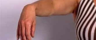
Severe weakness of the arms, a feeling of partial numbness and “pins and needles” in the upper body, difficulty breathing. In some cases, a person loses sensation throughout the entire body from the neck down. Cervical absolute stenosis is the most dangerous, since there is a possibility of not only disability of the patient, but also death.
- Thoracic region
A rare pathology that manifests itself as chest pain, disorders of the genitourinary system, intestines, numbness of the arms and shoulder girdle.
- Lumbar
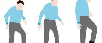
This is where stenosis develops most often. It is the lower back that is subject to the greatest stress in a person’s everyday life. Absolute stenosis manifests itself as severe pain – the pain radiates to the leg and buttock. So-called lumbago may be felt, walking is impossible or very difficult, and paralysis of the lower extremities develops - complete or partial. Stenosis also contributes to sexual dysfunction, spontaneous urination and defecation.
Treatment of the disease
In the case of spinal stenosis, conservative therapy is not always a priority. If the disease has started, or the progression of the pathology does not stop, then the doctor can immediately resort to surgery.
If the disease was detected at an early stage, then there is every chance of getting rid of the pathology with the help of conservative treatment.
Drug therapy
Drug treatment includes taking the following medications:
- Nonsteroidal anti-inflammatory drugs (NSAIDs). This type of medication is aimed at eliminating pain that interferes with the patient’s normal functioning. These drugs include Ibuprofen, Naproxen, Indomethacin, etc.;
- Painkillers analgesics. This group of drugs is also aimed at eliminating pain. The most common is Acetaminophen;
- Decongestant corticosteroid injections into tissue;
- Anti-inflammatory ointments, gels and patches. For example, Nanoplast forte, Finalgon, Voltaren, etc.;
- Drugs that optimize neuromuscular conduction (Succinylcholine, Pancuronium, etc.);
- Vitamin complexes.
Surgical intervention
As already mentioned, surgery can be prescribed first or last, depending on the clinical picture of a particular patient.
There are several types of surgical procedures used for stenosis:
- Resection of the lamina of the spine . With this type of surgery, part of the arch is removed to decompress the spinal nerve endings. The disadvantage of this method is instability of the spinal column after surgery;
- Stabilization of spinal segments . This type of surgery involves anterior and posterior stabilization of the spinal segments. However, this can lead to the development of other degenerative diseases (scoliosis, spondyloarthrosis, etc.);
- Interspinous fixation . This type of operation is used when the cause of narrowing of the spinal canal is a change in the height of the intervertebral disc and, as a result, increased pressure on its back. The support is strengthened using special implants. The operation is contraindicated in patients who have instability of spinal segments.
Physiotherapy
Important! Therapeutic exercise is at the same time the most effective and most dangerous way to treat stenosis, since incorrectly selected exercises or incorrect execution of a gymnastic complex can lead to complications and aggravation of the pathology. Be sure to consult with your doctor. A specialist will select the right set of exercises for you and monitor its implementation. Independent selection of a physical complex is prohibited!
In this article we present an approximate set of three exercises, each of which can be included in your treatment complex:
- Spread a gymnastics mat on the floor and lie on your back. Bend your legs at the knees and rest them on the mat. Place your knees bent at shoulder width. Take a deep breath, mentally count to 4, exhale while lifting your chest strongly. Repeat the exercise 8-10 times;
- Lie on the floor, spread your arms to the sides. Inhale deeply, then exhale. Raise your knees and press your chests together. Stay in this position for as long as you can, then lower your legs and relax. Repeat the exercise 8-10 times;
- Lie on your back, arms out to the side, legs bent. Now begin to turn your knees to the right, then to the left, while moving your head in the direction opposite to the movement of your legs. Do the exercise for 3-5 minutes.
Massage
Massage can be effective when the patient suffers from constant aching pain, as well as muscle spasms . Massage can help improve blood flow, metabolic processes, reduce pain, relieve swelling and muscle spasms.
Massage sessions can only be prescribed by the attending physician and carried out by an experienced master in order to avoid complications and injuries.
Treatment at home
To enhance the effectiveness of conservative treatment methods, you can use some folk recipes.
There are countless traditional methods of treating stenosis; here are some of the most effective :
- Honey mustard compress. Spread the affected area with honey, cover with a napkin or cotton cloth, and put 2-3 mustard plasters on top. Wrap up
the compress should be wrapped in cellophane; - Compress of horseradish, radish and sour cream . Grate the ingredients on a fine grater and mix with sour cream. Apply to the inflamed area;
- Infusion of thyme, elderberry flowers, St. John's wort, chamomile . Compresses from this infusion should be made at night;
- Frankincense compresses . Take 40-50 grams of frankincense, 50 grams of apple cider vinegar. Dissolve incense in vinegar, apply the resulting mixture to a woolen cloth and apply this compress to your back for three evenings in a row;
- Eucalyptus tincture should be used to wipe the inflamed area.
Disease prevention
The main ways to prevent spinal stenosis are:
- Regular physical activity;
- Selection of high-quality furniture for the workplace;
- Lift weights carefully (bend your knees and keep your hands level with the object);
- Choose a quality firm mattress;
- Monitor your body weight and your diet.
Of course, these are conditional and auxiliary recommendations, since no one is immune from age-related changes. If you follow at least these rules, then perhaps the disease will bypass you or will not bring serious complications in the future.
Video: “How is spinal stenosis treated?”
Diagnostics
- Studying the patient's complaints - the doctor asks about the location of the pain, its manifestation, and the presence of accompanying symptoms.
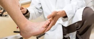
- Physical examination - the patient’s posture is studied, the presence of scoliosis, palpation and percussion diagnostics are performed.
- X-ray – X-ray will show bone formations and can also evaluate narrowing of the intervertebral canal to confirm the diagnosis of relative stenosis. If a narrowing of the lumen of less than 10 mm is detected, the patient is diagnosed with absolute stenosis.
- Multislice CT scan – detects degenerative processes of bone and cartilage tissue.
- A myelogram is an x-ray with contrast that allows you to evaluate the degree of pressure on the nerve roots.
- Magnetic resonance imaging – visualizes the condition of soft tissues and the spinal cord.
Causes of narrowing of the spinal canal
Narrowing of the spinal canal can occur due to various reasons. The leading ones are degenerative dystrophic changes associated with deformation of the intervertebral discs. This is osteochondrosis and its complications, such as protrusion and intervertebral hernia. The latter may be sequestered. In this case, part of the prolapsed nucleus pulposus separates and begins to move freely inside the spinal canal. This provokes its narrowing in different parts. Migrating, inconsistent clinical signs appear. It is very difficult for a doctor to make an accurate diagnosis. An MRI examination is required.
Other potential causes of narrowing of the spinal canal include:
- injuries (including compression and splintered fractures of vertebral bodies);
- displacement of the vertebral bodies (retrolisthesis and antelisthesis cause a significant narrowing of the canal lumen);
- subluxations of the vertebrae, their twisting around their axis;
- inflammatory processes leading to the formation of gross scar deformation of the dural membranes of the spinal cord;
- curvature of the spinal column (the most dangerous in this regard is considered to be lateral curvature, scoliosis);
- benign and malignant tumors (including metastases to the spinal column);
- increased cerebrospinal fluid pressure in brain pathologies.
All potential causes must be excluded at the diagnostic stage. By the time therapy begins, all negative factors should be eliminated if possible. You should bring your body weight into balance, engage in feasible types of physical activity on a regular basis, and properly organize your sleeping and working space.
Treatment of absolute stenosis
If the diagnosis of absolute spinal stenosis is confirmed, the patient must undergo surgical treatment as soon as possible. Drug therapy or exercise therapy are powerless in this case, and the time lost on them does not benefit the patient.
The operation protocol may be different:
- Discectomy – the disc at the level of which the maximum narrowing of the canal has formed is completely or partially removed. If a hernia is to blame for the narrowing, only it can be removed. Next, the patient receives a disc implant.
- Laminectomy - the surgeon removes part of the vertebral arch, resulting in the release of the spinal canal. Most often, such intervention is performed if the stenosis is of traumatic origin. After eliminating the compression factor, the patient is given an implant that ensures normal motor function of the spine and protects it.
Treatment of lateral spinal stenosis
Lateral spinal canal stenosis requires complex treatment. This includes both conservative and surgical methods.
Conservative treatment
- Painkillers – diclofenac 200 mg 1 – 2 times a day;
- Vascular drugs - Actovegin 5.0 ml diluted in 15.0 ml of physiological solution intravenously in a stream (slow) course for 10 days;
- B vitamins – neurorubin-forte-lactab, 1 tablet 1 time per day. The course of treatment is 30 days.
Surgical treatment
This treatment method is prescribed when conservative treatment is ineffective for 6 to 12 months, as well as in severe cases of stenosis and includes several types:
- Decompression laminectomy - excision of the bony part of the vertebra that compresses the spinal cord;
- Stabilizing operations – straightening the curvature of the spine by installing metal structures.
Possible complications if left untreated
Without timely and adequate treatment, absolute stenosis can have extremely serious consequences. And irreversible paralysis of the limbs is far from the only thing. Thus, due to impaired nutrition of spinal cord cells and deterioration of its oxygen supply, an ischemic stroke of the spinal cord may occur. And if the spinal canal is critically narrowed at the level of the thoracic or cervical region, death may occur due to respiratory distress or sudden cardiac arrest.
Causes
The causes of stenosis can be all types of degenerative pathologies of the spine, including pathology of the discs, facet joints, ligament hypertrophy, osteophytes, and spondylolysis. Degenerative spinal stenosis may occur with other manifestations of the disease, including spondylolisthesis or scoliosis. In addition to slowly progressive degenerative changes, spinal stenosis has a significant dynamic component that leads to the development of instability. The free space of the spinal canal decreases with loading and extension and increases with stretching and flexion.
Make an appointment with doctors at the Central Clinical Hospital of the Russian Academy of Sciences
The best specialists of the department of vertebrology
They receive patients with spinal canal stenoses of varying degrees of severity and origin.
The patient is examined directly on site, and an individual treatment program is developed for him, including drug therapy and physical therapy. In difficult cases, when absolute stenosis is accurately diagnosed, surgeons at the Central Clinical Hospital in Moscow
perform spinal surgeries, returning patients to motor activity and life without pain.
make an appointment
with
a vertebrologist, rheumatologist
or other musculoskeletal specialist by phone or using the online form.
Spinal stenosis - symptoms and treatment
Spinal canal stenosis is a condition when the size of the spinal canal on a cross section decreases, or the size of the intervertebral foramina decreases, as a result of which the contents of the canal (spinal cord, roots) are compressed. As a rule, spinal stenosis is detected at the level of the lower lumbar vertebrae, less often in the cervical and thoracic spine.
The vertebral (spinal) canal is a space inside the spinal column, which is formed in front by the vertebral bodies and intervertebral discs, and on the sides and back by vertebral arches connected by the ligamentum flavum. In cross section it is triangular or oval in shape.[1]
The spinal canal consists of: the spinal cord with roots surrounded by meninges, as well as fatty and loose connective tissue with arteries, veins and nerves. Paired nerve roots, surrounded by the dura mater, extend from the spinal cord, each of which extends beyond the spinal canal through its own opening. The spinal cord continues from the foramen magnum to the second lumbar vertebra. Below the second lumbar vertebra in the spinal canal there is a “cauda equina” - a bundle of roots of the four lower lumbar, five sacral and coccygeal roots of the spinal cord.
Functions of the spinal cord:
- conduction - conduction of a nerve impulse from the center to the periphery and back;
- reflex - the formation of a response of the nervous system to irritation.
Stenosis can be congenital or acquired. Congenital (primary) is formed at 3-6 weeks of intrauterine development of the human embryo. The causes of this disorder may be a genetic factor, as well as infectious and toxic factors affecting the formation of the spine.
Causes of congenital stenosis:
- Congenital chondrodystrophy (achondroplasia) is an intrauterine bone growth disorder in which the spinal canal narrows due to fusion of the vertebrae, shortening and thickening of the vertebral arches.
- Diastematomyelia is a division of the spinal canal by an internal septum, which consists of cartilage or bone tissue, bifurcation of the spinal cord.
Causes of acquired (secondary) stenosis:
- traumatic displacement of the vertebrae and their fragments, intracanal hematomas;
- degenerative-dystrophic changes in the intervertebral joints in the form of bone growths directed inside the spinal canal (facet arthropathy);
- prolapse of intervertebral hernia, its ossification or sequestration due to discopathy;
- anterior displacement of the vertebra (spondylolisthesis) due to an anatomical defect of the vertebral arch;
- thickening and calcification of the yellow ligaments of the spine due to their inflammation or degeneration;
- thickening of the capsule of the intervertebral joints due to their inflammation during ankylosing spondylitis and other inflammatory processes;
- coarsening of the anterior longitudinal ligament (Forestier disease);
- congestive congestion of the veins inside the spinal canal;
- scar changes and the introduction of steel structures inside the spinal canal due to spinal surgery;
- tumors and cysts inside the spinal canal, etc.
Often, the formation of spinal canal stenosis is influenced by both congenital and acquired factors. Mostly older people suffer from stenosis, as they have age-related degenerative diseases of the spine. The incidence of the disease increases sharply in people over 50 years of age and in this age group ranges from 1.8 to 8%.[2] The most common acquired spinal canal stenosis is the last stage of spinal osteochondrosis, when the bone tissue of the vertebral bodies and osteophytes grows.
Many people without congenital spinal disorders have a constitutionally anatomically narrower spinal canal than average. The normal depth of the spinal canal in the lumbar region is 13-25 mm, in the cervical region - 15-20 mm.
In the cervical spine, this feature of the bone structure of the canal can be detected on lateral radiographs by calculating and assessing the M.N. Tchaikovsky index. The Tchaikovsky index is the ratio of the sagittal size of the spinal canal to the sagittal size of the vertebral body at the level of this particular vertebra, without taking into account marginal bone growths. The radiograph measures the sagittal diameter of the spinal canal (a) and the sagittal size of the vertebral body (b), the first number is divided by the second (a:b).
Measuring the sagittal size of the spinal canal and vertebral body
- 0.9 to 1.1 - spinal canal of normal depth;
- less than 0.85 (according to some authors - 0.75) - constitutionally narrow spinal canal.
Signs of pathology development
Spinal stenosis leads to disability due to multiple impairments in the functionality of internal organs.
How do signs of spinal cord compression appear:
- Compression initially leads to disruption of the osteo-fibrous tissues surrounding the spinal canal;
- Infringement of neurovascular formations forms local edema at the site of injury;
- Impaired blood supply and innervation of internal organs leads to changes in the functionality of the abdominal organs, pelvis, and lower extremities;
- Pathology of the cerebrospinal fluid circulation forms cerebral hypoxia.
The pathogenetic signs of stenosis described above lead to disability if timely conservative treatment or surgery is not carried out. Without timely and competent treatment, the symptoms can provoke the death of a person due to the pathology of many organs.