Indications for evoked potential studies
Using this examination, pathologies are diagnosed:
- vascular diseases (stroke);
- lesions of the central, peripheral and autonomic nervous system;
- consequences of traumatic brain injuries;
- attention deficit hyperactivity disorder (ADHD) in children;
- sensory disorders, etc.
Using the study of evoked potentials, it is possible to study nervous and brain functions in completely healthy people. In this form, the method is in demand in sports, scientific research and in assessing the rate of development of children, especially in premature infants.
In medical practice, three types of brain evoked potential studies are most often used:
- Visual evoked potentials: make it possible to observe the visual pathway from the retina to the corresponding part of the cerebral cortex. This examination is one of the most informative methods for diagnosing patients with signs of pathologies such as multiple sclerosis, temporal arteritis, inflammatory and tumor diseases, diabetes mellitus, lesions of the autonomic nervous system, optic nerves and retina. Based on the results of the study, a specialist can make a forecast of visual impairment in a number of diseases of various etiologies (neurological, vascular, endocrine).
- Auditory evoked potentials are one of the ways of central, peripheral and autonomic lesions of the acoustic system. As a result of the examination, it is possible to quite accurately determine the nature, degree and localization of disorders of the human auditory and vestibular system. The result of the study is of high value in the study of multiple sclerosis (even in the absence of external symptoms), diseases of the facial and trigeminal nerve, acoustic neuritis, otitis media, otosclerosis, vascular pathologies of the brain, hidden and deep tumor pathologies.
- Somatosensory evoked potentials are the study of the path of a nerve signal from receptors in the skin of the hands and feet to the cerebral cortex. The purpose of the examination is to assess the sensory pathways, analyze the functioning and safety of the nervous structures of the spinal cord and brain, identify the degree of impairment and check the effects of medication. This technique is used to diagnose various pathologies of the spinal cord, multiple sclerosis, diseases of the peripheral and autonomic nervous system (neuropathies, traumatic lesions of nerve tissue, etc.) Somatosensory evoked potentials are one of the most informative methods for studying spinal cord diseases and the best way to monitor the effectiveness of treatment .
Story
The evoked potential method became widespread in the field of cognitive neuroscience more than 40 years ago, in the 60s of the last century. The first attempt to separate evoked potentials into components was made in the 70s using factor analysis and the principal components method. However, these techniques only revealed orthogonal (in a strictly mathematical sense) components of evoked potentials, which is clearly a limitation since it is clear that the components are not necessarily orthogonal. The development of new methods for objective separation of components (such as the method of independent components) has overcome the shortcomings of old approaches and is opening up new research opportunities in this direction. The accumulated knowledge demonstrates the high efficiency and diagnostic power of the method for assessing independent components of evoked potentials as endophenotypes of brain dysfunction.
Methodology for studying evoked potentials
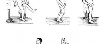
The patient is explained the essence and process of the examination - they are told that during the procedure he will be in a lying or reclining position. Depending on the nature of the study, electrodes are attached to the head, arms, legs, neck or lower back, which will not cause any harm or discomfort to the patient.
Such psychological preparation is necessary so that the patient is as relaxed and calm as possible. Any physical activity can lead to distorted results. All data from the sensors on the speed of the brain’s reaction is recorded, after which the doctor can compare the patient’s indicators with the norm and identify the nature of the lesion.
Behavioral paradigms
➥ Main article: Behavioral paradigms of VP
The vast majority of research paradigms designed to study the brain's responses to stimuli or actions involve the production of so-called evoked potentials, waveforms isolated from background EEG through an averaging procedure.
| System | Behavioral paradigm diagram | Description | Name | Component |
| Sensory systems and attention system | Standard (St) and deviant (Dev) stimuli are presented sequentially, in a random order with specified probabilities (usually 90% standard and 10% deviant). In the active modification of the task, subjects either press a button in response to the appearance of a deviant stimulus or count their number. In the passive modification (mainly for the auditory modality), subjects perform another parallel task (for example, reading a book), ignoring the presentation of standard and deviant stimuli | Oddball | Negativity of mismatch in passive modification and P3b in active modification | |
| Sensory systems and attention system | With different probabilities, standard ones are presented in random order and sequentially (St. 80%). deviant (Dev. 10%) and new (unexpected) stimuli (Nov. 10%). In the active modification of the task, subjects either press a button in response to the appearance of a deviant stimulus, or count their number. In the passive modification (mainly for the auditory modality), subjects perform another parallel task (for example, reading a book), ignoring the presentation of standard and deviant stimuli | Oddball - a paradigm with new incentives | Negativity of mismatch. РЗb and РЗа | |
| Sensory systems and attention system | Simultaneous presentation of two streams of stimulation of different spatial localization (for example, right or left ear). The subject’s task is to focus attention on one of the streams and respond to deviant stimuli presented in this stream | The dichotic listening paradigm and its visual analogue | Process negativity | |
| Sensory systems and attention system | Presentation of stimuli of different colors. Attention is concentrated on one of the stimuli of a certain color, and stimuli of a different color are ignored. EPs are recorded in response to target stimuli (on which attention is focused, relevant) and ignored (irrelevant) | Paradigm of selective (non-spatial) attention | The negativity of selection | |
| Sensory systems and attention system | In each trial, the target stimulus (in this case •+• can be presented both in the right and in the left part of the visual field. When a stimulus appears in a certain part of the visual field, the subject is preliminarily informed by a signal stimulus (in this example, an arrow). The appearance of target stimuli in may) be correctly or incorrectly predicted by the signal stimulus on different trials | Spatial signaling paradigm (Posner test) | The validity effect is the difference between EPs with a correct and incorrect signal stimulus. | |
| Executive system | First, a test stimulus (sample) is presented, followed by a pause and a test stimulus is presented. The subject's task is to compare the sample stimulus with the test stimulus and decide whether they correspond to each other | Delayed pattern matching paradigm | During a pause between two stimuli, a conditionally negative deviation is recorded | |
| Executive system | A series of stimuli is presented. The subject is required to determine whether the current stimulus corresponds to the previous one, which was presented a certain (N) number of trials before it | N-inverse problem | EP correlates of working memory | |
| Executive system | The subject's task is to respond most quickly and correctly to centrally located stimuli (in this case, letters) in a sequence of S stimuli. For example, with (H) you need to press the button with your left index finger, and with (S), respectively, with your right | Conflict detection paradigm (Erickson's flanker test, Stroop test not shown here) | Error-related negativity generated in invalid samples | |
| Executive system | Two types of stimuli (GO and NOGO) are presented in random order and with equal probability. The duration of the interstimulus interval is sufficient for the subject to have time to prepare the desired response. | GO/NOGO paradigm (the two-stimulus GO/NOGO test is a variant of this paradigm) | The N2 NOGO component is generated in response to NOGO stimuli. The P400 monitoring component is also generated by NOGO stimuli | |
| Affective system | First, stimuli related to the subject’s past and provoking the emergence of certain emotions are presented through headphones. After which images of faces expressing different emotions (joy, sadness or emotionally neutral) are presented. | Mood provocation paradigm | Difference in EP for emotions of joy and sadness | |
| Episodic memory | The day before the actual registration of the VP. The subject is presented with a list of words to memorize. During testing. The subject is presented with “old” (memorized the day before) and “new” stimuli. | Paradigm old - new | The old-new effect |
conclusions
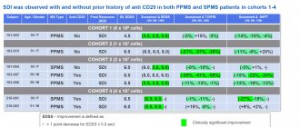
1. In case of BPPV of the PCP, dysfunction of the otolithic utriculus receptor is detected with preserved function of the otolithic sacculus receptor, which is confirmed by a decrease in amplitude on the affected side and clinically significant asymmetry of ocular MVEPs with intact cervical MVEPs.
2. With successful treatment of BPPV of the PCP and clinical resolution of otolithiasis of the PCP, the amplitude of ocular MVEPs on the affected side increases, the asymmetry of the response significantly decreases, which characterizes the restoration of the function of the otolithic utriculus receptor.
The authors declare no conflict of interest.
The essence of the method and the possibilities of its application
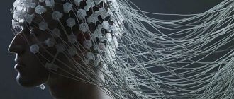
Evoked potential is an electrical signal that nerve cells respond to an external stimulus or to perform a mental task.
In 1929, HansBerger from Germany drew attention to the bioelectrical activity of the brain: when an electrical impulse is transmitted from one neuron to another, weak electrical waves arise, and an electroencephalograph device can record them.
The electroencephalogram reflects the general bioelectrical activity of brain activity. It was impossible to isolate from it the reaction to external stimulation of any individual visual or auditory analyzer at that time, since the biofield of the evoked potential (from 0.5 to 15 μV) is tens and hundreds of times weaker than the general activity of the brain (20 - 50 μV).
Only in the middle of the twentieth century did a device appear that made it possible to isolate weak amplitudes of evoked potential oscillations from the total amplitude of brain activity. This occurs using the summation method: stimulation that stimulates the potential being studied is repeated from 100 to 1000 times at precise time intervals.
The computer summarizes only those segments of the encephalogram (EEG) that immediately follow the sensory stimulation. If the total amplitude during this time can increase and decrease, take on positive and negative values and in total tend to zero, then the evoked potential has the same response form and accumulates depending on the number of stimuli given.
The more stimulating external influences, the lower the “noise level” of the overall activity. An evoked potential with a high intrinsic amplitude is isolated quite cleanly with the help of 50–60 repetitions, and a weak response to a stimulus requires more than 500 repetitions for its isolation.
To use the evoked potential method, the following equipment is required:
- stimulus generator - a device made from electrodes on the head;
- bioelectric impulse amplifier
- analog-to-digital converter;
- computer for data processing;
- printer for printing.