Pregnancy is always accompanied by risks, one of which is the pathological development of fetal organs. Special studies can exclude or confirm anomalies. Ultrasound is widely used during prenatal diagnosis. It allows you to see how the fetal organs develop and compare their parameters with normal fetometric indicators. One of the most important is the study of the lateral ventricles of the brain. What does their expansion mean, what consequences can it have?
What are the lateral ventricles of the brain and what are they responsible for?
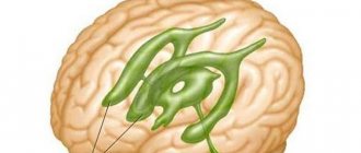
In the fetal brain there are four communicating cavities - the ventricles, which contain cerebrospinal fluid. A pair of them are symmetrical lateral, located in the thickness of the white matter. Each has an anterior, inferior and posterior horn, they are connected to the third and fourth ventricles, and through them to the spinal canal. The fluid in the ventricles protects the brain from mechanical influences and maintains stable intracranial pressure.
Each of the organs is responsible for the formation and accumulation of cerebrospinal fluid and consists of a single system of movement of cerebrospinal fluid. Liquor is necessary for stabilizing brain tissue, maintaining the correct acid-base balance, ensuring the activity of neurons. Thus, the main functions of the ventricles of the fetal brain are the production of cerebrospinal fluid and maintaining its continuous movement for active brain activity.
Ultrasound examination of the brain of newborn children (normal anatomy)
Ultrasound scanner HS60
Professional diagnostic tools.
Assessment of tissue elasticity, advanced 3D/4D/5D scanning capabilities, BI-RADS classifier, options for expert cardiological studies.
Indications for brain echography
- Prematurity.
- Neurological symptoms.
- Multiple stigmas of disembryogenesis.
- Indications of chronic intrauterine hypoxia in the anamnesis.
- Asphyxia during childbirth.
- Respiratory distress syndrome in the neonatal period.
- Infectious diseases in mother and child.
To assess the state of the brain in children with an open anterior fontanel, a sector or microconvex sensor with a frequency of 5-7.5 MHz is used. If the fontanel is closed, then you can use sensors with a lower frequency - 1.75-3.5 MHz, but the resolution will be low, which gives worse quality echograms. When studying premature babies, as well as for assessing superficial structures (furrows and convolutions on the convexital surface of the brain, extracerebral space), sensors with a frequency of 7.5-10 MHz are used.
Any natural opening in the skull can serve as an acoustic window for studying the brain, but in most cases the large fontanel is used, since it is the largest and the last to close. The small size of the fontanel significantly limits the field of view, especially when assessing the peripheral parts of the brain.
To conduct an echoencephalographic study, the sensor is placed over the anterior fontanel, orienting it so as to obtain a series of coronal (frontal) sections, and then turned 90° to perform sagittal and parasagittal scanning. Additional approaches include scanning through the temporal bone above the auricle (axial section), as well as scanning through open sutures, the posterior fontanelle and the atlanto-occipital joint.
Based on their echogenicity, the structures of the brain and skull can be divided into three categories:
- hyperechoic - bone, meninges, fissures, blood vessels, choroid plexuses, cerebellar vermis;
- medium echogenicity - parenchyma of the cerebral hemispheres and cerebellum;
- hypoechoic - corpus callosum, pons, cerebral peduncles, medulla oblongata;
- anechoic - liquor-containing cavities of the ventricles, cisterns, cavities of the transparent septum and Verge.
Normal variants of brain structures
Furrows and convolutions.
The fissures appear as echogenic linear structures separating the gyri. Active differentiation of the gyri begins from the 28th week of gestation; their anatomical appearance precedes echographic visualization by 2-6 weeks. Thus, the number and severity of the furrows can be used to judge the gestational age of the child.
Visualization of the insular complex structures also depends on the maturity of the newborn child. In very premature infants, it remains open and is presented in the form of a triangle, a flag - as a structure of increased echogenicity without identifying grooves in it. The closure of the Sylvian fissure occurs as the frontal, parietal, and occipital lobes form; complete closure of the islet of Reil with a clear Sylvian fissure and vascular formations in it ends by the 40th week of gestation.
Lateral ventricles.
The lateral ventricles, ventriculi lateralis, are cavities filled with cerebrospinal fluid, visible as anechoic zones. Each lateral ventricle consists of the anterior (frontal), posterior (occipital), inferior (temporal) horns, body and atrium (triangle) - Fig. 1. The atrium is located between the body, occipital and parietal horn. The occipital horns are difficult to visualize and their width is variable. The size of the ventricles depends on the degree of maturity of the child; with increasing gestational age, their width decreases; in mature children they are normally slit-like. Mild asymmetry of the lateral ventricles (difference in the sizes of the right and left lateral ventricles on a coronal section at the level of the foramen of Monro up to 2 mm) occurs quite often and is not a sign of pathology. Pathological expansion of the lateral ventricles most often begins with the occipital horns, so the lack of the ability to clearly visualize them is a serious argument against expansion. We can talk about expansion of the lateral ventricles when the diagonal size of the anterior horns on a coronal section through the foramen of Monroe exceeds 5 mm and the concavity of their bottom disappears.
Rice. 1.
Ventricular system of the brain. 1 - interthalamic ligament; 2 - supraoptic recess of the third ventricle; 3 - funnel-shaped pocket of the third ventricle; 4 - anterior horn of the lateral ventricle; 5 - Monroe hole; 6 - body of the lateral ventricle; 7 - III ventricle; 8 - pineal recess of the third ventricle; 9 - glomerulus of the choroid plexus; 10 - posterior horn of the lateral ventricle; 11 - lower horn of the lateral ventricle; 12 - Sylvian aqueduct; 13 - IV ventricle.
Choroid plexuses.
The choroid plexus (plexus chorioideus) is a richly vascularized organ that produces cerebrospinal fluid. Echographically, the plexus tissue appears as a hyperechoic structure. The plexuses pass from the roof of the third ventricle through the foramina of Monro (interventricular foramina) to the bottom of the bodies of the lateral ventricles and continue to the roof of the temporal horns (see Fig. 1); they are also present in the roof of the fourth ventricle, but are not detected echographically in this area. The anterior and occipital horns of the lateral ventricles do not contain choroid plexuses.
The plexuses usually have an even, smooth contour, but there may be irregularities and slight asymmetry. The choroid plexuses reach their greatest width at the level of the body and occipital horn (5-14 mm), forming a local compaction in the atrium area - the choroid glomerulus (glomus), which can have the shape of a finger-like outgrowth, be layered or fragmented. On coronal sections, the plexuses in the occipital horns appear as ellipsoidal densities, almost completely filling the lumen of the ventricles. In infants with a lower gestational age, the size of the plexuses is relatively larger than in full-term infants.
The choroid plexuses can be a source of intraventricular hemorrhages in full-term infants, then their clear asymmetry and local compactions are visible on echograms, in place of which cysts then form.
III ventricle.
The third ventricle (ventriculus tertius) appears as a thin slit-like vertical cavity filled with cerebrospinal fluid, located sagittally between the thalami above the sella turcica. It connects to the lateral ventricles through the foramen of Monroe (foramen interventriculare) and to the IV ventricle through the aqueduct of Sylvius (see Fig. 1). The supraoptic, infundibular and pineal processes give the third ventricle a triangular appearance on a sagittal section. On a coronal section, it is visible as a narrow gap between the echogenic visual nuclei, which are interconnected by the interthalamic commissure (massa intermedia), passing through the cavity of the third ventricle. In the neonatal period, the width of the third ventricle on a coronal section should not exceed 3 mm, in infancy - 3-4 mm. The clear outlines of the third ventricle on the sagittal section indicate its expansion.
Sylvian aqueduct and IV ventricle.
The Sylvian aqueduct (aquaeductus cerebri) is a thin canal connecting the third and fourth ventricles (see Fig. 1), rarely visible during ultrasound examination in standard positions. It can be visualized on an axial section in the form of two echogenic points against the background of hypoechoic cerebral peduncles.
The fourth ventricle (ventriculus quartus) is a small diamond-shaped cavity. On echograms in a strictly sagittal section, it appears as a small anechoic triangle in the middle of the echogenic medial contour of the cerebellar vermis (see Fig. 1). Its anterior border is not clearly visible due to the hypoechogenicity of the dorsal part of the bridge. The anteroposterior size of the IV ventricle in the neonatal period does not exceed 4 mm.
Corpus callosum.
The corpus callosum (corpus callosum) on a sagittal section looks like a thin horizontal arcuate hypoechoic structure (Fig. 2), limited above and below by thin echogenic stripes resulting from reflection from the peri-callosal groove (above) and the lower surface of the corpus callosum. Immediately below it are two leaves of a transparent septum, limiting its cavity. On a frontal section, the corpus callosum appears as a thin, narrow hypoechoic strip forming the roof of the lateral ventricles.
Rice. 2.
Location of the main brain structures on the midsagittal section. 1 - pons; 2 - prepontine cistern; 3 - interpeduncular cistern; 4 - transparent partition; 5 — legs of the arch; 6 - corpus callosum; 7 - III ventricle; 8 - quadrigeminal cistern; 9 - cerebral peduncles; 10 - IV ventricle; 11 - large tank; 12 - medulla oblongata.
Cavity of the septum pellucida and Verge's cavity.
These cavities are located directly under the corpus callosum between the layers of the transparent septum (septum pellucidum) and are limited by glia, not ependyma; they contain fluid but do not connect to either the ventricular system or the subarachnoid space. The cavity of the transparent septum (cavum cepti pellucidi) is located anterior to the fornix of the brain between the anterior horns of the lateral ventricles; Verge's cavity is located under the splenium of the corpus callosum between the bodies of the lateral ventricles. Sometimes, normally, dots and short linear signals originating from the subependymal median veins are visualized in the leaves of the septum pellucidum. On a coronal view, the cavity of the septum pellucidum appears as a square, triangular, or trapezoidal anechoic space with a base beneath the corpus callosum. The width of the cavity of the transparent septum does not exceed 10-12 mm and is wider in premature infants than in full-term infants. Verge's cavity, as a rule, is narrower than the cavity of the transparent septum and is rarely found in full-term children. These cavities begin to obliterate after 6 months of gestation in the dorsoventral direction, but there are no exact dates for their closure, and both of them can be detected in a mature child at the age of 2-3 months.
Basal ganglia, thalami and internal capsule.
The visual nuclei (thalami) are spherical hypoechoic structures located on the sides of the cavity of the septum pellucidum and forming the lateral borders of the third ventricle on coronal sections. The upper surface of the gangliothalamic complex is divided into two parts by the caudothalamic recess - the anterior one belongs to the caudate nucleus, the posterior one to the thalamus (Fig. 3). The optic nuclei are connected to each other by an interthalamic commissure, which becomes clearly visible only with the expansion of the third ventricle both on the frontal (in the form of a double echogenic transverse structure) and on the sagittal sections (in the form of a hyperechoic point structure).
Rice. 3.
The relative position of the structures of the basal-thalamic complex on a parasagittal section. 1 - shell of the lenticular nucleus; 2 - globus pallidus of the lenticular nucleus; 3 - caudate nucleus; 4 - thalamus; 5 - internal capsule.
The basal ganglia are subcortical accumulations of gray matter located between the thalamus and the insula of Reille. They have similar echogenicity, which makes their differentiation difficult. A parasagittal section through the caudothalamic notch is the most optimal approach for detecting the thalami, the lenticular nucleus consisting of the putamen, and the globus pallidus, and the caudate nucleus, as well as the internal capsule - a thin layer of white matter separating the nuclei of the striatum bodies from thalami. Clearer visualization of the basal nuclei is possible when using a 10 MHz sensor, as well as in case of pathology (hemorrhage or ischemia) - as a result of neuronal necrosis, the nuclei acquire increased echogenicity.
Germinal matrix
is an embryonic tissue with high metabolic and fibrinolytic activity that produces glioblasts. This subependymal plate is most active between the 24th and 34th weeks of gestation and is a cluster of fragile vessels, the walls of which are devoid of collagen and elastic fibers, are easily susceptible to rupture and are a source of peri-intraventricular hemorrhages in premature infants. The germinal matrix lies between the caudate nucleus and the lower wall of the lateral ventricle in the caudothalamic recess; on echograms it looks like a hyperechoic strip.
Brain cisterns.
Cisterns are spaces between brain structures containing cerebrospinal fluid (see Fig. 2), which may also contain large vessels and nerves. Normally, they are rarely visible on echograms. When enlarged, the cisterns appear as irregularly defined cavities, which indicates a proximally located obstruction to the flow of cerebrospinal fluid.
The cisterna magna (cisterna magna, c. cerebromedullaris) is located under the cerebellum and medulla oblongata above the occipital bone; normally, its superior-inferior size on a sagittal section does not exceed 10 mm. The pontine cistern is an echogenic zone above the pons in front of the cerebral peduncles, under the anterior recess of the third ventricle. It contains a bifurcation of the basilar artery, which causes its partial echo density and pulsation.
The basal (c. suprasellar) cistern includes the interpeduncular, c. interpeduncularis (between the cerebral peduncles) and chiasmatic, c. chiasmatis (between the optic chiasm and the frontal lobes) cisterns. The chiasm cistern appears as a pentagonal echo-dense zone, the angles of which correspond to the arteries of the circle of Willis.
The quadrigeminal cistern (c. quadrigeminalis) is an echogenic line between the plexus of the third ventricle and the cerebellar vermis. The thickness of this echogenic zone (normally not exceeding 3 mm) may increase with subarachnoid hemorrhage. Arachnoid cysts may also be located in the area of the quadrigeminal cistern.
Bypass (c. ambient) tank - carries out lateral communication between the prepontine and interpeduncular tanks in front and the quadrigeminal cistern behind.
Cerebellum
(cerebellum) can be visualized through both the anterior and posterior fontanel. When scanning through a large fontanel, the image quality is the worst due to the distance. The cerebellum consists of two hemispheres connected by the vermis. The hemispheres are weakly mid-echoic, the vermis is partially hyperechoic. On a sagittal section, the ventral part of the vermis looks like a hypoechoic letter “E” containing cerebrospinal fluid: at the top is the quadrigeminal cistern, in the center is the IV ventricle, at the bottom is the cistern magna. The transverse size of the cerebellum directly correlates with the biparietal diameter of the head, which makes it possible to determine the gestational age of the fetus and newborn based on its measurement.
The cerebral peduncles (pedunculus cerebri), bridge (pons) and medulla oblongata (medulla oblongata) are located longitudinally anterior to the cerebellum and appear as hypoechoic structures.
Parenchyma.
Normally, there is a difference in echogenicity between the cerebral cortex and the underlying white matter. The white matter is slightly more echogenic, possibly due to the relatively greater number of vessels. Normally, the thickness of the cortex does not exceed a few millimeters.
Around the lateral ventricles, predominantly over the occipital and less frequently over the anterior horns, in premature infants and some full-term infants there is a halo of increased echogenicity, the size and visualization of which depends on gestational age. It can persist up to 3-4 weeks of life. Normally, its intensity should be lower than that of the choroid plexus, the edges should be unclear, and the location should be symmetrical. If there is asymmetry or increased echogenicity in the periventricular region, an ultrasound examination of the brain should be performed over time to exclude periventricular leukomalacia.
Standard echoencephalographic sections
Coronal slices
(Fig. 4).
The first slice
passes through the frontal lobes in front of the lateral ventricles (Fig. 5). The interhemispheric fissure is determined in the middle in the form of a vertical echogenic strip separating the hemispheres. When it expands, a signal from the falx cerebri (falx) is visible in the center, which is not normally visualized separately (Fig. 6). The width of the interhemispheric fissure between the gyri does not normally exceed 3-4 mm. On the same section it is convenient to measure the size of the subarachnoid space - between the lateral wall of the superior sagittal sinus and the nearest gyrus (sinocortical width). To do this, it is advisable to use a sensor with a frequency of 7.5-10 MHz, a large amount of gel and very carefully touch the large fontanel without pressing on it. The normal size of the subarachnoid space in full-term babies is up to 3 mm, in premature babies – up to 4 mm.
Rice. 4.
Coronal scanning planes (1-6).
Rice. 5.
Echogram of the newborn's brain, first coronal slice through the frontal lobes. 1 - eye sockets; 2 - interhemispheric fissure (not widened).
Rice. 6.
Measurement of the width of the subarachnoid space and the width of the interhemispheric fissure on one or two coronal sections - diagram (a) and echogram of the brain (b). 1 - superior sagittal sinus; 2 — width of the subarachnoid space; 3 — width of the interhemispheric fissure; 4 - falx cerebri.
Second cut
is performed through the anterior horns of the lateral ventricles anterior to the foramina of Monroe at the level of the cavity of the septum pellucidum (Fig. 7). The frontal horns, which do not contain cerebrospinal fluid, are visualized on both sides of the interhemispheric fissure as echogenic stripes; if they contain cerebrospinal fluid, they look like anechoic structures, similar to boomerangs. The roof of the anterior horns of the lateral ventricles is represented by a hypoechoic strip of the corpus callosum, and between their medial walls there are layers of the transparent septum containing a cavity. On this section, the shape is assessed and the width of the cavity of the transparent septum is measured - the maximum distance between its walls. The lateral walls of the anterior horns form the basal nuclei - directly under the bottom of the horn - the head of the caudate nucleus, and laterally - the lentiform nucleus. Even more lateral on this section, the temporal lobes are identified on both sides of the chiasm cistern.
Rice. 7.
Echogram of the brain, second coronal section through the anterior horns of the lateral ventricles. 1 - temporal lobes; 2 - Sylvian fissure; 3 - cavity of the transparent septum; 4 - anterior horn of the lateral ventricle; 5 - corpus callosum; 6 - interhemispheric fissure; 7 - caudate nucleus; 8 - thalamus.
Third coronal slice
passes through the foramina of Monroe and the third ventricle (Fig. 8). At this level, the lateral ventricles connect to the third ventricle through the interventricular foramina (Monroe). The foramina themselves are not normally visible, but the choroid plexuses passing through them from the roof of the third ventricle to the bottom of the lateral ventricles appear as a hyperechoic Y-shaped structure located in the midline. Normally, the third ventricle may also not be visualized; when it enlarges, its width is measured between the medial surfaces of the thalami, which are its lateral walls. The lateral ventricles in this section are visible as slit-like or boomerang-shaped anechoic structures (Fig. 9), the width of which is measured diagonally (normally up to 5 mm). The cavity of the transparent septum on the third section in some cases still remains visible. Below the third ventricle, the brainstem and pons are visualized. Lateral to the third ventricle are the thalamus, basal ganglia and insula, above which a Y-shaped thin echogenic structure is defined - the Sylvian fissure, containing the pulsating middle cerebral artery.
Rice. 8.
Echogram of the brain, third coronal section through the foramina of Monroe. 1 - III ventricle; 2 - choroid plexuses in the interventricular canals and the roof of the third ventricle and the fornix of the brain; 3 - cavity of the lateral ventricle; 4 - corpus callosum; 5 - caudate nucleus; 6 - thalamus.
Rice. 9.
The relative position of the central brain structures on two to four coronal sections. 1 - III ventricle; 2 - cavity of the transparent septum; 3 - corpus callosum; 4 - lateral ventricle; 5 - caudate nucleus; 6 - peduncle of the cerebral vault; 7 - thalamus.
On the fourth cut
(through the bodies of the lateral ventricles and the posterior part of the third ventricle) the following are visible: interhemispheric fissure, corpus callosum, ventricular cavities with choroid plexuses in their bottom, thalamus, Sylvian fissures, vertically located hypoechoic cerebral peduncles (below the thalami), cerebellum separated from the cerebral peduncles by hyperechoic tentorium (Fig. 10). Inferior to the cerebellar vermis, the cistern magna can be visualized. In the area of the middle cranial fossa, an area of pulsation is visible, originating from the vessels of the circle of Willis.
Rice. 10.
Echogram of the brain, fourth coronal section through the bodies of the lateral ventricles. 1 - cerebellum; 2 - choroid plexuses in the lateral ventricles; 3 - bodies of the lateral ventricles; 4 - Verge's cavity.
Fifth cut
passes through the bodies of the lateral ventricles and the choroid plexuses in the region of the glomus, which on echograms almost completely fill the cavities of the lateral ventricles (Fig. 11). On this section, the density and size of the choroid plexuses on both sides are compared to exclude hemorrhages. If a Verge's cavity is present, it is visualized between the lateral ventricles in the form of a rounded anechoic formation. Inside the posterior cranial fossa, the cerebellum is visualized with medium echogenicity, and above its tentorium is the echogenic quadrigeminal cistern.
Rice. eleven.
Echogram of the brain, fifth coronal section through the glomus of the choroid plexuses - choroid plexuses in the atrium area, completely filling the lumen of the ventricles (1).
Sixth
, the last, coronal section is performed through the occipital lobes above the cavities of the lateral ventricles (Fig. 12). The interhemispheric fissure with grooves and convolutions is visualized in the middle, and on both sides there are cloud-like periventricular densities, which are more pronounced in premature infants. On this section, the symmetry of these seals is assessed.
Rice. 12.
Echogram of the brain, sixth coronal section through the occipital lobes above the lateral ventricles. 1 - normal periventricular seals; 2 - interhemispheric fissure.
Sagittal slices
(Fig. 13).
A midsagittal section
(Fig. 14) allows one to visualize the corpus callosum in the form of a hypoechoic arch, immediately below it is the cavity of the transparent septum (under its anterior sections) and the Verge's cavity connected to it (under the splenium). Near the genu of the corpus callosum there is a pulsating structure - the anterior cerebral artery, which goes around it and runs along the upper edge of the body. The pericallosal groove runs above the corpus callosum. Between the cavities of the transparent septum and Verge, an arcuate hyperechoic strip is determined, originating from the choroid plexus of the third ventricle and the fornix of the brain. Below is a hypoechoic triangular third ventricle, the contours of which are normally not clearly defined. When it expands in the center, you can see the interthalamic commissure in the form of a hyperechoic point. The posterior wall of the third ventricle consists of the pineal gland and the quadrigeminal plate, behind which the quadrigeminal cistern may be visible. Immediately below it, in the posterior cranial fossa, a hyperechoic cerebellar vermis is identified, on the anterior part of which there is a triangular notch - the fourth ventricle. The pons, cerebral peduncles and medulla oblongata are located anterior to the fourth ventricle and are visible as hypoechoic formations. On this section, the cisterna magna is measured - from the lower surface of the vermis to the inner surface of the occipital bone - and the depth of the fourth ventricle is measured.
Rice. 13.
Sagittal scanning planes (1-4).
Rice. 14.
Echograms of the brain, midsagittal section: 1 - cerebellum; 2 - IV ventricle; 3 - III ventricle; 4 - fornix and choroid plexus in the foramina of Monroe and the roof of the third ventricle; 5 - corpus callosum; 6 - cavity of the transparent septum; 7 - cerebral peduncles; 8 - large tank; 9 - Verge's cavity; 10 - corpus callosum; 11 — cavity of the transparent septum; 12 - III ventricle.
With a slight deviation of the sensor to the left and right, a parasagittal slice
through the caudothalamic recess (the location of the germinal matrix in premature infants), where its shape, as well as the structure and echogenicity of the gangliothalamic complex are assessed (Fig. 15).
Rice. 15.
Echogram of the brain, parasagittal section through the caudothalamic notch. 1 - choroid plexus of the lateral ventricle; 2 — cavity of the lateral ventricle; 3 - thalamus; 4 - caudate nucleus.
Next parasagittal slice
is performed through the lateral ventricle on each side so as to obtain its full image - the frontal horn, body, occipital and temporal horns (Fig. 16). In this plane, the height of various parts of the lateral ventricle is measured, and the thickness and shape of the choroid plexus are assessed. Above the body and occipital horn of the lateral ventricle, the homogeneity and density of the periventricular substance of the brain is assessed, comparing it with the density of the choroid plexus.
Rice. 16.
Echogram of the brain, parasagittal section through the lateral ventricle. 1 - lower horn; 2 - posterior horn; 3 - glomus choroid plexus; 4 - body; 5 - anterior horn.
Last parasagittal slice
are obtained by tilting the sensor even more laterally, which makes it possible to visualize the insula, its sulci and gyri and measure the Sylvian fissure in case of its expansion (Fig. 17). Normally, in full-term infants, it appears as an echogenic transverse structure; with subarachnoid hemorrhage or external hydrocephalus, a strip of cerebrospinal fluid is visualized between the insula and the parietal lobe.
Rice. 17.
Echogram of the brain, parasagittal section through the temporal lobe. 1 - temporal lobe of the brain; 2 - Sylvian fissure; 3 - parietal lobe.
If any deviations are determined on the obtained echograms in the coronal section, then they must be confirmed in the sagittal section, and vice versa, since artifacts can often occur.
Axial scanning.
An axial cut is made by placing the transducer horizontally above the ear. In this case, the cerebral peduncles are visualized as a hypoechoic structure shaped like a butterfly (Fig. 18). Between the legs (in contrast to coronal and sagittal sections) an echogenic structure is often visible, consisting of two points - the aqueduct of Sylvius, anterior to the legs - the slit-like third ventricle. On an axial section, the walls of the third ventricle are clearly visible, in contrast to the coronal one, which makes it possible to more accurately measure its size with slight expansion. When the sensor is tilted towards the calvarium, the lateral ventricles are visible, which makes it possible to estimate their size when the large fontanelle is closed. Normally, the parenchyma of the brain is closely adjacent to the bones of the skull in mature children, so the separation of echo signals from them on an axial section suggests the presence of pathological fluid in the subarachnoid or subdural spaces.
Rice. 18.
Echogram of the brain, axial section at the level of the base of the brain. 1 - cerebellum; 2 - Sylvian aqueduct; 3 - cerebral peduncles; 4 - Sylvian fissure; 5 - III ventricle.
Data from an echographic examination of the brain can be supplemented by the results of Doppler ultrasound assessment of cerebral blood flow. This is desirable, since in 40-65% of children, despite severe neurological disorders, echographic examination of the brain remains normal.
The brain is supplied with blood by the branches of the internal carotid and basilar arteries, which form the circle of Willis at the base of the brain. The direct continuation of the internal carotid artery is the middle cerebral artery, and its smaller branch is the anterior cerebral artery. The posterior cerebral arteries branch from the short basilar artery and communicate with the branches of the internal carotid through the posterior communicating arteries. The main cerebral arteries - the anterior, middle and posterior with their branches form an arterial network from which small vessels penetrate into the medulla, feeding the cortex and white matter of the brain.
Doppler examination of blood flow is carried out in the largest arteries and veins of the brain, trying to position the ultrasound sensor so that the angle between the ultrasound beam and the axis of the vessel is minimal.
Anterior cerebral artery
visualized on a sagittal section; To obtain blood flow measurements, a volumetric marker is placed in front of the knee of the corpus callosum or in the proximal part of the artery before it bends around this structure.
To study blood flow in the internal carotid artery
on the parasagittal section, its vertical part is used immediately after exiting the carotid canal above the level of the sella turcica.
Basilar artery
examined in a midsagittal section in the area of the base of the skull immediately in front of the pons, a few millimeters behind the location of the internal carotid artery.
Middle cerebral artery
determined in the Sylvian fissure. The best angle for its insonation is achieved with an axial approach. The vein of Galen is visualized on a coronal section under the corpus callosum along the roof of the third ventricle.
Ultrasound scanner HS60
Professional diagnostic tools.
Assessment of tissue elasticity, advanced 3D/4D/5D scanning capabilities, BI-RADS classifier, options for expert cardiological studies.
What size should the lateral ventricles of the fetal brain normally be?
The brain structures of the fetus are visualized already during the second ultrasound (at 18–21 weeks). The doctor evaluates many indicators, including the size of the lateral ventricles of the brain and the cistern magna in the fetus. The average size of the ventricles is about 6 mm; normally, their size should not exceed 10 mm. The basic norms of fetometric parameters by week are assessed according to the table:
| Gestation period, weeks | Large cylinder head tank, mm | Head circumference, mm | Fronto-occipital size, mm | Distance from the outer to the inner contour of the crown bones (BPR), mm |
| 17 | 2,1–4,3 | 121–149 | 46–54 | 34–42 |
| 18 | 2,8–4,3 | 131–161 | 49–59 | 37–47 |
| 19 | 2,8–6 | 142–174 | 53–63 | 41–49 |
| 20 | 3–6,2 | 154–186 | 56–68 | 43–53 |
| 21 | 3,2–6,4 | 166–200 | 60–72 | 46–56 |
| 22 | 3,4–6,8 | 177–212 | 64–76 | 48–60 |
| 23 | 3,6–7,2 | 190–224 | 67–81 | 52–64 |
| 24 | 3,9–7,5 | 201–237 | 71–85 | 55–67 |
| 25 | 4,1–7,9 | 214–250 | 73–89 | 58–70 |
| 26 | 4,2–8,2 | 224–262 | 77–93 | 61–73 |
| 27 | 4,4–8,4 | 236–283 | 80–96 | 64–76 |
| 28 | 4,6–8,6 | 245–285 | 83–99 | 70–79 |
| 29 | 4,8–8,8 | 255–295 | 86–102 | 67–82 |
| 30 | 5,0–9,0 | 265–305 | 89–105 | 71–86 |
| 31 | 5,5–9,2 | 273–315 | 93–109 | 73–87 |
| 32 | 5,8–9,4 | 283–325 | 95–113 | 75–89 |
| 33 | 6,0–9,6 | 289–333 | 98–116 | 77–91 |
| 34 | 7,0–9,9 | 293–339 | 101–119 | 79–93 |
| 35 | 7,5–9,9 | 299–345 | 103–121 | 81–95 |
| 36 | 7,5–9,9 | 303–349 | 104–124 | 83–97 |
| 37 | 7,5–9,9 | 307–353 | 106–126 | 85–98 |
| 38 | 7,5–9,9 | 309–357 | 106–128 | 86–100 |
| 39 | 7,5–9,9 | 311–359 | 109–129 | 88–102 |
| 40 | 7,5–9,9 | 312–362 | 110–130 | 89–103 |
Choroid plexuses of the ventricles of the brain
As noted, the function of the abdominal system is the production of cerebrospinal fluid. But how is this liquid formed? The only brain structure that provides the synthesis of cerebrospinal fluid is the choroid plexus. These are small villous formations belonging to vertebrates.
The choroid plexus is a derivative of the pia mater. They contain a huge number of vessels and carry a large number of nerve endings.
What is ventriculomegaly and what types of it occurs?
If, according to the results of an ultrasound at 16–35 weeks of pregnancy, an increase in the lateral ventricles to 10–15 mm is recorded, and the size of the child’s head is within normal limits, the sonologist questions ventriculomegaly. One study is not enough to make an accurate diagnosis. Changes are assessed over time, for which at least two more ultrasounds are performed with an interval of 2–3 weeks. This pathology is caused by chromosomal abnormalities, intrauterine hypoxia, and infectious diseases that the mother suffered during pregnancy.
Ventriculomegaly can be isolated asymmetric (expansion of one ventricle or its horns without changes in the brain parenchyma), symmetric (observed in both hemispheres) or diagnosed in combination with other pathologies of fetal development. Ventricular pathology is divided into three types:
- mild - organ dilatation is 10.1–12 mm, usually detected at 20 weeks and needs to be monitored before birth;
- moderate – the size of the ventricles reaches 12–15 mm, which impairs the outflow of cerebrospinal fluid;
- pronounced - ultrasound readings exceed 15 mm, which disrupts brain function and negatively affects the vital functions of the fetus.
If the lateral ventricles are enlarged to 10.1–15 mm (deviation from the table standards by 1–5 mm), borderline ventriculomegaly is diagnosed. It may be asymptomatic up to a certain point, but in fact it indicates the emergence of a complex pathological process that gradually changes the functioning of many important organs.
Causes of enlargement of the cerebral ventricles
Throughout life, there is a gradual increase in the spaces containing cerebrospinal fluid. Some pathological conditions lead to compression of the ventricular structures:
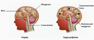
- Hydrocephalus - accumulation of fluid inside the brain with the development of dropsy;
- Birth injury;
- Long-term inflammation of the meninges with the formation of adhesions;
- Hemorrhagic hemorrhages;
- Strokes, hemorrhages.
There is a fundamental difference between the concepts of asymmetry and ventricular dilatation. The second nosology is an independent disease. It is formed under the influence of intrauterine infections and acquired pathology. Asymmetry is caused by additional cerebral formations (tumors, limited accumulation of blood).
The clinical picture of the asymmetrical arrangement of cerebral structures depends on the location and degree of compression. Small imbalances in the accumulation of cerebrospinal fluid are compensated by the reverse absorption of cerebrospinal fluid.
The cause of dilation may be overproduction of cerebrospinal fluid. In most cases, the etiological factor of the nosology is a violation of oxygen supply (hypoxia). The condition provokes increased formation of cerebrospinal fluid in order to improve the supply of nutrients and vitamins to the brain formations.
At the moment, many factors are known that influence the appearance of pathologies of the ventricles of the brain in children. All of them can be divided into two categories: acquired and congenital. Acquired causes include those reasons that could arise during the pregnancy of the child’s mother:
- Infectious diseases that a woman suffers from during pregnancy.
- Infections and sepsis inside the womb.
- Penetration of foreign bodies into the brain.
- Chronic diseases of the mother that affect the normal course of pregnancy.
- Delivery ahead of schedule.
- Hypoxia of the fetus inside the womb (insufficient or, conversely, increased blood supply to the placenta).
- Abnormal duration of the dry period.
- Injury to the baby during childbirth (suffocation by the umbilical cord or deformation of the skull).
- Stormy birth.
Congenital causes include a genetic predisposition to enlarged ventricles; abnormalities occurring in chromosomes, as well as various neoplasms (cysts, malignant or benign tumors, hematomas). Along with the listed reasons, characteristic changes in the size of the ventricles of the brain can be provoked by traumatic brain injury, cerebral hemorrhage, or stroke.
The human brain is a very complex structure, in which each substructure and each component part is responsible for fulfilling certain goals. In humans, there is a special structure in the brain that contains cerebrospinal fluid (CSF). The purpose of this structure is the circulation and production of cerebrospinal fluid. Each child and adult has 3 types of brain ventricles, and their total number is 4. They are connected to each other through channels and openings, valves. So, the ventricles are distinguished:
- Lateral.
- Third.
- Fourth.
The lateral ventricles are located symmetrically relative to each other. The left is designated first, the right is designated second, they are connected to the third. The third ventricle is the anterior one and houses the centers of the autonomic nervous system. The fourth is the posterior one, it is shaped like a pyramid and is connected to the spinal cord.
Treatment of pathology
When treating a pathology, the doctor sets two goals: finding and eliminating the cause of abnormal organ enlargement and neutralizing its consequences for the newborn. In case of a mild isolated form, which is not caused by chromosomal abnormalities, the expectant mother is prescribed drug therapy: taking diuretics, vitamins, injections of drugs that prevent hypoxia and placental insufficiency. Exercise therapy has a good effect (the emphasis is on the pelvic floor muscles). To prevent neurological changes in the baby's body, the expectant mother is prescribed medications to retain potassium.
In the postpartum period, infants are given several courses of massage aimed at relieving muscle tone, strengthening, and eliminating neurological symptoms. Monitoring by a neurologist is required in the first weeks, months and years of life. Severe forms of pathology require surgical treatment after the birth of the baby. In the brain, neurosurgeons install a tube that has a drainage function to maintain proper circulation of cerebrospinal fluid.
The prognosis for a child with a severe form and chromosomal abnormalities is unfavorable.
Doctors often decide to terminate a pregnancy due to severe brain damage to the fetus. For this reason, it is very important to identify pathology at 17–25 weeks and prevent its progression.
Possible consequences of ventriculomegaly
Abnormal development of the ventricles can lead to serious defects of the fetus, newborn, and death of the child. Such tragedies are a consequence of impaired outflow of cerebrospinal fluid, which interferes with the development and functioning of the nervous system. Pathology often provokes early labor, impaired brain activity, heart defects, and infections. In severe stages, the disease ends in hydrocephalus of the brain in children. The earlier such a change occurs, the worse the doctors' prognosis.
If the cause of the deviations is chromosomal abnormalities or complicated heredity, ventriculomegaly occurs together with the following pathologies:
- abnormal connection of blood vessels and lymphatic systems;
- Patau, Down syndrome;
- delay in mental and physical abilities;
- pathologies of the musculoskeletal system.
The prognosis for mild and moderate ventriculomegaly is favorable if the pathology is not accompanied by other disorders of the nervous system. This is confirmed by clinical assessments of the neurological status of babies who were given a similar diagnosis in fetal development. 82% of children do not have serious abnormalities, 8% have certain problems, and 10% have severe impairments with disability. The pathology requires constant monitoring by a neurologist and timely correction.
What diseases can affect the ventricles
Despite the small size of the ventricular system, the process of liquor circulation is extremely important for the functioning of the central nervous system and the whole organism. Like any organ, the cavities in the brain are often susceptible to various diseases. Hydrocephalus of the brain is considered the most common disease that has no age restrictions.
The lateral cisterns are characterized by asymmetry, which means that the cause of the anomaly lies in the malfunction of the choroid plexuses. The accumulation of cerebrospinal fluid in the cavities is not always caused by increased secretion production; blockage of the conductor channels often leads to a pathological condition.
Hydrocephalus syndrome
The expansion of the ventricles of the brain manifests itself in a bright clinic, since the central nervous system ceases to receive balanced nutrition and oxygen. Neurological symptoms appear when the brain is unable to perform its functions productively.
Pathologies in the ventricular system have primary or secondary causes and treatment is aimed at restoring all stages of liquor circulation. Diseases of the ventricles require immediate treatment, since delays threaten degenerative changes in the brain matter.
Enlargement of the ventricles of the brain in the fetus is considered a congenital disorder; early diagnosis helps to carry out emergency neurosurgical intervention immediately after the birth of the child.
Suspicion of enlargement of the lateral ventricles of the brain is always subject to a thorough diagnostic examination. Visualization of brain structures allows you to evaluate the entire ventricular system and identify the cause of the failure.
Diagnostic methods:
- Physical examination by a neurologist - neurological status is assessed;
- Consultation with an ophthalmologist – examination of the fundus;
- Neurosonography - the study is carried out through the central fontanelle for children under 1 - 1.5 years old;
- MRI or CT with contrast – layer-by-layer visualization of structures, performed on adults and children from 2 years of age;
- Doppler ultrasound – evaluates the vessels of the head and neck.
Highly accurate methods allow the assessment of all parameters in the ventricular system. An index is calculated for each cavity; if the percentage values are higher than normal, then a diagnosis of hydrocele is made. The degree of pathology is determined by the compliance of the ventricles with age criteria and the individual index.
The absorption of cerebrospinal fluid in the subarachnoid space into the vascular network directly depends on the constancy of intracranial pressure. Failure of the cerebrospinal fluid circulation leads to unstable pressure; when it decreases, fluid absorption is impossible.
Liquid is continuously produced and if there is no free outflow, then the cisterns grow, increasing in volume and width; the pressure on nearby structures depends on how enlarged the ventricles of the brain are.
Thus, during diagnosis it is revealed at what stage in the cerebrospinal fluid circulation the failure occurred and what was the cause. The primary causes always relate to congenital anomalies.
Ventricular dilatation (MRI image)
Secondary disorders in the ventricular system are a consequence of the influence of various negative factors and associated pathologies:
- Traumatic brain injuries (including birth injuries);
- Cystic formations;
- Intracranial neoplasms - tumors inside the cavity can be of intraventricular origin, that is, the primary focus, and the origin of cancer cells occurs precisely in the storage area. When atypical cells grow through the walls of the ventricle, we are talking about a secondary focus (metastasis). Ependymoma and choroidpapilloma are formed by abnormal cell division in the choroid plexus;
- Penetration of viruses and bacteria - the entry of pathogenic agents causes inflammation of the meninges and a suppurative process, as a result of which a structural change in blood vessels occurs;
- Vascular thrombosis;
- Cerebral hemorrhage – more common in older people.
Bleeding in the ventricles is a life-threatening condition. The pathology has a high mortality rate, since the clinical picture develops rapidly and the patient cannot always be transported to the hospital on time.
The disease more often affects older people (atraumatic hemorrhage), as age-related changes occur in the structure of blood vessels.
In the presence of concomitant diseases (atherosclerosis, cardiovascular pathology, blood diseases), this leads to various internal damage to the blood vessels, resulting in rupture and effusion of blood into the cavity. Intraventricular hemorrhage can also be traumatic.
A burst vessel leads to acute cerebrovascular accident. If the hemorrhage occurs in the lateral cisterns, then the blood flows through the channels to the lower ventricles. Blood clots form in the cavities, thus causing ventricular tamponade and rapidly increasing cerebral edema.
Blockage of the rhomboid cistern quickly leads to death, since the work of the vegetative centers that control vital mechanisms is disrupted.
With a timely neurosurgical operation, a drainage system is installed, with the help of which it is possible to remove intracerebral hematomas.
Poor circulation of the cerebrospinal fluid causes the accumulation of fluid in the cavities, as a result of which the ventricles enlarge and put pressure on neighboring structures. An increase in the total volume of fluid in the brain leads to an increase in ICP and the development of intracranial hypertension.
As a result of increased pressure, nerve cells die, which is dangerous for the development of encephalopathy - dystrophic damage to brain tissue is irreversible.
In children, there are cases where the lateral ventricles are dilated, but parents do not notice changes in behavior or other pathological symptoms. Perhaps this will be a variant of the norm (individual) and we are not talking about a congenital disease (ventriculomegaly), but provided that the CMD is also normal.
To do this, ICP is measured; there are several methods - non-invasive measurements using CT or MRI diagnostics, a minimally invasive technique in the form of a puncture in the lumbar spinal canal.
But neurosurgeons more often resort to the technique of puncturing cavities; when puncturing the brain, a special needle is inserted and the anterior horns of the lateral ventricles are punctured. Direct collection of cerebrospinal fluid from cavities is considered a highly informative technique.
If the ICP exceeds the indicators, then in all cases of failure of the cerebrospinal fluid circulation, emergency surgery is indicated. Installation of shunt systems allows you to restore fluid circulation, which leads to stabilization of cerebral pressure and relief of pathological symptoms.
A shunt in the brain is a silicone conductor - a tube. Through the drainage system, cerebrospinal fluid from the ventricles flows into the abdominal (or thoracic) cavity; the pump in the system takes on the function of regulating drainage and CMD.
How not to miss ventricular diseases? The causes of CNS pathologies are different, but all of them are manifested by general cerebral symptoms, which should be paid special attention to and promptly seek medical help.
In infants, liquorodynamic disturbances are often visible visually. Unossified vaults in the skull provide additional space for the accumulation of excess fluid, thus making the skull voluminous and asymmetrical. There is a delay in psychomotor development and a number of neuralgic syndromes.
But quite often hydrocephalus occurs in a compensatory manner and does not have pronounced symptoms; the signs appear gradually as fluid accumulates.
General cerebral symptoms in cases of impaired cerebrospinal fluid circulation in children and adults:
- Severe headaches. In children they manifest themselves in the form of incessant crying and anxiety, the baby refuses to eat;
- Vomiting that does not bring relief. Infants experience excessive regurgitation after feeding;
- Dizziness, nausea;
- Decreased performance. Children are characterized by lethargy or, conversely, hyperactivity;
- Loss of coordination - unsteadiness of gait;
- Decreased vision;
- Constant feeling of fatigue, drowsiness;
- Sleep disturbance;
- Violations in the psycho-emotional sphere – mood swings, up to the development of mental illness.
When a certain brain structure is compressed, additional symptoms appear - focal ones. With timely diagnosis of the pathology, hydrocephalus can be successfully treated with conservative methods (except for congenital forms).
Severe forms of dropsy or advanced cases are accompanied by convulsions and seizures. Pressure on the brain stem causes paralysis.
In case of chronic disturbance of liquor circulation, the prognosis for life is often unfavorable. Installing a shunt may be ineffective because the brain tissue already has degenerative changes.
Since various pathologies of the central nervous system have similar symptoms, a comprehensive diagnosis is important to differentiate diseases of the ventricular system.