Without a study such as EEG, today it is impossible to imagine diagnosing most neurological and mental diseases, which are based on convulsive readiness of the brain, as well as identifying the causes of insomnia. In children, the initial stages of the disease often manifest as seizures accompanied by high fever. In adults, this may be a consequence of a traumatic brain injury, stroke, or developing tumor.
What is EEG sleep
EEG (electroencephalography) is a study of the bioelectric potentials of the brain. This diagnostic method gives the most informative results when carried out in a dream. This allows:
- identify hidden or obvious convulsive readiness;
- determine the affected part of the central nervous system (CNS);
- establish how far the disease has progressed (stage);
- monitor the prescribed drug treatment (its effectiveness).
EEG of sleep is the most informative way that allows you to study all phases of this state, starting with falling asleep; This way you can identify hidden changes in the functioning of various parts of the brain.
What does an electroencephalogram show in epilepsy?
- Registration of EEG during an epileptic seizure allows us to record high-amplitude paroxysmal activity in the form of peak waves and sharp waves
- Outside of an attack, the convulsive readiness of the brain may not appear, so various tests are used to provoke epileptic activity. Often evidence of paroxysmal activity is the presence of high-voltage theta and delta waves
- For long-term recording of the brain encephalogram, you can use EEG monitoring or video-EEG monitoring (registration of an electroencephalogram and video recording of the patient’s behavior for 3-8 hours, sometimes throughout the day) with subsequent decoding.
Recording methods
There are several ways to record electroencephalography:
- Short-term (routine) recording while awake for 15 - 20 minutes. This is how primary diagnostics are carried out to identify obvious pathology. Sometimes the procedure is accompanied by additional provoking elements (light, rapid breathing, etc.) that promote the activation of the central nervous system.
- EEG with night sleep deprivation . The indication for this is the absence of pathology during the initial diagnosis. The patient is deprived of the opportunity to fall asleep for the whole night or 2 - 3 hours - this makes it possible to identify hidden convulsive readiness.
- Recording during a long nap . It is carried out if there is a suspicion of increased convulsive readiness while falling asleep.
- Recording during night sleep (monitoring) . It is considered the most effective way to identify hidden convulsive readiness and related diseases. This method is especially effective in identifying neurological pathology in childhood. Sometimes monitoring is accompanied by video surveillance, a study of the degree of muscle relaxation (electromyography) and the state of cardiac activity (ECG).
The essence of the study
The encephalograph processes the received impulses and records them on paper.
During an EEG, data is collected on the total electrical activity of nerve cells in the brain by removing leads from the scalp and recording potentials.
Thanks to its high sensitivity, the method is able to display even small changes in brain activity and, most importantly, deep structures that are almost impossible to study in other ways.
The technique provides a high level of temporal resolution of the recorded signal, so EEG is superior to studies such as PET (positron emission tomography) or fMRI (functional magnetic resonance imaging). Since the study is carried out without intervention in the body, with the help of EEG it is possible to carry out not only a quantitative, but also a qualitative analysis of the functional state of the brain and its normal reaction to common stimuli.
Recording these parameters is often used in diagnostic and research work, as it allows you to evaluate things such as perception, adaptation processes, sleep and memory. By assessing rhythmic activity, doctors are able to assess how many stimuli are perceived by the nervous system and the effect of the stimuli on the brain.
At the same time, the encephalogram is recorded extremely simply and does not require a lot of additional equipment. The main condition is that the device is placed in a separate closed and well-lit room.
Who is the procedure prescribed for?
The procedure is prescribed:
- adults - after suffering traumatic brain injuries, strokes, neuroinfections, against the background of age-related metabolic-dystrophic brain lesions, Alzheimer's disease, epileptic seizures;
- children - with perinatal (from the 28th week of intrauterine development to the 4th week after birth) lesions of the central nervous system, retardation in neuropsychic development, hyperactivity, inappropriate behavior, stuttering, nocturnal enuresis (urinary incontinence);
- for various types of insomnia.
Interpretation of results
When diagnosing the brain, 4 basic oscillation rhythms are recorded. The amplitude and frequency of the waves is determined by the level of human activity.
- α-waves are associated with oscillations when the patient arrives in a state of rest but is not asleep.
- β-waves refer to the waking state when a person is active.
- δ waves are recorded during periods of deep sleep.
- θ waves occur during the process of falling asleep, when patients are no longer active but are not yet asleep.
When interpreting EEG data, the recorded waves are analyzed. Normally, both hemispheres of the brain have equal electrical activity. During wakefulness, alpha and beta waves are recorded without sudden bursts. After taking a large number of sedatives, beta waves are in the lead. In people who are in a coma or after taking narcotic drugs, delta waves predominate.
The frequency of electrical impulses should not be zero; such a state means physical death. Sudden bursts of activity and asymmetry of curves in certain areas may indicate the presence of cysts and tumor processes. When metabolic functions are disrupted, infectious processes and intoxications of the body, a large difference in activity is found in different parts of the brain.
Why is overnight EEG monitoring needed?
This study allows us to identify the causes of disorders: falling asleep, insomnia, sleepwalking, snoring (including those accompanied by stopping breathing for more than 10 seconds - apnea), etc. This makes it possible to make a correct diagnosis in a timely manner and prescribe the necessary treatment. Often, overnight monitoring is performed to confirm the effectiveness of the prescribed drug. If during the study it is revealed that the treatment is ineffective, the drug is changed.
You might be interested in learning about effective techniques and exercises for falling asleep quickly
Sleep deprivation in children and adults
Sleep deprivation or chronic lack of sleep occurs in both children and adults. In young children (from birth to 3 years) this is most often associated with perinatal lesions of the central nervous system, including birth trauma. In preschool children, deprivation is often associated with a lack of attention from adults, when the child is showered with toys but has little interaction with him. And at school age, increased anxiety associated with mastering the school curriculum and communicating with peers comes to the fore.
In adults, lack of sleep can be associated both with a sedentary lifestyle and with a conscious restriction of sleep (evening activities with work, study, entertainment, etc.), as well as with various neuropsychiatric diseases.
Constant lack of sleep leads to the formation of neuroses and depression, so it is important to promptly identify the causes of lack of sleep and eliminate them.
The problem is best dealt with by a neurologist who specializes in such issues (somnologist).
Remote EEG interpretation
Next, click save, archive it and send it to our doctor through your personal account on the remote EEG interpretation server.
Connect to our service for remote EEG interpretation
and forget about the problems associated with finding a qualified functional diagnostics doctor!
By cooperating with us, you receive not only a high-quality expert’s opinion, but also a free consultation for staff
How to properly perform an electroencephalogram on your patient.
Carrying out the procedure
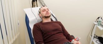
Before the procedure, the patient sits in a comfortable chair. Then sensors are installed in the head area (their attachment points are wiped with alcohol to degrease). The sensors are mounted on the head using special helmets consisting of harnesses. Sometimes sensors are installed in the nasal cavity. Each sensor is connected to a numbered socket on the encephalograph. This makes it possible to record the bioelectrical activity of individual areas of the brain.
As prescribed by the doctor, additional loads that provoke seizures are added (bright light, frequent blinking, deep rapid breathing, etc.). If day or night monitoring is carried out, the patient is constantly monitored by a medical professional.
The video demonstrates the procedure for performing electroencephalography:
Purpose of the test
EEG procedure
EEG is most often used to assess the presence or risk of developing seizures - abnormal electrical discharges in the brain that can manifest as confusion, agitation, uncontrolled movements, hallucinations and even collapse. If you are being evaluated for epilepsy, the neurologist will look for characteristic patterns on the EEG called epileptiform patterns, which may appear as spikes, sharp waves, or spikes and waves. In this case, you can find out the exact location of the seizure.
For example, if you have generalized seizures, which means they affect both hemispheres of your brain, you will likely have spike-and-wave complexes throughout your brain. If you have focal seizures, meaning they only affect one area of your brain and are localized, there will be spikes or sharp waves in that specific location.
Although the main reason an EEG is performed is to diagnose epilepsy, the test has many other uses. These include looking for abnormal brain activity, which may be caused by:
- Traumatic brain injury (TBI);
- Brain tumor;
- Infection (encephalitis);
- Stroke;
- Sleep disturbances caused by seizures. For this purpose, an EEG may be performed in conjunction with a standard sleep study called polysomnography, which tracks sleep stages and cycles to identify disturbances in sleep patterns and the causes of them.
Also, according to the Decree of the Government of the Russian Federation of April 28, 1993 No. 377 on the implementation of the Law of the Russian Federation “On psychiatric care and guarantees of the rights of citizens in its provision”1 when undergoing a psychiatric (narcological) examination in connection with preliminary and / or periodic medical examinations, for example, when applying for a certain job, an EEG is a mandatory general functional study.
Types of EEG
There are several types of electroencephalography, as well as different variations. The use of one type or another will depend on the specific situation. However, EEG can basically be divided into three types:
- Routine EEG;
- EEG monitoring;
- Outpatient EEG.
In what cases does an EEG help make a diagnosis?
The method helps to identify the causes of insomnia and eliminate them with the help of medication.
Allows you to make the correct diagnosis for epilepsy, vascular, inflammatory and metabolic-dystrophic disorders of the central nervous system. This diagnostic method is of particular importance for identifying tumors, as it allows one to determine their location.
In children, the degree of brain damage is revealed, which allows adequate treatment to be prescribed.
Learn more about which medications will help you cope with insomnia
There are no contraindications for EEG
- EEG for epilepsy:
This is the most important test in the diagnosis of epilepsy.
carried out immediately after the appearance of the first attacks.
Based on examination data, a specialist can determine what changes have occurred in the brain, clarify the type of seizures, and, based on this, determine which drugs will be most effective in treatment. The effectiveness of the treatment is also monitored and the issue of stopping treatment is decided.
How often to do an EEG for epilepsy: the number of EEG examinations and their frequency depends on what the attending physician needs to identify. If there are no attacks (for example, if they are successfully treated), then an EEG can be done approximately 1-2 times a year. In the presence of attacks, changes in treatment or dosage of drugs, the frequency of EEG increases.