Subcortical structures of the brain
The brain is a particularly specialized part of the central nervous system.
In humans, its mass averages 1375 g. It is here that huge clusters of interneurons store the experience of actions gained throughout life. The brain is represented by 5 sections. Three of them - the medulla oblongata, the pons and the midbrain - are collectively called the brainstem (or stem part) of the brain. The brainstem is fundamentally different from the other two parts of the brain, as it is equipped with cranial nerves, through which the brainstem directly controls the head region and part of the neck. The other two sections - the diencephalon and the telencephalon - do not directly influence the structures of the human body; they regulate their functions and affect the centers of the trunk and spinal cord. The latter, equipped with cranial and spinal nerves, transmit through them generalized commands to the performers - muscles and glands.
In addition to clusters of neurons that are directly related to nerves, the brain stem also contains other nerve centers that are similar in nature to the centers of the diencephalon and telencephalon (reticular formation, red nuclei, substantia nigra), which significantly distinguishes it from the spinal cord. A special place is occupied by the cerebellum, which performs the most important tasks of maintaining the degree of muscle tension (tone), coordinating their work in performing movements, and maintaining balance. The cerebellum has a huge number of interneurons, which it accommodates solely because they are located not only in its thickness, but also as part of the extreme folded surface, making up the cerebellar cortex. This phenomenon manifests itself, in addition to it, only in the cerebral cortex.
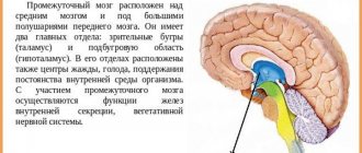
In front and above the trunk is the diencephalon and the main components in the form of the visual thalamus (thalamus) - an important intermediate center along the sensitive paths to the telencephalon, the subthalamic region (hypothalamus) - it contains a lot of centers important for the regulation of metabolism in the body and its behavior and is closely related to the functioning of the pituitary gland, with which it is connected by a stalk. Behind the visual thalamus is the pineal gland, an endocrine gland involved in the regulation of pigment metabolism in the skin and puberty.
The largest portion of the brain's mass is the telencephalon, usually described as two cerebral hemispheres connected by the corpus callosum. Its surface is sharply folded due to the mass of grooves (lateral, central, etc.) separating the gyri. Many of them are permanent in nature, which makes it possible to distinguish between areas of the cortex.
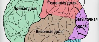
The hemispheres are divided into 4 main lobes. The frontal lobe is largely associated with determining the personal qualities of a person, and all the motor centers of the trunk and spinal cord are subordinate to its posterior part. Therefore, when it is damaged, muscle paralysis appears. In the parietal lobe, sensations of heat, cold, touch, and the position of body parts in space are mainly formed. The occipital lobe contains the visual centers, the temporal lobe contains the auditory and olfactory centers.
In the depths of the hemispheres, neurons are concentrated in the form of nodes (subcortex). They, together with other centers and the cerebellum, ensure coordination of muscle work when performing motor programs of varying complexity. The brain is surrounded by a complex system of membranes. The soft shell is fused with its substance and contains vessels that supply the brain, the branches of which penetrate into the thickness of the brain. Between it and the more superficial arachnoid membrane, very thin and avascular, there is a subarachnoid space with cerebrospinal fluid. Most of it is produced in the cavities of the brain (ventricles) and through the openings between the medulla oblongata and the cerebellum it enters this space, forming a protective hydraulic cushion around the brain. The outermost hard shell connects to the bones of the skull.
SUBCORTAL STRUCTURES OF THE BRAIN - parts of the brain located between the cerebral cortex and the medulla oblongata. They have an activating effect on the cortex, participate in the formation of all behavioral reactions in humans and animals, in maintaining muscle tone, etc.
Subcortical formations include structures located between the cerebral cortex and the medulla oblongata: thalamus, hypothalamus, basal ganglia, a complex of formations united in the limbic system of the brain, as well as the reticular formation of the brain stem and thalamus. Any afferent excitation that arises from stimulation of receptors in the periphery is transformed at the level of the brain stem into two streams of excitations. One flow along specific paths reaches the projection area of the cortex specific for a given stimulation; the other, through collaterals, enters the reticular formation, from where, in the form of an ascending flow of excitations, it is directed to the cerebral cortex, activating it. The reticular formation has close functional and anatomical connections with the hypothalamus, thalamus, medulla oblongata, limbic system, cerebellum, therefore many types of body activities (breathing, food and pain reactions, motor acts, etc.) are carried out with its mandatory participation.
Afferent flows of excitations from peripheral receptors on the way to the cerebral cortex have numerous synaptic switches in the thalamus. From the lateral group of thalamic nuclei (specific nuclei), excitations are directed along two paths: to the subcortical ganglia and to specific projection zones of the cerebral cortex. The medial group of thalamic nuclei (nonspecific nuclei) serves as a switching point for ascending activating influences that are directed from the stem reticular formation to the cerebral cortex. Close functional relationships between the specific and nonspecific nuclei of the thalamus provide the primary analysis and synthesis of all afferent excitations entering the brain. In animals at low stages of phylogenetic development, the thalamus and limbic formations play the role of the highest center for the integration of behavior, providing all the necessary motor acts of the animal aimed at preserving its life. In higher animals and humans, the highest center of integration is the cerebral cortex.
The limbic system includes a complex of brain structures that plays a leading role in the formation of the basic innate reactions of humans and animals: food, sexual and defensive. It includes the lumbar gyrus, hippocampus, piriformis gyrus, olfactory tubercle, amygdala complex and septal area. The central place among the formations of the limbic system is given to the hippocampus. The hippocampal circle is anatomically established (hippocampus - fornix - mammillary bodies - anterior nuclei of the thalamus - cingulate gyrus - hippocampus), which, together with the hypothalamus, plays a leading role in the formation of emotions. The regulatory influences of the limbic system widely extend to autonomic functions (maintaining a constant internal environment of the body, regulation of blood pressure, respiration, vascular tone, gastrointestinal motility, sexual functions).
The cerebral cortex has constant descending (inhibitory and facilitating) influences on subcortical structures. There are various forms of cyclic interaction between the cortex and subcortical structures, expressed in the circulation of excitations between them. The most pronounced closed cyclic connection exists between the thalamus and the somatosensory area of the cerebral cortex, which functionally constitute a single whole. Cortical-subcortical circulation of excitations can serve as the basis for the formation of conditioned reflex activity of the body.
The thalamus (thalamus, visual thalamus) is a structure in which the processing and integration of almost all signals going to the cerebral cortex from the spinal cord, midbrain, cerebellum, and basal ganglia of the brain occurs.
Morphofunctional organization. In the nuclei of the thalamus, information coming from extero-, proprioceptors and interoceptors is switched and thalamocortical pathways begin.
Considering that the geniculate bodies of the thalamus are the subcortical centers of vision and hearing, and the frenulum node and the anterior visual nucleus are involved in the analysis of olfactory signals, it can be argued that the visual thalamus as a whole is a subcortical “station” for all types of sensitivity. Here, irritations from the external and internal environment are integrated and then enter the cerebral cortex.
The visual thalamus is the center of organization and implementation of instincts, drives, and emotions. The ability to receive information about the state of many body systems allows the thalamus to participate in the regulation and determination of the functional state of the body as a whole (this is confirmed by the presence of about 120 multifunctional nuclei in the thalamus). The nuclei form unique complexes that can be divided based on their projection into the cortex into 3 groups: the anterior one projects the axons of its neurons into the cingulate gyrus of the cerebral cortex; medial - in the frontal lobe of the cortex; lateral - into the parietal, temporal, occipital lobes of the cortex. The function of the nuclei is also determined from the projections. This division is not absolute, since one part of the fibers from the thalamic nuclei goes to strictly limited cortical formations, the other to different areas of the cerebral cortex.
The nuclei of the thalamus are functionally divided into specific, nonspecific and associative according to the nature of the pathways entering and exiting them.
Specific nuclei include the anterior ventral, medial, ventrolateral, postlateral, postmedial, lateral and medial geniculate bodies. The latter belong to the subcortical centers of vision and hearing, respectively.
The main functional unit of specific thalamic nuclei are “relay” neurons, which have few dendrites and a long axon; their function is to switch information going to the cerebral cortex from skin, muscle and other receptors.
From specific nuclei, information about the nature of sensory stimuli comes to strictly defined areas of the III-IV layers of the cerebral cortex (somatotopic localization). Dysfunction of specific nuclei leads to loss of specific types of sensitivity, since the nuclei of the thalamus, like the cerebral cortex, have a somatotopic localization. Individual neurons of specific thalamic nuclei are excited by receptors only of their own type. Signals from receptors in the skin, eyes, ear, and muscular system go to specific nuclei of the thalamus. Signals from the interoceptors of the projection zones of the vagus and celiac nerves and the hypothalamus also converge here.
The lateral geniculate body has direct efferent connections with the occipital lobe of the cerebral cortex and afferent connections with the retina and the anterior colliculus. Neurons of the lateral geniculate bodies react differently to color stimulation, turning on and off the light, i.e., they can perform a detector function.
The medial geniculate body (MCC) receives afferent impulses from the lateral lemniscus and from the inferior colliculi. Efferent pathways from the medial geniculate bodies go to the temporal zone of the cerebral cortex, reaching there the primary auditory area of the cortex. MCT has a clear tonotopic pattern. Consequently, already at the level of the thalamus, the spatial distribution of sensitivity of all sensory systems of the body is ensured, including sensory messages from interoreceptors of blood vessels, abdominal organs, and thoracic cavities.
The association nuclei of the thalamus are represented by the anterior mediodorsal, lateral dorsal nuclei and the cushion. The anterior nucleus is connected with the limbic cortex (cingulate gyrus), the mediodorsal - with the frontal lobe of the cortex, the lateral dorsal - with the parietal, the pillow - with the associative zones of the parietal and temporal lobes of the cerebral cortex.
The main cellular structures of these nuclei are multipolar, bipolar triprocess neurons, i.e. neurons capable of performing polysensory functions. A number of neurons change activity only with simultaneous complex stimulation. On polysensory neurons, excitations of different modalities converge, an integrated signal is formed, which is then transmitted to the associative cortex of the brain. Pillow neurons are connected mainly with the associative zones of the parietal and temporal lobes of the cerebral cortex, neurons of the lateral nucleus - with the parietal nucleus, neurons of the medial nucleus - with the frontal lobe of the cerebral cortex.
Nonspecific nuclei of the thalamus are represented by the median center, paracentral nucleus, central medial and lateral, submedial, ventral anterior, parafascicular complexes, reticular nucleus, periventricular and central gray mass. The neurons of these nuclei form their connections according to the reticular type. Their axons rise into the cerebral cortex and contact all its layers, forming not local, but diffuse connections. Nonspecific nuclei receive connections from the RF of the brainstem, hypothalamus, limbic system, basal ganglia, and specific nuclei of the thalamus.
Excitation of nonspecific nuclei causes the generation of specific spindle-shaped electrical activity in the cortex, indicating the development of a sleepy state. Dysfunction of nonspecific nuclei makes it difficult for spindle-shaped activity to appear, i.e., the development of a sleepy state.
Projection fibers are those that connect the cerebral hemispheres with the underlying parts of the brain - the brainstem and spinal cord. The projection fibers contain pathways carrying afferent (sensitive) and efferent (motor) information.
So, the main sections, grooves and convolutions of the brain are presented in Fig. 5.6.
Rice. 5. Brain, left hemisphere (side view):
1 – precentral gyrus; 2 – precentral sulcus; 3 – superior frontal gyrus; 4 – central groove; 5 – middle frontal gyrus;
b – inferior frontal gyrus; 7 – ascending branch of the lateral sulcus;
8 – horizontal branch of the lateral sulcus; 9 – posterior branch of the lateral sulcus; 10 – superior temporal gyrus; 11 – middle temporal gyrus;
12 – inferior temporal gyrus; 13 – parietal lobule; 14 – post-central sulcus; 15 – postcentral gyrus; 16 – supramarginal gyrus;
17 – angular gyrus; 18 – occipital lobe; 19 – cerebellum; 20 – horizontal fissure of the cerebellum; 21 – medulla oblongata
Anatomy of the human subcortex - information:
In addition to the gray cortex on the surface of the hemisphere, there are also accumulations of gray matter in its thickness, called the basal ganglia and constituting what is called the subcortex . Unlike the cortex, which has the structure of screen centers, the subcortical nuclei have the structure of nuclear centers.
There are three clusters of subcortical nuclei : corpus striatum, claustrum and corpus amygdaloideum.
1. Coprus striatum, striatum , consists of two parts that are not completely separated from each other - the nucleus caudatus and the nucleus lentiformis.
A. Nucleus caudatus, caudate nucleus , lies above and medial to the nucleus lentiformis, separated from the latter by a layer of white matter called the internal capsule, capsula interna. The thickened anterior part of the caudate nucleus, its head, caput nuclei caudati, forms the lateral wall of the anterior horn of the lateral ventricle, while the posterior thinned section of the caudate nucleus, corpus et cauda nuclei caudati, stretches back along the bottom of the central part of the lateral ventricle; The cauda wraps around the upper wall of the lower horn. On the medial side, the nucleus caudatus is adjacent to the thalamus, separated from it by a strip of white matter, stria terminalis. In front and below, the head of the caudate nucleus reaches the substantia perforata anterior, where it connects with the nucleus lentiformis (with the part of the latter called putamen). In addition to this wide connection of both nuclei on the ventral side, there are also thin strips of gray matter, interspersed with white bundles of the internal capsule. They gave rise to the name “striatum”, corpus striatum.
B. Nucleus lentiformis, lentiform nucleus , lies lateral to the nucleus caudatus and thalamus, separated from them by the capsula interna. On a horizontal section of the hemisphere, the medial surface of the lenticular nucleus, facing the internal capsule, has the shape of an angle with the apex directed towards the middle; the anterior side of the angle is parallel to the caudate nucleus, and the posterior side is parallel to the thalamus. The lateral surface is slightly convex and faces the lateral side of the hemisphere in the region of the insula. Anteriorly and ventrally, as already indicated, the lenticular nucleus merges with the head of the nucleus caudatus. In a frontal section, the lentiform nucleus has the shape of a wedge, the apex of which faces the medial side and the base faces the lateral side.
The lenticular nucleus is divided into three segments by two parallel white layers, laminae medullares, of which the lateral, dark gray, is called the putamen, and the two medial, lighter ones, are collectively called the globus pallidus , globus pallidus. Differing already in its macroscopic appearance, the globus pallidus also has a histological structure that is different from other parts of the striatum.
Phylogenetically, globus pallidus is an older formation (paleostriatum) than putamen and nucleus caudatus (neostriatum). In view of all these features, globus pallidus is currently classified into a special morphological unit called pallidum, while the designation striatum is reserved only for putamen and nucleus caudatus. As a result, the term “lenticular nucleus” loses its previous meaning and can only be used in a purely topographical sense, and instead of the previous name corpus striatum, the caudate and lenticular nucleus are called the striopallidal system.
The striopallidal system is the main part of the extrapyramidal system, and in addition, it is the highest regulatory center of autonomic functions in relation to heat regulation and carbohydrate metabolism, dominating over similar autonomic centers in the hypothalamus.
2. Claustrum, fence , is a thin plate of gray matter located in the region of the insula, between it and the putamen. It is separated from the latter by a layer of white matter, capsula externa, and from the insular cortex by a layer called capsula extrema.
3. Corpus amygdaloideum, the amygdala, is located under the putamen at the anterior end of the temporal lobe. Corpus amygdaloideum apparently belongs to the subcortical olfactory centers and to the limbic system. It ends in a bundle of fibers coming from the olfactory lobe and substantia perforata anterior, noted in the description of the thalamus under the name stria terminalis.
The limbic system is a complex of formations of the telencephalon, diencephalon and mesencephalon, involved in the regulation of various autonomic functions, maintaining the constancy of the internal environment of the body (homeostasis) and in the formation of emotionally charged behavioral reactions. Therefore, some authors designate the limbic system as the “visceral brain.” The main part of it consists of the structures of the cerebral cortex, located mainly on the medial surface of its hemispheres, and subcortical formations closely associated with them, namely: amygdaloid region, stria terminalis, hypothalamus, hippocampus, fornix, septal region, mammillary bodies, mastoid-thalamic fascicle , thalamus, cingulate gyrus. On the medial surface of the cerebral hemispheres, the limbic system is represented by the cingulate and parahippocampal gyri.
The structure of the subcortical region of the brain. Diencephalon
Striopallidar system
In the thickness of the white matter of the cerebral hemispheres there are clusters of gray matter called the subcortical nuclei (basal ganglia). These include the caudate nucleus, the lenticular nucleus, the cervical nucleus, and the amygdala (Fig. 6). The lenticular nucleus, located outside the caudate nucleus, is divided into three parts. It contains a shell and two pale balls.
Rice. 6. Subcortical nuclei:
1 – caudate nucleus; 2 – lentiform nucleus; 3 – visual thalamus.
A – horizontal section: a – fence; b – shell; c and d – globus pallidus;
B-frontal section: a – globus pallidus; b – shell
Functionally, the caudate nucleus and putamen are united into the striatum (striatum), and the globus pallidus, together with the substantia nigra and red nuclei located in the cerebral peduncles, form the corpus pallidus (pallidum).
Together they represent a very functionally important formation - the striopallidal system. In terms of morphological features and phylogenetic origin (their appearance at a certain stage of evolutionary development), the pallidum is a more ancient formation than the striatum.
The striopallidal system is an important part of the motor system. It is part of the so-called pyramid system. In the motor zone of the cerebral cortex, the motor - pyramidal - path begins, along which the order to perform this or that movement follows.
The extrapyramidal system, an important part of which is the striopallidum, being included in the motor pyramidal system, takes an auxiliary part in ensuring voluntary movements.
At a time when the cerebral cortex was not yet developed, the striopallidal system was the main motor center that determined the behavior of the animal. Due to the striopallidal motor apparatus, diffuse, mass movements of the body were carried out, ensuring movement, swimming, etc.
With the development of the cerebral cortex, the striopallidal system passed into a subordinate state. The main motor center became the cerebral cortex.
The striopallidal system began to provide the background, readiness for movement; Against this background, fast, precise, strictly differentiated movements are carried out, controlled by the cerebral cortex.
To perform a movement, it is necessary that some muscles contract and others relax, in other words, an accurate and coordinated redistribution of muscle tone is needed.
This redistribution of muscle tone is precisely carried out by the striopallidal system. This system ensures the most economical consumption of muscle energy during movement. Improving movements in the process of learning to perform them (for example, practicing to the limit the perfect running of a musician’s fingers, the swing of a mower’s hand, the precise movements of a car driver) leads to gradual economization and automation.
This possibility is provided by the striopallidar system.
It was noted above that, phylogenetically, the striatum is a younger formation than the corpus pallidus. An example of pallidal organisms is fish.
They move in the water using powerful throwing movements of the body, without “concerning” about saving muscle energy. These movements are relatively precise and powerful. However, they are energetically wasteful. In birds, the striatum is already well defined, which helps them more carefully regulate the quality, accuracy and quantity of movements. Thus, the pallidum inhibits and regulates the activity of the pallidal system (since phylogenetically younger formations control and inhibit more ancient ones).
Motor acts of a newborn are pallidal in nature: they are uncoordinated, jerky and often unnecessary. With age, as the striatum matures, the child’s movements become more economical, sparing, and automated.
The striopallidal system has connections with the cerebral cortex, the cortical motor system (pyramidal) and muscles, formations of the extrapyramidal system, the spinal cord and the visual thalamus.
The other basal ganglia (the cervical nucleus and the amygdala) are located lateral to the lenticular nucleus. The amygdala is part of another functional system - the limbic-reticular complex.
Optic thalamus
The optic thalamus and the subtubercular region (hypothalamus) develop from the intermediate vesicle, and the third ventricle develops from the cavity of the intermediate vesicle.
optic thalamus,
or
thalamus,
located on the sides of the third ventricle and consists of a powerful accumulation of gray matter.
The optic thalamus is divided into the visual thalamus itself, the suprathalamic area (the suprathalamic area, or epilamus), and the suprathalamic area (the metathalamic area, or metathalus). The bulk of the gray tubercle is the thalamus (see Fig. 7).
Rice. 7. Topography of the thalamus
1 – thalamus; 2 – body of the caudate nucleus; 3 – body of the lateral ventricle; 4 – corpus callosum; 5 – medulla oblongata.
In it there is a protrusion of the cushion, behind which there are two elevations - the external and internal geniculate bodies (they enter the subcutaneous region).
There are several nuclear groups in the thalamus.
The epithalamus region, or epithalamus, consists of the pineal gland and the posterior commissure of the brain.
The subcutaneous region, or metathalamus, includes the geniculate bodies, which are the elevation of the thalamus. They lie outward and inferior to the thalamic cushion.
The subcutaneous region, or hypothalamus, lies downward from the thalamus, has a number of nuclei lying in the walls of the third ventricle.
The visual thalamus is an important stage on the path to all types of sensitivity. Sensitive pathways approach it and are concentrated in it - touch, pain, temperature, visual tracts, auditory pathways, olfactory pathways and fibers from the extrapyramidal system. The next stage of transmission of sensitive impulses begins from the neurons of the visual thalamus - to the cerebral cortex.
At a certain stage in the evolution of the nervous system, the thalamus was a center of sensitivity, just as the striopallidal system was a mechanism of movement. As the cerebral cortex appeared and developed, the main role in the function of the sensitive sphere passed to the cerebral cortex, and the visual thalamus remained only a transfer station of sensitive impulses from the periphery to the cerebral cortex.
Main nerve centers, their characteristics
Clusters of brain neurons in the hypothalamus are responsible for maintaining a constant internal environment - homeostasis. To do this, they analyze the composition of the blood flowing in the arteries and issue commands to the endocrine glands through the pituitary gland. The hormones released change the intensity of metabolism, shape human behavior, and the hypothalamus directly controls the autonomic nervous system.
Highest vegetative regulator
The hypothalamic centers are considered the main coordinator of the body's autonomic reactions. Thanks to their work, a person can live in a comatose state without activity of the cerebral cortex. This level of life is called vegetative, as well as the part of the nervous system that supports independent, independent life.
The relationship between irritation of the posterior region of the hypothalamus and the reactions of the sympathetic nervous system was discovered a long time ago. Its activation is accompanied by:
- increased breathing;
- acceleration and increased heart rate;
- increased blood pressure;
- dilated pupils;
- inhibition of intestinal contractions.
It has also been established that the anterior part is responsible for the activity of the parasympathetic department. Her excitement causes exactly the opposite effects.
Further study revealed that there is no clear division of functions between them, and the hypothalamus uses sympathetic and parasympathetic reactions depending on the body’s need for them. That is, figuratively speaking, he chooses which thread to pull in order to get the desired result.
Lateral hunger center and satiety nucleus
The hypothalamus controls eating behavior. Its nuclei can “measure” the concentration of blood sugar, insulin levels, and the degree of stomach filling. If the lateral (side) zones of hunger are destroyed, this leads to a complete refusal of food, and when exposed to a weak electric current, they force a person to eat after a heavy meal.
The medial (middle) centers are responsible for satiety; if they are damaged (inflammation, stroke), then the patient ceases to experience satiety, no matter how much food is consumed. The centers of hunger and satiety have an inhibitory effect on each other.
Thirst and drinking behavior centers of the hypothalamus
When exposed to nuclei located near the cells that produce vasopressin, a feeling of thirst occurs. If there is damage in this area, then the person completely stops experiencing it, regardless of the blood concentration. It has been established that the sensitivity of neurons weakens with age, and older people rarely feel very thirsty.
Normally, the neurons of the center evaluate the salt content in the bloodstream, the level of vasopressin (regulates water-salt metabolism), and also receive impulses from osmoreceptors. This is the name given to proteins that respond to the osmotic pressure of the blood, that is, its density.
Thermoregulation
The hypothalamus ensures a balance between the processes of heat production and heat transfer. For this purpose, it has two corresponding centers. When overheated, the anterior zone is activated, it stimulates:
- dilation of skin blood vessels;
- sweating;
- frequent breathing.
Information about the external environment comes to the center from thermoreceptors, which are located on the very surface of the skin, as well as in deeper layers. Internal temperature is assessed by such “detectors” in the vessel wall and located directly in the brain tissue.
They can also react to bacterial toxins and substances that cause inflammation (cytokines). When these nuclei are destroyed, the body will overheat. The heat production zone belongs to the posterior region of the hypothalamus.
When the temperature drops:
- the rate of energy generation increases;
- muscle tone increases;
- skin blood vessels constrict.
If the cells are damaged, then the person loses the ability to tolerate body cooling.
Sleep and awakening centers
With a tumor, inflammation of the brain tissue and vascular diseases in the hypothalamic region, the duration of sleep, the ratio of short and long phases are disrupted, and it is difficult to fall asleep. Some cell damage can cause lethargy.
In the front part of the hypothalamus there is a sleep center, and in the back there are cells responsible for wakefulness and awakening. The hypothalamus acts as the body's internal clock. To do this, it interacts with the pineal gland, which secretes the sleep hormone melatonin.
Sexual behavior centers of the hypothalamus
In men, in the middle part of the hypothalamus, cells synthesize gonadoliberins in a constant rhythm. These hormones stimulate sexual activity through the pituitary gland. In women, in addition, there is a cyclic center in the anterior zone, which is responsible for the rhythm of menstruation.
A pleasure center is found in the posterior hypothalamic region. Its activity causes a feeling of joy and pleasure during sexual intercourse. Impulses to these centers come from the gonads (testes and ovaries), as well as from erogenous zones when they are stimulated.