We use the concepts “nerves” and “nervous system” quite often and for a variety of reasons. Someone has ruined all our nerves, and someone is constantly getting on our nerves, good for those who have a stable nervous system. And we often remember the words from the song: “Talking on this topic spoils the nervous system.” How many people understand the essence of these concepts? As a small survey among my acquaintances and first-year students showed, the proposal to explain what the “nervous system” is has puzzled many. It is unlikely that the answer: “The nervous system is in the head” can be considered satisfactory.
What is the nervous system
The simplest living organisms, such as amoebas, do not have any nervous system. They do not have specialized organs - the processes of nutrition, reproduction, contact with the environment, etc. are carried out by the same cell.
But evolution is the development of organisms along the path of their complexity. And the more complex a creature becomes, the more organs it appears that specialize in performing different functions. This process must be somehow controlled, subordinated to a common goal for the entire organism. It is equally important to preserve the experience of interacting with the environment, so that you can use it later when necessary. That is, a single coordination center is required, which becomes the nervous system, which is formed in the process of evolution in highly developed creatures.
The nervous system (NS) is a complex of various structures that ensure the functioning and interaction of all parts of the body. And the more complex this organism and its contacts with the outside world, the more complex the NS. It is most complex in humans, as the most highly organized being on planet Earth. The main elements of our nervous system are the central and peripheral nervous system, connected to each other by numerous nerve fibers. The operation of this entire complex structure is ensured by specialized nerve cells - neurons. We’ll talk about all this in more detail now.
central nervous system
The CNS is like a master processor that controls all body functions. It receives and processes signals coming both from the outside world and from organs, muscles, ligaments, receptors, etc. The processed signals become the basis for feedback - reflex reactions that occur at the command of the “main processor” and conscious actions and actions . It is the central nervous system that sends commands to the heart, lungs, gastrointestinal tract, speech apparatus, motor nerves and many, many other systems.
The central nervous system consists of two main sections - the brain and the spinal cord.
Structure and functions of the brain
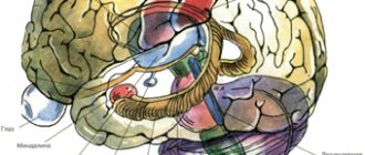
The brain is a very complex mechanism consisting of several sections. This is the result of a long evolution, therefore its departments were formed at different times and are responsible for performing tasks of different levels of complexity. The youngest layer of the brain is the top layer, consisting primarily of gray matter - neurons. This is the cerebral cortex.
Hemispheres of the brain and their work
If you look at the brain from above, it resembles a walnut kernel. Firstly, because it is divided into two parts, as if tied with a thread. Secondly, the surface of the brain is as wrinkled as a walnut. This is due to the fact that the new cortex of the cerebral hemispheres (neocortex) is much larger than the hemispheres themselves, and if it is stretched, the area will be about 2 m2. Although the thickness of the neocortex does not exceed 4 mm, there are at least 14 billion neurons in the cerebral cortex. Imagine how many nerve connections can arise between them.
Along with neurons (gray matter), many other cells, including glial cells, are responsible for brain function. They protect neurons, provide them with nutrition and remove metabolic products.
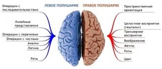
The hemispheres of the brain are specialized. The left is responsible for the work of the organs of the right half of the body, and the right, accordingly, supervises the activity of what is on our left. In addition, the center of speech is located in the left hemisphere; it is responsible for operations with signs (counting, writing) and for rational, logical thinking. And the right hemisphere contains the center of imaginative thinking; it controls a person’s abilities for imagination and creativity.
The cerebral cortex has a complex structure. The following zones are distinguished:
- frontal lobes;
- parietal lobes;
- temporal lobes;
- occipital lobes.
Different zones are associated with different systems and types of mental activity and perform their functions. Thus, the frontal lobes play a major role in thinking. This is the most “human” part of the brain also because the frontal lobes have an inhibitory effect on behavior, making it more conscious and controlled. They are the ones who suffer primarily from alcohol intoxication. Well, and also the cerebellum, which is responsible for the balance of the body.
Thus, the cerebral cortex is the source of what is called higher mental functions, that is, thinking, operations with signs, imagination and speech activity.
Under the upper, relatively thin layer of the neocortex, covering the cerebral hemispheres, there are two more layers:
- The limbic system, which includes part of the ancient cortex, plays an important role in the primary processing of signals entering the brain. Even in the limbic system, more precisely in the hypothalamus, there is a center for pleasure and control of sexual behavior.
- The R-complex or “reptile brain” is the most ancient part of our brain, formed more than 100 million years ago. This area controls primitive reflexes and ancient instincts, such as the instinct to reproduce, protect one's territory, and the desire to dominate.
The cerebral hemispheres are the most famous part of the brain, but it has a much more complex structure.
Brain structure
The hemispheres, covered with a layer of cortex and connected by the corpus callosum, are called the cerebrum or telencephalon. In addition to this, there are other departments:
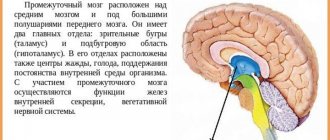
- The diencephalon, which includes the thalamus, epithalamus and hypothalamus, is a filter that allows only necessary information to pass through.
- The midbrain regulates perception processes, primarily auditory and visual reflexes. It is here that images of the objects we see appear.
- The medulla oblongata is one of the ancient sections responsible for breathing and heart function. Damage to it is fatal.
- The hindbrain, which includes the cerebellum and the pons, connects the brain to the spinal cord and carries signals to and from the peripheral nervous system. The cerebellum is responsible for the coordination of movements.
Such a complex structure of the brain is the result of long evolution. This is the control panel for our entire body, so nature has tried to reliably protect the brain from possible damage by hiding it behind a durable skull.
Spinal cord
The second section of the central nervous system is structured more simply, and its origin is more ancient. However, the role of the spinal cord in controlling our body should not be underestimated.
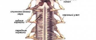
The spinal cord is like a cord about 10 mm wide and about 45 cm long. It is also well protected and runs in a special canal of the spine. The central part of the spinal cord is formed by gray matter, that is, the same neurons that ensure the functioning of the brain. The membrane of the spinal cord consists of white matter of conducting nerve fibers - axons.
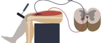
The spinal cord performs the function of delivering signals from the brain to organs and muscles. Also, this section of the central nervous system provides a number of simple, but very important reflexes, primarily motor ones. For example, when we withdraw our hand from a hot frying pan, the spinal cord gives the command. And thanks to this brain, we can walk, dance, ride a bike, that is, learn motor skills.
CONDUCTING PATHWAYS OF THE BRAIN AND SPINAL CORD
The doctrine of the nervous system (neurology)
Impulses that arise when exposed to receptors are transmitted through neurons that contact each other, forming chains. Along these circuits, nerve impulses travel only in a certain direction. Along some chains of neurons, the impulse propagates centripetally - from the place of its origin in the skin, mucous membranes, organs of movement, blood vessels to the spinal cord or brain. Through other chains of neurons, the impulse is carried centrifugally from the brain to the periphery to the working organ, muscle, gland. A chain of nerve cells, including afferent (sensitive) and effector (motor or secretory) neurons, along which a nerve impulse moves from its place of origin (from the receptor) to the working organ (effector), is called a reflex arc (Fig. 176).
The processes of neurons, directed from the spinal cord to various structures of the brain, and from them in the opposite direction, to the spinal cord, form bundles that connect the nerve centers. These bundles make up the pathways. Pathways are a collection of nerve fibers passing through certain areas of the white matter of the brain and spinal cord, united by a common structure and function.
In the spinal cord and brain there are three groups of nerve fibers: associative, commissural and projection.
Associative pathways (short and long) connect nerve centers located in one half of the brain. Short ones connect nearby areas of gray matter and are located, as a rule, within one lobe of the brain. Among them, arcuate fibers of the cerebrum are distinguished, which connect the gray matter of neighboring gyri and do not extend beyond the cortex - intracortical and extracortical, passing in the white matter of the hemisphere. Long (interlobar) associative pathways connect areas of gray matter located in different lobes of the brain. These include the superior longitudinal fasciculus, connecting the cortex of the frontal lobe with the parietal and occipital lobes, the lower longitudinal fasciculus, connecting the gray matter of the temporal lobe with the occipital lobe, the uncinate fasciculus, connecting the cortex in the frontal pole with the anterior part of the temporal lobe (Fig. 177, 178) .
In the spinal cord, associative pathways connect neurons located in different segments and form their own bundles of the spinal cord (intersegmental bundles), which are located near the gray matter. Short bundles spread across 2–3 segments, while long bundles connect widely separated segments of the spinal cord.
Commissural (commissural) pathways connect the same centers (gray matter) of the right and left hemispheres of the cerebrum, forming the corpus callosum, commissure of the fornix and anterior commissure. The corpus callosum connects the cerebral cortex of the right and left hemispheres, in which the fibers of the corpus callosum diverge in a fan-shaped manner, forming the radiance of the corpus callosum. The anterior bundles of fibers, passing in the knee and beak of the corpus callosum, connect the cortex of the anterior parts of the frontal lobes, forming the “frontal forceps”. The cortex of the occipital and posterior parts of the parietal lobes of the hemispheres is connected by bundles of fibers passing in the splenium of the corpus callosum. They form the so-called “nuchal forceps”. Fibers passing in the central sections of the corpus callosum connect the cortex of the central gyri, parietal and temporal lobes of the cerebral hemispheres.
In the anterior commissure there are fibers that connect the areas of the cortex of the temporal lobes of both hemispheres, belonging to the olfactory brain. The commissure fibers of the fornix connect the gray matter areas of the hippocampus and temporal lobes of both hemispheres.
Projection pathways include ascending and descending systems of nerve fibers. The ascending tracts connect the spinal cord with the brain, as well as the nuclei of the brain stem with the basal ganglia and the cerebral cortex. Descending pathways (projection) go from the cerebral cortex, subcortical centers and from the nuclei of the brain stem to the lower nuclei of the brain and to the spinal cord.
Ascending projection pathways, afferent, or sensory, conduct nerve impulses that arise in different organs under the influence of various factors. There are three groups of ascending pathways: exteroceptive, proprioceptive and interoceptive pathways.
Exteroceptive pathways carry impulses that arise when the body is exposed to environmental factors. These are impulses from the skin (pain, temperature sensitivity, touch and pressure) (Table 19), from the senses (vision, hearing, taste and smell). Proprioceptive pathways conduct impulses from the organs of the musculoskeletal system (muscles, tendons, joints, etc.) (Table 20). Interoceptive pathways conduct impulses from internal organs, the heart, blood vessels, from the receptors located in them (mechano-, baro-, chemo-, which perceive information about the intensity of metabolic processes, the chemical composition of blood and lymph, pressure in blood vessels, etc.) . Impulses enter the cerebral cortex along direct ascending sensory pathways and from subcortical centers.
Exteroceptive pathways include the pathways of pain and temperature sensitivity, touch and pressure, as well as the pathways of the sensory organs.
The pathway for pain and temperature sensitivity (lateral spinothalamic tract) consists of three neurons (Fig. 179). The receptors of the first (sensitive) neurons are located in the skin and mucous membranes, and their bodies lie in the spinal ganglion. The central processes within the dorsal roots are directed to the dorsal horn of the spinal cord, where they end at synapses on the cells of the second neuron. All axons of the second neurons pass through the anterior commissure to the opposite side of the spinal cord, enter the lateral cord, and become part of the lateral spinothalamic tract. These fibers ascend into the medulla oblongata, pass through the tegmentum of the pons and the tegmentum of the midbrain and end in the thalamus (III neuron).
Rice. 179. Conducting pathways of pain and temperature sensitivity, touch and pressure (diagram): 1 – lateral spinothalamic tract; 2 – anterior spinothalamic tract; 3 – thalamus; 4 – medial loop; 5 – cross section of the midbrain; 6 – cross section of the bridge; 7 – cross section of the medulla oblongata; 8 – spinal node; 9 – transverse section of the spinal cord. The arrows show the direction of movement of nerve impulses
The axons of thalamic cells are directed to layer IV (internal granular plate) of the cortex of the postcentral gyrus, where the cortical end of general sensitivity is located.
The pathway of touch and pressure (anterior spinothalamic tract) carries impulses from the skin to the cells of the cortex of the postcentral gyrus. The fibers of the first neuron of this pathway pass to the opposite side of the spinal cord through the anterior commissure, enter the anterior cord and, as part of it, follow upward to the same brain structures as the lateral spinothalamic tract. Some of the fibers carrying impulses of touch and pressure do not pass to the opposite side; they go as part of the posterior cord of the spinal cord on their side along with the axons of the proprioceptive sensitivity pathway in the cortical direction.
The proprioceptive pathway conducts impulses from muscles, tendons, joint capsules, ligaments; it carries information about the position of body parts and range of motion.
Rice. 180. Conducting pathway of proprioceptive sensitivity in the cortical direction (diagram): 1 – spinal node; 2 – cross section of the spinal cord; 3 – posterior cord of the spinal cord; 4 – anterior external arcuate fibers; 5 – medial loop; 6 – thalamus; 7 – cross section of the midbrain; 8 – cross section of the bridge; 9 – cross section of the medulla oblongata; 10 – posterior external arcuate fibers. The arrows show the direction of movement of nerve impulses
The pathway of proprioceptive sensitivity in the cortical direction carries impulses of the muscular-articular sense to the cortex of the postcentral gyrus (Fig. 180). The receptors of the first neuron, located in muscles, tendons, joint capsules, and ligaments, perceive signals about the state of the musculoskeletal system as a whole, muscle tone, and the degree of tendon tension. Proprioceptive sensitivity allows a person to assess the position of parts of his body in space, analyze his own complex movements and makes it possible to carry out their targeted correction. The bodies of the first neuron of this pathway also lie in the spinal ganglia; their axons, as part of the dorsal roots, without entering the dorsal horn, are directed to the posterior funiculus of the spinal cord, where they form the thin and wedge-shaped bundles, and follow up into the medulla oblongata to the thin and wedge-shaped nuclei, where they end with synapses on the bodies of second neurons. The axons of the second neurons emerging from these nuclei pass to the opposite side, forming a medial loop, pass through the pontine tegmentum and the midbrain tegmentum and end in the thalamus with synapses on the bodies of the third neurons. The axons of the latter are sent to the cortex of the postcentral gyrus, where they end in synapses with neurons of the IV layer of the cortex. Part of the fibers of the second neurons, upon exiting the thin and cuneate nuclei, is directed to the inferior cerebellar peduncle and ends in the cortex of the vermis on its side. Another part of the fibers of the second neurons passes to the opposite side and also goes through the inferior cerebellar peduncle to the vermis cortex of the opposite side. These fibers carry proprioceptive impulses to the cerebellum to correct subconscious movements of the musculoskeletal system.
There are also proprioceptive anterior and posterior spinocerebellar tracts, which carry information to the cerebellum about the state of the musculoskeletal system and motor centers of the spinal cord.
The cerebral cortex (with the participation of consciousness) controls the motor functions of the body directly through descending pathways. Descending motor pathways conduct impulses from the cerebral cortex and subcortical centers to the underlying parts of the central nervous system, to the nuclei of the brain stem and the motor nuclei of the anterior horns of the spinal cord. These pathways are divided into two groups: pyramidal and extrapyramidal (Table 21). The pyramidal tracts are the major motor pathways. They carry impulses from the cerebral cortex to the skeletal muscles of the head, neck, trunk, and limbs through the corresponding motor nuclei of the brain and spinal cord.
Extrapyramidal tracts carry impulses from the subcortical centers and various parts of the cortex to the motor nuclei of the cranial and spinal nerves, and then to the muscles, as well as to other nerve centers of the brain stem and spinal cord.
The main motor, or pyramidal, path goes from gigantopyramidal neurocytes (Bertz pyramidal cells), located in the cortex of the precentral gyrus (layer V), to the motor nuclei of the cranial nerves and anterior horns of the spinal cord, and from them to the skeletal muscles. Depending on the direction and location of the fibers, the path is divided into three parts: the corticonuclear path, going to the nuclei of the cranial nerves, the lateral and anterior corticospinal (pyramidal) paths, going to the nuclei of the anterior horns of the spinal cord (Fig. 181).
Rice. 181. Pyramid paths (diagram):
1 – precentral gyrus; 2 – thalamus; 3 – cortical-nuclear pathway; 4 – cross section of the midbrain; 5 – cross section of the bridge; 6 – cross section of the medulla oblongata; 7 – intersection of pyramids; 8 – lateral corticospinal tract; 9 – cross section of the spinal cord; 10 – anterior corticospinal tract. The arrows show the direction of movement of nerve impulses
The corticonuclear tract is a bundle of axons of giant pyramidal cells located in the lower third of the precentral gyrus, which passes through the knee of the internal capsule and the base of the cerebral peduncle. The fibers of the cortical-nuclear tract pass to the opposite side to the motor nuclei of the cranial nerves: oculomotor and trochlear - in the midbrain; trigeminal, abducens, facial – in the pons; glossopharyngeal, vagus, accessory, sublingual - in the medulla oblongata, where they end with synapses on their neurons. The axons of the motor neurons of the nuclei of these nerves emerge from the pons as part of the corresponding cranial nerves and are directed to the skeletal muscles of the head and neck.
The lateral and anterior corticospinal (pyramidal) tracts begin from giant pyramidal neurocytes located in layer V of the cortex of the middle and upper third of the precentral gyrus. The axons of these cells are directed to the internal capsule, pass through the anterior part of its posterior leg, then the base of the cerebral peduncle and pons, and pass into the medulla oblongata, forming its pyramids. At the border of the medulla oblongata with the spinal cord, part of the fibers of the corticospinal tract passes to the opposite side, continues in the lateral cord of the spinal cord (lateral corticospinal tract) and gradually ends in the anterior horns of the spinal cord with synapses on the motor cells of the anterior horns. The fibers of the corticospinal tract, which do not pass to the opposite side at the border of the medulla oblongata with the spinal cord, descend down as part of the anterior cord of the spinal cord, forming the anterior corticospinal tract. They pass segment by segment to the opposite side through the white commissure of the spinal cord and end with synapses on the motor (radicular) neurocytes of the anterior horns of the opposite side of the spinal cord. The axons of the anterior horn cells emerge from the spinal cord as part of the anterior roots and, being part of the spinal nerves, innervate skeletal muscles. So, all pyramid paths are crossed.
The extrapyramidal pathways have many connections with the brain stem and with the cerebral cortex, which controls and controls the extrapyramidal system. The common beginning of the extrapyramidal tracts can be considered the cerebral cortex, and the place where they end is the motor nuclei of the brain stem and the anterior horns of the spinal cord. The influence of the cerebral cortex on the human body is carried out through the cerebellum, red nuclei, reticular formation, and through the vestibular nuclei. One of the functions of the red nucleus is to maintain muscle tone, necessary to keep the body in balance. This nucleus, in turn, receives impulses from the cerebral cortex, from the cerebellum (through the cerebellar proprioceptive pathways). From the red nuclei, nerve impulses are sent to the motor nuclei of the anterior horns of the spinal cord (red nucleus-spinal tract) (Fig. 182).
In the coordination of human movements in case of imbalance, the vestibular tract plays an important role, which connects the vestibular nuclei with the anterior horns of the spinal cord, as well as with the cerebellum, and, through the posterior longitudinal fasciculus, with the motor nuclei of the oculomotor, trochlear and abducens cranial nerves. This ensures that the position of the eyeball is maintained during movements of the head and neck. The axons of the first neurons of the vestibulospinal tract descend as part of the anterior funiculus of the spinal cord and end at synapses on the motor cells of the anterior horns of the spinal cord. Neurons of the cells of the reticular formation provide communication between the vestibulospinal tract and the basal nuclei of the telencephalon.
The cerebral cortex also controls the functions of the cerebellum, which is involved in the coordination of movements, through the bridge along the corticopontine-cerebellar pathway. The cell bodies of the first neurons lie in the cortex of the frontal, temporal, parietal and occipital lobes, their axons end with synapses on the cells of the own pons nuclei of their side (second neurons). The axons of these neurons form bundles of transverse pontine fibers, which pass to the opposite side and are directed through the middle cerebellar peduncle to the cerebellar hemisphere of the opposite side. In turn, the cerebellum is connected with the red nuclei and the vestibular apparatus.
Thus, the pathways of the brain and spinal cord establish connections between afferent and efferent (effector) centers and close complex reflex arcs in the human body. Some of them are closed on nuclei that lie in the brain stem and provide functions that have a certain automaticity, without the participation of consciousness, although under the control of the cerebral hemispheres. Other pathways are closed with the participation of the functions of the cerebral cortex (the higher parts of the central nervous system) and provide voluntary actions of organs and organ systems. The pathways functionally unite the body into a single whole and ensure the consistency of its actions.
Peripheral nervous system
Nerve fibers stretch from the brain and spinal cord to all organs, muscles and ligaments, making up an extensive nervous network that entangles our entire body. This complex of nerve fibers and ganglia is called the peripheral nervous system. Its main task is to maintain two-way communication between the central nervous system and all parts of our body.
Clusters of sensitive nerve cells - receptors - receive information from the outside world or from internal organs. This information is then sent along receiving nerve fibers (afferent) for processing to the spinal cord and brain. If the most primitive but quick reaction is needed (withdrawing the hand), then the spinal cord is triggered and directs the nerve impulse to the muscles. If more complex information processing and decision-making are required, then the signal enters the brain, where a decision is made and a command is sent to the muscles and ligaments.
For example, there is candy on the table. We see it, that is, the signal reaches the sensitive nerve endings of the visual receptor, then the impulse reaches the visual part of the brain along the afferent nerves, an image appears, the brain comprehends it, makes a decision and gives a command. This signal reaches the muscles of the hand along the transmitting (efferent) nerve fibers, and the hand grabs the candy. Everything is very simple and at the same time incredibly complex, since it is the result of the exchange of nerve impulses between millions of nerve cells, various biochemical reactions, and the activity of different parts of the brain and spinal cord.
The peripheral nervous system is of two types:
- Vegetative, responsible for the functioning of glands, blood vessels and all internal organs.
- Somatic, controlling muscles, reflex and automatic movements.
The peripheral nervous system, unlike the central one, is practically not protected by anything, and therefore suffers more often. For example, for colds and mechanical damage. A variety of neuralgia are fairly common diseases that cause a lot of discomfort.
From which part of the neural tube is the GM formed? What stages does it go through during the early stages of development?
The GM is formed from the anterior part of the neural tube.
At the end of the 3rd week, at the anterior end of the neural tube there are 3 thickenings (vesicles) - stage 3 brain vesicles:
- Prosencephalon – most anterior position,
- Mesencephalon – middle position,
- Rombencephalon – posterior position.
At week 4, the anterior and posterior medullary vesicles are divided in half by a constriction - stage 5 medullary vesicles:
- Prosencephalon: Telencephalon – telencephalon,
- Diencephalon – diencephalon;
- Metencephalon - hindbrain,
