What is the BBB?
One of the most interesting and mysterious histohematic barriers is the blood-brain barrier, or the barrier between capillary blood and neurons of the central nervous system. In modern, informational language, there is a completely “secure connection” between the capillaries and the substance of the brain.
The meaning of the blood-brain barrier (abbreviation - BBB) is that neurons do not come into direct contact with the capillary network, but interact with the supplying capillaries through “intermediaries”. These mediators are astrocytes, or neuroglial cells.
Neuroglia is an auxiliary tissue of the central nervous system that performs many functions, such as supporting, supporting neurons, and trophic, nourishing them. In this case, astrocytes directly take from the capillary everything that the neurons need and transfer it to them. At the same time, they control that harmful and foreign substances do not enter the brain.
Thus, not only various toxins, but also many drugs do not pass through the blood-brain barrier, and this is the subject of research in modern medicine, since every day the number of drugs that are registered for the treatment of brain diseases, as well as antibacterial and antiviral drugs, is increasing .
A little history
The famous physician and microbiologist, Paul Ehrlich, became a world celebrity thanks to the invention of salvarsan, or drug No. 606, which became the first, albeit toxic, but effective drug for the treatment of chronic syphilis. This medicine contained arsenic.
But Ehrlich also experimented a lot with dyes. He was sure that just as the dye sticks tightly to fabric (indigo, purple, carmine), it will stick to a pathogenic microorganism, as soon as such a substance is found. Of course, it must not only be firmly fixed to the microbial cell, but also be lethal to microbes. Undoubtedly, the fact that he married the daughter of a famous and wealthy textile manufacturer “added fuel to the fire.”
And Ehrlich began experimenting with various and very poisonous dyes: aniline and trypan.
By dissecting laboratory animals, he was convinced that the dye penetrated into all organs and tissues, but was not able to diffuse (penetrate) into the brain, which remained pale.
At first, his conclusions were incorrect: he assumed that the dye simply did not stain the brain because it contained a lot of fat, and it repelled the dye.
And then the discoveries preceding the discovery of the blood-brain barrier rained down as if from a cornucopia, and the idea itself gradually began to take shape in the minds of scientists. The following experiments were of greatest importance :
- if the dye is injected intravenously, the maximum that it can stain is the choroidal choroid plexuses of the ventricles of the brain. Then “the path is closed” to him;
- if the dye was forcibly injected into the cerebrospinal fluid by performing a lumbar puncture, the brain was stained. However, the dye did not get “out” from the cerebrospinal fluid, and the remaining tissues remained colorless.
After this, it was completely logical to assume that cerebrospinal fluid is a liquid that is located “on the other side” of the barrier, the main task of which is to protect the central nervous system.
The term BBB first appeared in 1900, one hundred and sixteen years ago. In the English-language medical literature it is called the “blood-brain barrier”, and in Russian the name has taken root in the form of the “blood-brain barrier”.
Subsequently, this phenomenon was studied in sufficient detail. Before the Second World War, evidence appeared that there is a blood-brain and blood-CSF barrier, and there is also a hematoneural variant, which is not in the central nervous system, but is located in the peripheral nerves.
Structure and functions of the barrier
Our life depends on the uninterrupted operation of the blood-brain barrier. After all, our brain consumes a fifth of the total amount of oxygen and glucose, and at the same time its weight is not 20% of the total body weight, but about 2%, that is, the brain’s consumption of nutrients and oxygen is 10 times higher than the arithmetic average.
Unlike, for example, liver cells, the brain works only “on oxygen,” and aerobic glycolysis is the only possible option for the existence of all neurons without exception . If the supply of neurons stops within 10-12 seconds, the person loses consciousness, and after blood circulation stops, being in a state of clinical death, the chances of complete restoration of brain function exist only for 5-6 minutes.
This time increases with strong cooling of the body, but at normal body temperature, the final death of the brain occurs after 8-10 minutes, so only intense activity of the BBB allows us to be “in shape.”
It is known that many neurological diseases develop only due to the fact that the permeability of the blood-brain barrier is impaired, in the direction of its increase.
We will not go into detail about the histology and biochemistry of the structures that make up the barrier. Let us only note that the structure of the blood-brain barrier includes a special structure of capillaries. The following features are known that lead to the appearance of a barrier:
- tight junctions between endothelial cells lining the capillaries from the inside.
In other organs and tissues, the capillary endothelium is made “carelessly”, and there are large gaps between the cells through which there is a free exchange of tissue fluid with the perivascular space. Where the capillaries form the blood-brain barrier, the endothelial cells are located very tightly, and the tightness is not broken;
- energy stations - mitochondria in capillaries exceed the physiological need for those in other places, since the blood-brain barrier requires large amounts of energy;
- the height of endothelial cells is significantly lower than in vessels of other localizations, and the number of transport enzymes in the cell cytoplasm is much higher. This allows us to assign a large role to transmembrane cytoplasmic transport;
- the vascular endothelium in its depth contains a dense, skeletal-forming basement membrane, to which the processes of astrocytes are externally adjacent;
In addition to the characteristics of the endothelium, outside the capillaries there are special auxiliary cells - pericytes. What is pericyte? This is a cell that can regulate the lumen of the capillary from the outside, and, if necessary, can have the functions of a macrophage to capture and destroy harmful cells.
Therefore, before reaching neurons, we can note two lines of defense of the blood-brain barrier : the first is tight junctions of endothelial cells and active transport, and the second is macrophage activity of pericytes.
Further, the blood-brain barrier includes a large number of astrocytes, which make up the largest mass of this histohematic barrier. These are small cells that surround neurons and, by definition of their role, can do “almost everything.”
They constantly exchange substances with the endothelium, control the safety of tight junctions, the activity of pericytes and the lumen of capillaries. In addition, the brain needs cholesterol, but it cannot penetrate from the blood into the cerebrospinal fluid or pass through the blood-brain barrier. Therefore, astrocytes take over its synthesis, in addition to the main functions.
By the way, one of the factors in the pathogenesis of multiple sclerosis is impaired myelination of dendrites and axons. And for the formation of myelin, cholesterol is needed. Therefore, the role of BBB dysfunction in the development of demyelinating diseases is established and has recently been studied.
Where there are no barriers
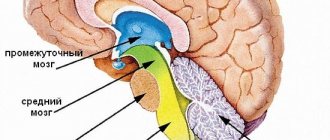
Are there places in the central nervous system where the blood-brain barrier does not exist? It would seem impossible: so much effort has been put into creating several levels of protection from external harmful substances. But it turns out that in some places the BBB does not constitute a single “wall” of protection, but there are holes in it. They are needed for those substances that are produced by the brain and sent to the periphery as commands: these are pituitary hormones. Therefore, there are free areas, just in the zone of the pituitary gland and epiphysis. They exist to allow hormones and neurotransmitters to pass freely into the blood.
There is another zone free from the BBB, which is located in the area of the rhomboid fossa or the bottom of the 4th ventricle of the brain. The vomiting center is located there. It is known that vomiting can occur not only due to mechanical irritation of the back wall of the pharynx, but also in the presence of toxins that have entered the blood . Therefore, it is in this area that there are special neurons that constantly “monitor” the quality of the blood for the presence of harmful substances.
As soon as their concentration reaches a certain value, these neurons are activated, causing a feeling of nausea and then vomiting. To be fair, it must be said that vomiting is not always associated with the concentration of harmful substances. Sometimes, with a significant increase in intracranial pressure (with hydrocephalus, meningitis), the vomiting center is activated due to direct excess pressure with the development of intracranial hypertension syndrome. Therefore, so-called central or cerebral vomiting develops, which can occur suddenly, and without any signs of nausea.
Functions
The blood-brain barrier is a formation within the brain that performs a protective function, which is expressed in preventing the passage of neurotoxic substances that negatively affect the structures of the central nervous system. Other functions are associated with the regulation of cerebral blood flow and metabolic processes in neurons and synaptic contacts.
One of the main tasks of the BBB is to retain substances coming from outside or formed in the body that can damage nerve cells. The blood-brain barrier cannot be called an insurmountable barrier to neurotoxic substances; the structure relatively protects the nervous tissue. The degree of reliability of the protective function depends on the following factors:
- The amount (level) of neurotoxic substances contained in the blood.
- General state of human health.
- The duration of presence of harmful substances in the body.
- Influence of the external environment.
The principle of operation of the BBB is based on the combined participation in the defense of cellular elements and intercellular contacts, which limit the movement of water-soluble compounds, slow down endocytosis (the process of capture of external material by the cell) and transcytosis (transfer of substances through the cell).
The blood-brain barrier coordinates the activity of brain structures, provides nutrition to nerve cells and removes secondary waste products. In physiology, the main function of the BBB is defined as maintaining homeostasis (self-regulation, the ability to ensure the constancy of the physiological state) of nervous tissue.
The BBB prevents endotoxins and exotoxins from entering the brain and ensures the transport of nutrients necessary for the functional activity of neurons, maintaining the energy balance of nerve cells and their plastic potential. The function of the BBB is to resist the effects of toxic compounds, preventing their penetration from the capillaries into the medulla.